![Immunohistochemistry-Paraffin: DC8 Antibody [NB100-2575]](https://resources.rndsystems.com/images/products/DC8-Antibody-Immunohistochemistry-Paraffin-NB100-2575-img0008.jpg "Immunohistochemistry-Paraffin: DC8 Antibody [NB100-2575]")
Key Product Details
Validated by
Independent Antibodies
Species Reactivity
Human
Applications
Immunohistochemistry, Immunohistochemistry-Paraffin, Immunoprecipitation, Western Blot (Negative)
Label
Unconjugated
Antibody Source
Polyclonal Rabbit IgG
Format
BSA Free
Loading...
Product Specifications
Immunogen
The immunogen recognized by this antibody maps to a region between residue 150 and 200 of human DC8 (C1orf48) using the numbering given in entry NP_056286.2 (GeneID 25936 ).
Clonality
Polyclonal
Host
Rabbit
Isotype
IgG
Scientific Data Images for DC8 Antibody - BSA Free
Immunohistochemistry-Paraffin: DC8 Antibody [NB100-2575]
Immunohistochemistry-Paraffin: DC8 Antibody [NB100-2575] - Detection of human DC8 by immunohistochemistry. Sample: FFPE section of human ovarian carcinoma. Antibody: Affinity purified rabbit anti-DC8 (NB100-2575) used at a dilution of 1:5,000 (0.2ug/ml). Detection: DAB![Immunohistochemistry-Paraffin: DC8 Antibody [NB100-2575]](https://resources.rndsystems.com/images/products/DC8-Antibody-Immunohistochemistry-Paraffin-NB100-2575-img0001.jpg "Immunohistochemistry-Paraffin: DC8 Antibody [NB100-2575]")
Immunohistochemistry-Paraffin: DC8 Antibody [NB100-2575]
Immunohistochemistry-Paraffin: DC8 Antibody [NB100-2575] - FFPE section of human breast carcinoma. Affinity purified rabbit anti-DC8 used at a dilution of 1:250.![Immunohistochemistry: DC8 Antibody [NB100-2575]](https://resources.rndsystems.com/images/products/DC8-Antibody-Immunohistochemistry-NB100-2575-img0005.jpg "Immunohistochemistry: DC8 Antibody [NB100-2575]")
Immunohistochemistry: DC8 Antibody [NB100-2575]
Immunohistochemistry: DC8 Antibody [NB100-2575] - Sample: FFPE section of human breast carcinoma. Antibody: Affinity purified rabbit anti-DC8 used at a dilution of 1:1,000 (1ug/ml). Detection: DAB![Immunohistochemistry: DC8 Antibody [NB100-2575]](https://resources.rndsystems.com/images/products/DC8-Antibody-Immunohistochemistry-NB100-2575-img0006.jpg "Immunohistochemistry: DC8 Antibody [NB100-2575]")
Immunohistochemistry: DC8 Antibody [NB100-2575]
Immunohistochemistry: DC8 Antibody [NB100-2575] - Sample: FFPE section of human breast carcinoma. Antibody: Affinity purified rabbit anti-DC8 used at a dilution of 1:1,000 (1ug/ml). Detection: DAB![Immunoprecipitation: DC8 Antibody [NB100-2575]](https://resources.rndsystems.com/images/products/DC8-Antibody-Immunoprecipitation-NB100-2575-img0004.jpg "Immunoprecipitation: DC8 Antibody [NB100-2575]")
Immunoprecipitation: DC8 Antibody [NB100-2575]
Immunoprecipitation: DC8 Antibody [NB100-2575] - Whole cell lysate (1 mg for IP, 20% of IP loaded) from HeLa cells. NB100-2575 used for IP at 3 mcg/mg lysate. DC8 was also immunoprecipitated by rabbit anti-DC8 antibodies NB100-2576.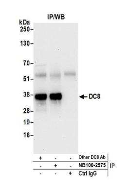
DC8 Antibody [NB100-2575] - Detection of human DC8 by western blot of immunoprecipitates. Samples: Whole cell lysate (0.5 or 1.0 mg per IP reaction; 20% of IP loaded) from HeLa cells prepared using NETN lysis buffer. Antibodies: Affinity purified rabbit anti- DC8 antibody NB100-2575 used for IP at 6 ug per reaction. DC8 was also immunoprecipitated by another rabbit anti-DC8 antibody. For blotting immunoprecipitated DC8, NB100-2575 was used at 1 ug/ml. Detection: Chemiluminescence with an exposure time of 30 seconds.
Applications for DC8 Antibody - BSA Free
Application
Recommended Usage
Immunohistochemistry
1:10-1:500
Immunohistochemistry-Paraffin
1:10-1:500
Immunoprecipitation
2-5 ug/mg lysate
Formulation, Preparation, and Storage
Purification
Immunogen affinity purified
Formulation
Tris-Citrate/Phosphate (pH 7.0 - 8.0)
Format
BSA Free
Preservative
0.09% Sodium Azide
Concentration
1.0 mg/ml
Shipping
The product is shipped with polar packs. Upon receipt, store it immediately at the temperature recommended below.
Stability & Storage
Store at 4C. Do not freeze.
Background: DC8
Alternate Names
C1orf48, chromosome 1 open reading frame 48, DC8, DKFZP566O1646, kinetochore-associated protein NSL1 homolog, MIS14, NSL1, MIND kinetochore complex component, homolog (S. cerevisiae)
Entrez Gene IDs
25936 (Human)
Gene Symbol
NSL1
UniProt
Additional DC8 Products
Product Documents for DC8 Antibody - BSA Free
Certificate of Analysis
To download a Certificate of Analysis, please enter a lot or batch number in the search box below.
Product Specific Notices for DC8 Antibody - BSA Free
This product is for research use only and is not approved for use in humans or in clinical diagnosis. Primary Antibodies are guaranteed for 1 year from date of receipt.
Customer Reviews for DC8 Antibody - BSA Free
There are currently no reviews for this product. Be the first to review DC8 Antibody - BSA Free and earn rewards!
Have you used DC8 Antibody - BSA Free?
Submit a review and receive an Amazon gift card!
$25/€18/£15/$25CAN/¥2500 Yen for a review with an image
$10/€7/£6/$10CAN/¥1110 Yen for a review without an image
Submit a review
Protocols
Find general support by application which include: protocols, troubleshooting, illustrated assays, videos and webinars.
- Antigen Retrieval Protocol (PIER)
- Antigen Retrieval for Frozen Sections Protocol
- Appropriate Fixation of IHC/ICC Samples
- Cellular Response to Hypoxia Protocols
- Chromogenic IHC Staining of Formalin-Fixed Paraffin-Embedded (FFPE) Tissue Protocol
- Chromogenic Immunohistochemistry Staining of Frozen Tissue
- ClariTSA™ Fluorophore Kits
- Detection & Visualization of Antibody Binding
- Fluorescent IHC Staining of Frozen Tissue Protocol
- Graphic Protocol for Heat-induced Epitope Retrieval
- Graphic Protocol for the Preparation and Fluorescent IHC Staining of Frozen Tissue Sections
- Graphic Protocol for the Preparation and Fluorescent IHC Staining of Paraffin-embedded Tissue Sections
- Graphic Protocol for the Preparation of Gelatin-coated Slides for Histological Tissue Sections
- IHC Sample Preparation (Frozen sections vs Paraffin)
- Immunofluorescent IHC Staining of Formalin-Fixed Paraffin-Embedded (FFPE) Tissue Protocol
- Immunohistochemistry (IHC) and Immunocytochemistry (ICC) Protocols
- Immunohistochemistry Frozen Troubleshooting
- Immunohistochemistry Paraffin Troubleshooting
- Immunoprecipitation Protocol
- Preparing Samples for IHC/ICC Experiments
- Preventing Non-Specific Staining (Non-Specific Binding)
- Primary Antibody Selection & Optimization
- Protocol for Heat-Induced Epitope Retrieval (HIER)
- Protocol for Making a 4% Formaldehyde Solution in PBS
- Protocol for VisUCyte™ HRP Polymer Detection Reagent
- Protocol for the Preparation & Fixation of Cells on Coverslips
- Protocol for the Preparation and Chromogenic IHC Staining of Frozen Tissue Sections
- Protocol for the Preparation and Chromogenic IHC Staining of Frozen Tissue Sections - Graphic
- Protocol for the Preparation and Chromogenic IHC Staining of Paraffin-embedded Tissue Sections
- Protocol for the Preparation and Chromogenic IHC Staining of Paraffin-embedded Tissue Sections - Graphic
- Protocol for the Preparation and Fluorescent IHC Staining of Frozen Tissue Sections
- Protocol for the Preparation and Fluorescent IHC Staining of Paraffin-embedded Tissue Sections
- Protocol for the Preparation of Gelatin-coated Slides for Histological Tissue Sections
- TUNEL and Active Caspase-3 Detection by IHC/ICC Protocol
- The Importance of IHC/ICC Controls
- Troubleshooting Guide: Immunohistochemistry
- View all Protocols, Troubleshooting, Illustrated assays and Webinars
Loading...