DDX21 Antibody - BSA Free
Novus Biologicals | Catalog # NB100-1718
![Immunocytochemistry/ Immunofluorescence: DDX21 Antibody - BSA Free [NB100-1718]](https://resources.rndsystems.com/images/products/DDX21-Antibody-Immunocytochemistry-Immunofluorescence-NB100-1718-img0015.jpg "Immunocytochemistry/ Immunofluorescence: DDX21 Antibody - BSA Free [NB100-1718]")
Key Product Details
Species Reactivity
Validated:
Human, Mouse, Zebrafish
Cited:
Human, Mouse, Porcine, Fish - Danio rerio (Zebrafish)
Predicted:
Canine (100%), Equine (100%), Porcine (100%), Primate (100%), Rat (100%), Rhesus Macaque (100%), Squirrel (100%). Backed by our 100% Guarantee.
Applications
Validated:
Immunohistochemistry, Immunohistochemistry-Paraffin, Western Blot, Flow Cytometry, Immunocytochemistry/ Immunofluorescence, Immunoprecipitation, Chromatin Immunoprecipitation, Chromatin Immunoprecipitation (ChIP), Knockdown Validated
Cited:
Western Blot, Immunocytochemistry/ Immunofluorescence, Chemotaxis, IF/IHC, Knockdown Validated
Label
Unconjugated
Antibody Source
Polyclonal Rabbit IgG
Format
BSA Free
Loading...
Product Specifications
Immunogen
The immunogen recognized by this antibody maps to a region between residue 725 and the C-terminus (residue 783) of human DEAD/H (Asp-Glu-Ala-Asp/His) Box Polypeptide 21 using the numbering given in entry NP_004719.2 (GeneID 9188).
Reactivity Notes
Based on 100% sequence identity, this antibody is predicted to react with Panda, Orangutan, Gorilla, Chimpanzee, African elephant, Northern white-cheeked gibbon, and Thirteen-lined ground squirrel. Use in Zebrafish reported in scientific literature (PMID:32231306).
Clonality
Polyclonal
Host
Rabbit
Isotype
IgG
Theoretical MW
87 kDa.
Disclaimer note: The observed molecular weight of the protein may vary from the listed predicted molecular weight due to post translational modifications, post translation cleavages, relative charges, and other experimental factors.
Disclaimer note: The observed molecular weight of the protein may vary from the listed predicted molecular weight due to post translational modifications, post translation cleavages, relative charges, and other experimental factors.
Scientific Data Images for DDX21 Antibody - BSA Free
Immunocytochemistry/ Immunofluorescence: DDX21 Antibody - BSA Free [NB100-1718]
Immunocytochemistry/Immunofluorescence: DDX21 Antibody [NB100-1718] - HeLa cells were fixed in 4% paraformaldehyde for 10 minutes and permeabilized in 0.5% Triton X-100 in PBS for 5 minutes. The cells were incubated with anti-DDX21 Antibody NB100-1718 at 2 ug/ml overnight at 4C and detected with an anti-rabbit Dylight 488 (Green) at a 1:1000 dilution for 60 minutes. Nuclei were counterstained with DAPI (Blue). Cells were imaged using a 100X objective and digitally deconvolved.![Flow Cytometry: DDX21 Antibody - BSA Free [NB100-1718]](https://resources.rndsystems.com/images/products/DDX21-Antibody-Flow-Cytometry-NB100-1718-img0011.jpg "Flow Cytometry: DDX21 Antibody - BSA Free [NB100-1718]")
Flow Cytometry: DDX21 Antibody - BSA Free [NB100-1718]
Flow Cytometry: DDX21 Antibody [NB100-1718] - An intracellular stain was performed on Jurkat Cells with DDX21Antibody NB100-1718 and a matched isotype control. Cells were fixed with 4% PFA and then permeablized with 0.1% saponin. Cells were incubated in an antibody dilution of 2.5 ug/mL for 30 minutes at room temperature, followed by Rabbit IgG APC-conjugated Secondary Antibody (R&D Systems, F0111).![Immunocytochemistry/ Immunofluorescence: DDX21 Antibody - BSA Free [NB100-1718]](https://resources.rndsystems.com/images/products/DDX21-Antibody-Immunocytochemistry-Immunofluorescence-NB100-1718-img0016.jpg "Immunocytochemistry/ Immunofluorescence: DDX21 Antibody - BSA Free [NB100-1718]")
Immunocytochemistry/ Immunofluorescence: DDX21 Antibody - BSA Free [NB100-1718]
Immunocytochemistry/Immunofluorescence: DDX21 Antibody [NB100-1718] - HeLa cells were fixed in 4% paraformaldehyde for 10 minutes and permeabilized in 0.05% Triton X-100 in PBS for 5 minutes. The cells were incubated with DDX21 Antibody conjugated to Alexa Fluor 488 (NB100-1718AF488) at 5 ug/ml for 1 hour at room temperature. Nuclei were counterstained with DAPI (Blue). Cells were imaged using a 100X objective and digitally deconvolved.![Immunocytochemistry/ Immunofluorescence: DDX21 Antibody - BSA Free [NB100-1718]](https://resources.rndsystems.com/images/products/DDX21-Antibody-Immunocytochemistry-Immunofluorescence-NB100-1718-img0010.jpg "Immunocytochemistry/ Immunofluorescence: DDX21 Antibody - BSA Free [NB100-1718]")
Immunocytochemistry/ Immunofluorescence: DDX21 Antibody - BSA Free [NB100-1718]
Immunocytochemistry/Immunofluorescence: DDX21 Antibody [NB100-1718] - ICC/IF detection of DDX21 in HeLa cells which were fixed for 10 minutes using 10% formalin and then permeabilized for 5 minutes using 1X TBS + 0.5% Triton X-100. The cells were incubated with anti-DDX21 [NB100-1718] at a 1:200 dilution overnight at 4C and detected with an anti-rabbit DylightTM 488 (green) at a 1:500 dilution. Alpha tubulin (DM1A) [NB100-690] was used as a co-stain at a 1:1000 dilution and detected with an anti-mouse DylightTM 550 (red) at a 1:500 dilution. Nuclei were counterstained with DAPI (blue) [NBP2-31156].![Immunocytochemistry/ Immunofluorescence: DDX21 Antibody - BSA Free [NB100-1718]](https://resources.rndsystems.com/images/products/DDX21-Antibody-Immunocytochemistry-Immunofluorescence-NB100-1718-img0017.jpg "Immunocytochemistry/ Immunofluorescence: DDX21 Antibody - BSA Free [NB100-1718]")
Immunocytochemistry/ Immunofluorescence: DDX21 Antibody - BSA Free [NB100-1718]
Immunocytochemistry/Immunofluorescence: DDX21 Antibody [NB100-1718] - HeLa cells were fixed in 4% paraformaldehyde for 10 minutes and permeabilized in 0.05% Triton X-100 in PBS for 5 minutes. The cells were incubated with conjugated to Alexa Fluor 647 (NB100-1718AF647) at 2 ug/ml for 1 hour at room temperature. Nuclei were counterstained with DAPI (Blue). Cells were imaged using a 100X objective and digitally deconvolved.![Immunocytochemistry/ Immunofluorescence: DDX21 Antibody - BSA Free [NB100-1718]](https://resources.rndsystems.com/images/products/DDX21-Antibody-Immunocytochemistry-Immunofluorescence-NB100-1718-img0013.jpg "Immunocytochemistry/ Immunofluorescence: DDX21 Antibody - BSA Free [NB100-1718]")
Immunocytochemistry/ Immunofluorescence: DDX21 Antibody - BSA Free [NB100-1718]
Immunocytochemistry/Immunofluorescence: DDX21 Antibody [NB100-1718] - PC12 cells were fixed for 10 minutes using 10% formalin and then permeabilized for 5 minutes using 1X PBS + 0.5% Triton-X100. The cells were incubated with anti-DDX21 at 2 ug/ml overnight at 4C and detected with an anti-rabbit Dylight 488 (Green) at a 1:500 dilution. Nuclei were counterstained with DAPI (Blue). Cells were imaged using a 40X objective.![Immunocytochemistry/ Immunofluorescence: DDX21 Antibody - BSA Free [NB100-1718]](https://resources.rndsystems.com/images/products/DDX21-Antibody-Immunocytochemistry-Immunofluorescence-NB100-1718-img0014.jpg "Immunocytochemistry/ Immunofluorescence: DDX21 Antibody - BSA Free [NB100-1718]")
Immunocytochemistry/ Immunofluorescence: DDX21 Antibody - BSA Free [NB100-1718]
Immunocytochemistry/Immunofluorescence: DDX21 Antibody [NB100-1718] - NIH-3T3 cells were fixed for 10 minutes using 10% formalin and then permeabilized for 5 minutes using 1X PBS + 0.5% Triton-X100. The cells were incubated with anti-DDX21 at 2 ug/ml overnight at 4C and detected with an anti-rabbit Dylight 488 (Green) at a 1:500 dilution. Nuclei were counterstained with DAPI (Blue). Cells were imaged using a 40X objective.![Western Blot: DDX21 AntibodyBSA Free [NB100-1718]](https://resources.rndsystems.com/images/products/DDX21-Antibody-Western-Blot-NB100-1718-img0004.jpg "Western Blot: DDX21 AntibodyBSA Free [NB100-1718]")
Western Blot: DDX21 AntibodyBSA Free [NB100-1718]
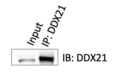
Western Blot: DDX21 Antibody [NB100-1718] - Detection of Human DDX21 on HeLa whole cell lystate using NB100-1718. DDX21 was also immunoprecipitated using rabbit anti-DDX21 antibodies NB100-1716 and NB100-1717.![Immunohistochemistry: DDX21 Antibody - BSA Free [NB100-1718]](https://resources.rndsystems.com/images/products/DDX21-Antibody-Immunohistochemistry-NB100-1718-img0007.jpg "Immunohistochemistry: DDX21 Antibody - BSA Free [NB100-1718]")
Immunohistochemistry: DDX21 Antibody - BSA Free [NB100-1718]
Immunohistochemistry: DDX21 Antibody [NB100-1718] - Sample: FFPE section of human breast carcinoma (left) and mouse squamous cell carcinoma (right). Antibody: Affinity purified rabbit anti-DDX21 used at a dilution of 1:200 (1ug/ml). Detection: DAB.![Flow Cytometry: DDX21 Antibody - BSA Free [NB100-1718]](https://resources.rndsystems.com/images/products/DDX21-Antibody---BSA-Free-Flow-Cytometry-NB100-1718-img0019.jpg "Flow Cytometry: DDX21 Antibody - BSA Free [NB100-1718]")
Flow Cytometry: DDX21 Antibody - BSA Free [NB100-1718]
Flow Cytometry: DDX21 Antibody - BSA Free [NB100-1718] - An intracellular stain was performed on A431 cells with DDX21 NB100-1718AF647 (blue) and a matched isotype control NBP2-24891 (orange). Cells were fixed with 4% PFA and then permeabilized with 0.1% saponin. Cells were incubated in an antibody dilution of 2.5 ug/mL for 30 minutes at room temperature. Both antibodies were conjugated to Alexa Fluor 647.![Immunohistochemistry-Paraffin: DDX21 Antibody - BSA Free [NB100-1718]](https://resources.rndsystems.com/images/products/DDX21-Antibody-Immunohistochemistry-Paraffin-NB100-1718-img0005.jpg "Immunohistochemistry-Paraffin: DDX21 Antibody - BSA Free [NB100-1718]")
Immunohistochemistry-Paraffin: DDX21 Antibody - BSA Free [NB100-1718]
Immunohistochemistry-Paraffin: DDX21 Antibody [NB100-1718] - FFPE section of human larynx squamous cell carcinoma. Antibody used at a dilution of 1:250. Detection: DAB staining using Immunohistochemistry Accessory Kit.![Immunohistochemistry-Paraffin: DDX21 Antibody - BSA Free [NB100-1718]](https://resources.rndsystems.com/images/products/DDX21-Antibody-Immunohistochemistry-Paraffin-NB100-1718-img0006.jpg "Immunohistochemistry-Paraffin: DDX21 Antibody - BSA Free [NB100-1718]")
Immunohistochemistry-Paraffin: DDX21 Antibody - BSA Free [NB100-1718]
Immunohistochemistry-Paraffin: DDX21 Antibody [NB100-1718] - FFPE section of mouse squamous cell carcinoma. Antibody used at a dilution of 1:250. Detection: DAB staining using Immunohistochemistry Accessory Kit.![Immunohistochemistry: DDX21 Antibody - BSA Free [NB100-1718]](https://resources.rndsystems.com/images/products/DDX21-Antibody-Immunohistochemistry-NB100-1718-img0008.jpg "Immunohistochemistry: DDX21 Antibody - BSA Free [NB100-1718]")
Immunohistochemistry: DDX21 Antibody - BSA Free [NB100-1718]
Immunohistochemistry: DDX21 Antibody [NB100-1718] - Sample: FFPE section of human breast carcinoma (left) and mouse squamous cell carcinoma (right). Antibody: Affinity purified rabbit anti-DDX21 used at a dilution of 1:200 (1ug/ml). Detection: DAB.![Flow Cytometry: DDX21 Antibody - BSA Free [NB100-1718]](https://resources.rndsystems.com/images/products/DDX21-Antibody-Flow-Cytometry-NB100-1718-img0018.jpg "Flow Cytometry: DDX21 Antibody - BSA Free [NB100-1718]")
Flow Cytometry: DDX21 Antibody - BSA Free [NB100-1718]
Flow Cytometry: DDX21 Antibody [NB100-1718] - An intracellular stain was performed on HeLa cells with DDX21 Antibody NB100-1718AF488 (blue) and a matched isotype control (orange). Cells were fixed with 4% PFA and then permeabilized with 0.1% saponin. Cells were incubated in an antibody dilution of 5 ug/mL for 30 minutes at room temperature. Both antibodies were conjugated to Alexa Fluor 488.
DDX21 in A431 Human Cell Line.
DDX21 was detected in immersion fixed A431 human skin carcinoma cell line using Rabbit anti-DDX21 Antigen Affinity Purified Polyclonal Antibody conjugated to Alexa Fluor® 647 (Catalog # NB100-1718AF647) (light blue) at 5 µg/mL overnight at 4C. Cells were counterstained with DAPI (blue). Cells were imaged using a 100X objective and digitally deconvolved.
DDX21 in NIH-3T3 Mouse Cell Line.
DDX21 was detected in immersion fixed NIH3T3 Mouse fibroblast cell line using Rabbit anti-DDX21 Antigen Affinity-purified Polyclonal Antibody conjugated to Alexa Fluor® 647 (Catalog # NB100-1718AF647) (light blue) at 5 µg/mL overnight at 4C. Cells were counterstained with DAPI (blue). Cells were imaged using a 100X objective and digitally deconvolved.
Western Blot: DDX21 Antibody - BSA Free [NB100-1718] -
PRRSV nsp1 beta upregulates DDX21 transcription and expression. (a,b) HEK-293T cells were transfected with 3 μg of plasmids expressing HA-tagged nsp1 alpha, nsp1 beta, nsp4, nsp12, or N protein. At 30 h post-transfection, the cells were collected to detect the mRNA expression of DDX21 by qRT-PCR (a) or DDX21 protein expression by Western blotting (b). (c–h) HEK-293T cells (c,d) and MARC-145 cells (e,f) were transfected with increasing amounts (0.1875 μg, 0.375 μg, 0.75 μg, 1.5 μg, 3 μg) of plasmid encoding HA-tagged nsp1 beta or empty vector (3 μg). iPAM cells (g,h) were transfected with increasing amounts (0.375 μg, 0.75 μg, 1.5 μg, 3 μg) of plasmid encoding HA-tagged nsp1 beta or empty vector (3 μg). At 30 h post-transfection, the cells were collected for use in qRT-PCR (c,e,g) or Western blotting (d,f,h). All experiments were performed in triplicate. The data are shown as the means +/- SD of three independent experiments (n.s, no significant differences; * p < 0.05; ** p < 0.01). The relative levels of DDX21 in experimentally transfected cells as compared with in empty vector-transfected cells were analyzed by ImageJ software, and the resulting ratio is displayed below the images as the fold change. Image collected and cropped by CiteAb from the following open publication (https://pubmed.ncbi.nlm.nih.gov/35336874), licensed under a CC-BY license. Not internally tested by Novus Biologicals.
Western Blot: DDX21 Antibody - BSA Free [NB100-1718] -
PRRSV infection promotes pDDX21 translocation from the nucleus to the cytoplasm. (a,b) iPAM cells were infected with PRRSV (MOI = 1). At 12, 24, and 36 hpi, these cells were collected for use in qRT-PCR to determine the number of mRNA copies of pDDX21 (a) or in Western blotting with antibodies against DDX21, PRRSV N protein, and beta -actin (b). (c,d) iPAM cells were infected with increasing doses of PRRSV (0.01 MOI, 0.1 MOI, or 1.0 MOI). At 24 hpi, these cells were collected for use in qRT-PCR (c) and Western blotting (d) as described in (a,b), respectively. (e) iPAM cells were grown until they formed a monolayer on 100-mm plates and then infected with PRRSV (MOI = 0.1). These cells were collected at 24 hpi, and a nuclear cytosol fractionation assay was performed to detect the pDDX21 expression in the nucleus or cytoplasm. Data are presented as the means +/- SD of three independent experiments (* p < 0.05; *** p < 0.001). The relative levels of pDDX21 in PRRSV-infected cells as compared with in mock-infected cells were analyzed by ImageJ software, and the ratio is displayed below the images as the fold change. Image collected and cropped by CiteAb from the following open publication (https://pubmed.ncbi.nlm.nih.gov/35336874), licensed under a CC-BY license. Not internally tested by Novus Biologicals.
Immunocytochemistry/ Immunofluorescence: DDX21 Antibody - BSA Free [NB100-1718] -
Survival motor neuron (SMN)–deficient motor neurons exhibit reduced DDX21 levels in a cell autonomous manner.(A) p75 enriched motor neurons derived from SMN delta 7 and wild type E13 embryos were labelled with DDX21 antibody, at DIV7. Scale bars represent 5 μm. (B, C, D) Nuclear, (C) nucleoplasmic and (D) foci fluorescence intensity of DDX21 staining normalised to control. Bar graphs of mean +/- s.d. *P < 0.05; paired two-tailed t test (P = 0.0417). (E) iPSC-derived motor neurons isolated from healthy individuals (csi4i and miff) and spinal muscular atrophy (SMA) type I patients (SMA86 and SMA32) were labelled with DDX21 antibody. Scale bars represent 5 μm. (F) Nuclear fluorescence intensity of DDX21 staining normalised to the average of control samples. (G) Nuclear fluorescence intensity where control samples (csi4i and miff) and SMA samples (SMA68 and SMA32) have been pulled together. (H) Nucleoplasmic fluorescence intensity of DDX21 staining normalised to the average of control samples. (I) Nucleoplasmic fluorescence intensity where control samples (csi4i and miff) and SMA samples (SMA68 and SMA32) have been pulled together. (J) Foci fluorescence intensity of DDX21 staining normalised to the average of control samples. (K) Foci fluorescence intensity where control samples (csi4i, miff) and SMA samples (SMA68, SMA32) have been pulled together. Bar graphs of mean +/- s.d. *P < 0.05; paired two-tailed t test (P = 0.0344). Image collected and cropped by CiteAb from the following open publication (https://pubmed.ncbi.nlm.nih.gov/35440492), licensed under a CC-BY license. Not internally tested by Novus Biologicals.
Immunocytochemistry/ Immunofluorescence: DDX21 Antibody - BSA Free [NB100-1718] -
DDX21 levels in spinal muscular atrophy (SMA) embryonic cortical neurons and fibroblasts derived from SMA patients.(A) SMA and control embryonic cortical neurons were labelled with DDX21 antibody, at DIV7. Scale bars represent 5 μm. (B, C, D) Nuclear (B), nucleoplasmic (C), and foci (D) fluorescence intensity of DDX21 staining is presented normalised to control. Bar graphs of mean +/- s.d (N = 3). ns, not significant (P > 0.05). (E) Fibroblasts derived from SMA type I patient (SMA) and a healthy individual (control) were doubled-stained with DDX21 and cyclin A1. Scale bars represent 10 μm. Cyclin A1–positive cells were excluded from the analysis. (F, G, H) Nuclear (F), nucleoplasmic (G), and foci (H) fluorescence intensity of DDX21 staining is presented normalised to control. Bar graphs of mean +/- s.d (N = 3). ns, not significant (P > 0.05). Image collected and cropped by CiteAb from the following open publication (https://pubmed.ncbi.nlm.nih.gov/35440492), licensed under a CC-BY license. Not internally tested by Novus Biologicals.Applications for DDX21 Antibody - BSA Free
Application
Recommended Usage
Chromatin Immunoprecipitation
reported in scientific literature (PMID 32231306)
Flow Cytometry
1:1000
Immunocytochemistry/ Immunofluorescence
1:50 - 1:500
Immunohistochemistry
1:10-1:500
Immunohistochemistry-Paraffin
1:10-1:500
Immunoprecipitation
2 - 10 ug/mg of lysate
Knockdown Validated
reported in scientific literature (PMID 35440492)
Western Blot
1:2000-1:10000
Reviewed Applications
Read 1 review rated 4 using NB100-1718 in the following applications:
Flow Cytometry Panel Builder
Bio-Techne Knows Flow Cytometry
Save time and reduce costly mistakes by quickly finding compatible reagents using the Panel Builder Tool.
Advanced Features
- Spectra Viewer - Custom analysis of spectra from multiple fluorochromes
- Spillover Popups - Visualize the spectra of individual fluorochromes
- Antigen Density Selector - Match fluorochrome brightness with antigen density

Formulation, Preparation, and Storage
Purification
Immunogen affinity purified
Formulation
PBS
Format
BSA Free
Preservative
0.02% Sodium Azide
Concentration
1.0 mg/ml
Shipping
The product is shipped with polar packs. Upon receipt, store it immediately at the temperature recommended below.
Stability & Storage
Store at 4C short term. Store at -20C long term. Avoid freeze-thaw cycles.
Background: DDX21
Alternate Names
DEAD (Asp-Glu-Ala-Asp) box polypeptide 21, DEAD box protein 21, DEAD/H (Asp-Glu-Ala-Asp/His) box polypeptide 21, DKFZp686F21172, EC 3.6.1, EC 3.6.4.13, Gu protein, GUA, Gu-alpha, GURDB, nucleolar RNA helicase 2, Nucleolar RNA helicase Gu, Nucleolar RNA helicase II, RH II/Gu, RH-II/GU, RH-II/GuA, RNA helicase II/Gu alpha
Gene Symbol
DDX21
UniProt
Additional DDX21 Products
Product Documents for DDX21 Antibody - BSA Free
Certificate of Analysis
To download a Certificate of Analysis, please enter a lot or batch number in the search box below.
Product Specific Notices for DDX21 Antibody - BSA Free
This product is for research use only and is not approved for use in humans or in clinical diagnosis. Primary Antibodies are guaranteed for 1 year from date of receipt.
Citations for DDX21 Antibody - BSA Free
 Powered by Bioz
Customer Reviews for DDX21 Antibody - BSA Free (1)
4 out of 5
1 Customer Rating
Have you used DDX21 Antibody - BSA Free?
Submit a review and receive an Amazon gift card!
$25/€18/£15/$25CAN/¥2500 Yen for a review with an image
$10/€7/£6/$10CAN/¥1110 Yen for a review without an image
Submit a review
Customer Images
Showing
1
-
1 of
1 review
Showing All
Filter By:
-
Application: Western BlotSample Tested: hTERT RPE-1 cell line from ATCCSpecies: HumanVerified Customer | Posted 03/13/2024NB100-1718 antibody for WB to check the IP DDX21 efficiency in WT hTERT RPE1 cell lysates.

There are no reviews that match your criteria.
Protocols
View specific protocols for DDX21 Antibody - BSA Free (NB100-1718):
Protocol for Flow Cytometry Intracellular Staining
Sample Preparation.
1. Grow cells to 60-85% confluency. Flow cytometry requires between 2 x 10⁵ and 1 x 10⁶ cells for optimal performance.
2. If cells are adherent, harvest gently by washing once with staining buffer and then scraping. Avoid using trypsin as this can disrupt certain epitopes of interest. If enzymatic harvest is required, use Accutase, Collagenase, or TrypLE Express for a less damaging option.
3. Reserve 100 uL for counting, then transfer cell volume into a 50 mL conical tube and centrifuge for 8 minutes at 400 RCF.
a. Count cells using a hemocytometer and a 1:1 trypan blue exclusion stain to determine cell viability before starting the flow protocol. If cells appear blue, do not proceed.
4. Re-suspend cells to a concentration of 1 x 10⁶ cells/mL in staining buffer (NBP2-26247).
5. Aliquot out 1 mL samples in accordance with your experimental samples.
Tip: When cell surface and intracellular staining are required in the same sample, it is advisable that the cell surface staining be performed first since the fixation and permeablization steps might reduce the availability of surface antigens.
Intracellular Staining.
Tip: When performing intracellular staining, it is important to use appropriate fixation and permeabilization reagents based upon the target and its subcellular location. Generally, our Intracellular Flow Assay Kit (NBP2-29450) is a good place to start as it contains an optimized combination of reagents for intracellular staining as well as an inhibitor of intracellular protein transport (necessary if staining secreted proteins). Certain targets may require more gentle or transient permeabilization protocols such as the commonly employed methanol or saponin-based methods.
Protocol for Cytoplasmic Targets:
Optional: Perform cell surface staining as described in the previous section.
1. Fix the cells by adding 100 uL fixation solution (such as 4% PFA) to each sample for 10-15 minutes.
2. Permeabilize cells by adding 100 uL of a permeabization buffer to every 1 x 10⁶ cells present in the sample. Mix well and incubate at room temperature for 15 minutes.
a. For cytoplasmic targets, use a gentle permeabilization solution such as 1X PBS + 0.5% Saponin or 1X PBS + 0.5% Tween-20.
b. To maintain the permeabilized state throughout your experiment, use staining buffer + 0.1% of the permeabilization reagent (i.e. 0.1% Tween-20 or 0.1% Saponin).
3. Following the 15 minute incubation, add 2 mL of the staining buffer + 0.1% permeabilizer to each sample.
4. Centrifuge for 5 minutes at 400 RCF.
5. Discard supernatant and re-suspend in 1 mL of staining buffer + 0.1% permeabilizer.
6. Stain each sample at 1 uL/ 1 x 10⁶ cells of primary antibody or 1-3 uL/ 1 x 10⁶ cells for directly conjugated antibodies. Mix well and incubate at room temperature for 30 minutes- 1 hour. Gently mix samples every 10-15 minutes.
7. Following the primary/conjugate incubation, add 2 mL/sample of staining buffer +0.1% permeabilizer and centrifuge for 5 minutes at 400 RCF.
8. Remove supernatant and re-suspend each sample in 2 mL staining buffer + 0.1% permeabilizer, repeat wash for 5 minutes at 400 RCF.
9. If using a directly conjugated antibody, after the second wash, re-suspend cell pellet to a final volume of 500 uL per sample and proceed with flow analysis.
Immunocytochemistry Protocol
Culture cells to appropriate density in 35 mm culture dishes or 6-well plates.
1. Remove culture medium and wash the cells briefly in PBS. Add 10% formalin to the dish and fix at room temperature for 10 minutes.
2. Remove the formalin and wash the cells in PBS.
3. Permeablize the cells with 0.1% Triton X100 or other suitable detergent for 10 min.
4. Remove the permeablization buffer and wash three times for 10 minutes each in PBS. Be sure to not let the specimen dry out.
5. To block nonspecific antibody binding, incubate in 10% normal goat serum from 1 hour to overnight at room temperature.
6. Add primary antibody at appropriate dilution and incubate overnight at 4C.
7. Remove primary antibody and replace with PBS. Wash three times for 10 minutes each.
8. Add secondary antibody at appropriate dilution. Incubate for 1 hour at room temperature.
9. Remove secondary antibody and replace with PBS. Wash three times for 10 minutes each.
10. Counter stain DNA with DAPi if required.
Immunohistochemistry-Paraffin Embedded Sections
Antigen Unmasking:
Bring slides to a boil in 10 mM sodium citrate buffer (pH 6.0) then maintain at a sub-boiling temperature for 10 minutes. Cool slides on bench-top for 30 minutes (keep slides in the sodium citrate buffer at all times).
Staining:
1. Wash sections in deionized water three times for 5 minutes each.
2. Wash sections in PBS for 5 minutes.
3. Block each section with 100-400 ul blocking solution (1% BSA in PBS) for 1 hour at room temperature.
4. Remove blocking solution and add 100-400 ul diluted primary antibody. Incubate overnight at 4 C.
5. Remove antibody solution and wash sections in wash buffer three times for 5 minutes each.
6. Add 100-400 ul HRP polymer conjugated secondary antibody. Incubate 30 minutes at room temperature.
7. Wash sections three times in wash buffer for 5 minutes each.
8. Add 100-400 ul DAB substrate to each section and monitor staining closely.
9. As soon as the sections develop, immerse slides in deionized water.
10. Counterstain sections in hematoxylin.
11. Wash sections in deionized water two times for 5 minutes each.
12. Dehydrate sections.
13. Mount coverslips.
Western Blot Protocol
1. Perform SDS-PAGE on samples to be analyzed, loading 10-25 ug of total protein per lane.
2. Transfer proteins to PVDF membrane according to the instructions provided by the manufacturer of the membrane and transfer apparatus.
3. Stain the membrane with Ponceau S (or similar product) to assess transfer success, and mark molecular weight standards where appropriate.
4. Rinse the blot TBS -0.05% Tween 20 (TBST).
5. Block the membrane in 5% Non-fat milk in TBST (blocking buffer) for at least 1 hour.
6. Wash the membrane in TBST three times for 10 minutes each.
7. Dilute primary antibody in blocking buffer and incubate overnight at 4C with gentle rocking.
8. Wash the membrane in TBST three times for 10 minutes each.
9. Incubate the membrane in diluted HRP conjugated secondary antibody in blocking buffer (as per manufacturer's instructions) for 1 hour at room temperature.
10. Wash the blot in TBST three times for 10 minutes each (this step can be repeated as required to reduce background).
11. Apply the detection reagent of choice in accordance with the manufacturer's instructions.
Find general support by application which include: protocols, troubleshooting, illustrated assays, videos and webinars.
- 7-Amino Actinomycin D (7-AAD) Cell Viability Flow Cytometry Protocol
- Antigen Retrieval Protocol (PIER)
- Antigen Retrieval for Frozen Sections Protocol
- Appropriate Fixation of IHC/ICC Samples
- Cellular Response to Hypoxia Protocols
- ChIP Protocol Video
- Chromatin Immunoprecipitation (ChIP) Protocol
- Chromatin Immunoprecipitation Protocol
- Chromogenic IHC Staining of Formalin-Fixed Paraffin-Embedded (FFPE) Tissue Protocol
- Chromogenic Immunohistochemistry Staining of Frozen Tissue
- ClariTSA™ Fluorophore Kits
- Detection & Visualization of Antibody Binding
- Extracellular Membrane Flow Cytometry Protocol
- Flow Cytometry Protocol for Cell Surface Markers
- Flow Cytometry Protocol for Staining Membrane Associated Proteins
- Flow Cytometry Staining Protocols
- Flow Cytometry Troubleshooting Guide
- Fluorescent IHC Staining of Frozen Tissue Protocol
- Graphic Protocol for Heat-induced Epitope Retrieval
- Graphic Protocol for the Preparation and Fluorescent IHC Staining of Frozen Tissue Sections
- Graphic Protocol for the Preparation and Fluorescent IHC Staining of Paraffin-embedded Tissue Sections
- Graphic Protocol for the Preparation of Gelatin-coated Slides for Histological Tissue Sections
- ICC Cell Smear Protocol for Suspension Cells
- ICC Immunocytochemistry Protocol Videos
- ICC for Adherent Cells
- IHC Sample Preparation (Frozen sections vs Paraffin)
- Immunocytochemistry (ICC) Protocol
- Immunocytochemistry Troubleshooting
- Immunofluorescence of Organoids Embedded in Cultrex Basement Membrane Extract
- Immunofluorescent IHC Staining of Formalin-Fixed Paraffin-Embedded (FFPE) Tissue Protocol
- Immunohistochemistry (IHC) and Immunocytochemistry (ICC) Protocols
- Immunohistochemistry Frozen Troubleshooting
- Immunohistochemistry Paraffin Troubleshooting
- Immunoprecipitation Protocol
- Intracellular Flow Cytometry Protocol Using Alcohol (Methanol)
- Intracellular Flow Cytometry Protocol Using Detergents
- Intracellular Nuclear Staining Flow Cytometry Protocol Using Detergents
- Intracellular Staining Flow Cytometry Protocol Using Alcohol Permeabilization
- Intracellular Staining Flow Cytometry Protocol Using Detergents to Permeabilize Cells
- Preparing Samples for IHC/ICC Experiments
- Preventing Non-Specific Staining (Non-Specific Binding)
- Primary Antibody Selection & Optimization
- Propidium Iodide Cell Viability Flow Cytometry Protocol
- Protocol for Heat-Induced Epitope Retrieval (HIER)
- Protocol for Liperfluo
- Protocol for Making a 4% Formaldehyde Solution in PBS
- Protocol for VisUCyte™ HRP Polymer Detection Reagent
- Protocol for the Characterization of Human Th22 Cells
- Protocol for the Characterization of Human Th9 Cells
- Protocol for the Fluorescent ICC Staining of Cell Smears - Graphic
- Protocol for the Fluorescent ICC Staining of Cultured Cells on Coverslips - Graphic
- Protocol for the Preparation & Fixation of Cells on Coverslips
- Protocol for the Preparation and Chromogenic IHC Staining of Frozen Tissue Sections
- Protocol for the Preparation and Chromogenic IHC Staining of Frozen Tissue Sections - Graphic
- Protocol for the Preparation and Chromogenic IHC Staining of Paraffin-embedded Tissue Sections
- Protocol for the Preparation and Chromogenic IHC Staining of Paraffin-embedded Tissue Sections - Graphic
- Protocol for the Preparation and Fluorescent ICC Staining of Cells on Coverslips
- Protocol for the Preparation and Fluorescent ICC Staining of Non-adherent Cells
- Protocol for the Preparation and Fluorescent ICC Staining of Stem Cells on Coverslips
- Protocol for the Preparation and Fluorescent IHC Staining of Frozen Tissue Sections
- Protocol for the Preparation and Fluorescent IHC Staining of Paraffin-embedded Tissue Sections
- Protocol for the Preparation of Gelatin-coated Slides for Histological Tissue Sections
- Protocol for the Preparation of a Cell Smear for Non-adherent Cell ICC - Graphic
- Protocol: Annexin V and PI Staining by Flow Cytometry
- Protocol: Annexin V and PI Staining for Apoptosis by Flow Cytometry
- R&D Systems Quality Control Western Blot Protocol
- TUNEL and Active Caspase-3 Detection by IHC/ICC Protocol
- The Importance of IHC/ICC Controls
- Troubleshooting Guide: Fluorokine Flow Cytometry Kits
- Troubleshooting Guide: Immunohistochemistry
- Troubleshooting Guide: Western Blot Figures
- Western Blot Conditions
- Western Blot Protocol
- Western Blot Protocol for Cell Lysates
- Western Blot Troubleshooting
- Western Blot Troubleshooting Guide
- View all Protocols, Troubleshooting, Illustrated assays and Webinars
Loading...