Decorin Antibody - BSA Free
Novus Biologicals | Catalog # NBP1-57923
![Western Blot: Decorin Antibody [NBP1-57923]](https://resources.rndsystems.com/images/products/Decorin-Antibody-Western-Blot-NBP1-57923-img0006.jpg "Western Blot: Decorin Antibody [NBP1-57923]")
Loading...
Key Product Details
Species Reactivity
Validated:
Human, Canine
Cited:
Canine
Applications
Validated:
Immunohistochemistry, Immunohistochemistry-Paraffin, Western Blot, Immunocytochemistry/ Immunofluorescence
Cited:
Western Blot
Label
Unconjugated
Antibody Source
Polyclonal Rabbit IgG
Format
BSA Free
Loading...
Product Specifications
Immunogen
Synthetic peptides corresponding to DCN(decorin) The peptide sequence was selected from the N terminal of DCN. Peptide sequence IGPEVPDDRDFEPSLGPVCPFRCQCHLRVVQCSDLGLDKVPKDLPPDTTL. The peptide sequence for this immunogen was taken from within the described region.
Reactivity Notes
Canine reactivity reported in scientific literature (PMID: 26341258).
Clonality
Polyclonal
Host
Rabbit
Isotype
IgG
Theoretical MW
36 kDa.
Disclaimer note: The observed molecular weight of the protein may vary from the listed predicted molecular weight due to post translational modifications, post translation cleavages, relative charges, and other experimental factors.
Disclaimer note: The observed molecular weight of the protein may vary from the listed predicted molecular weight due to post translational modifications, post translation cleavages, relative charges, and other experimental factors.
Description
The addition of 50% glycerol is optional for those storing this antibody at -20C and not aliquoting smaller units. However, please note that glycerol may interrupt some downstream antibody applications and should be added with caution.
Scientific Data Images for Decorin Antibody - BSA Free
Western Blot: Decorin Antibody [NBP1-57923]
Western Blot: Decorin Antibody [NBP1-57923] - Antibody Titration: 0.2-1 ug/ml ELISA Titer: 1:312500 Positive Control: Human Placenta![Immunohistochemistry: Decorin Antibody [NBP1-57923]](https://resources.rndsystems.com/images/products/Decorin-Antibody-Immunohistochemistry-NBP1-57923-img0010.jpg "Immunohistochemistry: Decorin Antibody [NBP1-57923]")
Immunohistochemistry: Decorin Antibody [NBP1-57923]
Decorin-Antibody-Immunohistochemistry-NBP1-57923-img0010.jpg![Western Blot: Decorin Antibody [NBP1-57923]](https://resources.rndsystems.com/images/products/Decorin-Antibody-Western-Blot-NBP1-57923-img0008.jpg "Western Blot: Decorin Antibody [NBP1-57923]")
Western Blot: Decorin Antibody [NBP1-57923]
Decorin-Antibody-Western-Blot-NBP1-57923-img0008.jpg![Immunocytochemistry/ Immunofluorescence: Decorin Antibody [NBP1-57923]](https://resources.rndsystems.com/images/products/Decorin-Antibody-Immunocytochemistry-Immunofluorescence-NBP1-57923-img0012.jpg "Immunocytochemistry/ Immunofluorescence: Decorin Antibody [NBP1-57923]")
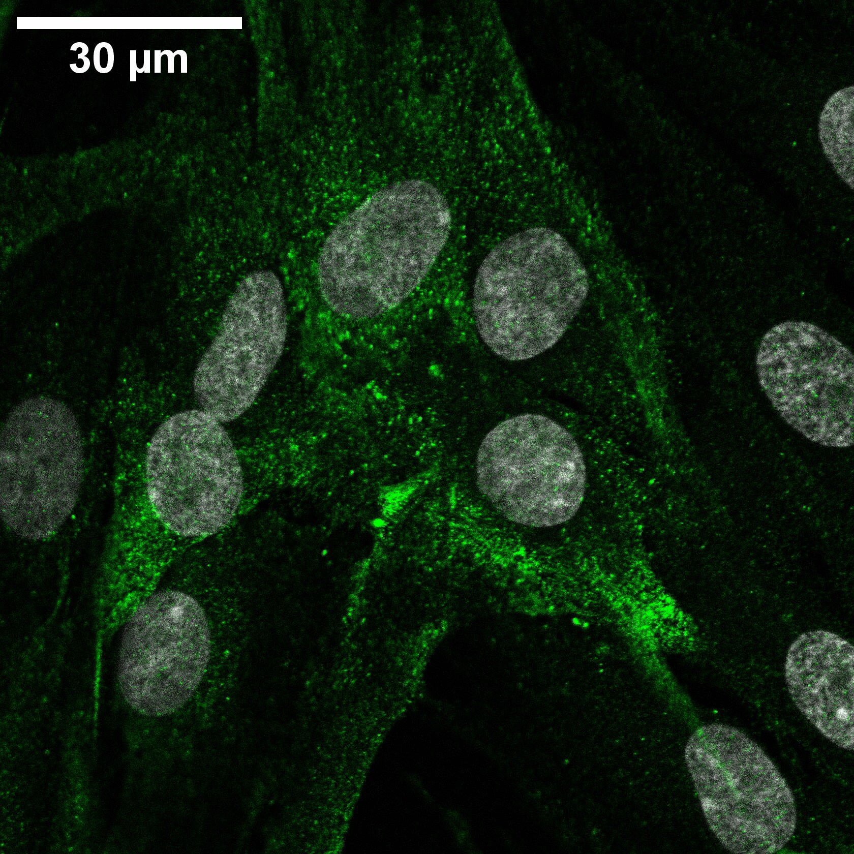
Immunocytochemistry/ Immunofluorescence: Decorin Antibody [NBP1-57923]
Immunocytochemistry/Immunofluorescence: Decorin Antibody [NBP1-57923] - Human periodontal ligament cells stained with rabbit anti-decorin 1:300 and goat anti-rabbit Alexa 488 1:500. Nuclei stained with DAPI. ICC/IF image submitted by a verified customer review.![Immunohistochemistry-Paraffin: Decorin Antibody [NBP1-57923]](https://resources.rndsystems.com/images/products/Decorin-Antibody-Immunohistochemistry-Paraffin-NBP1-57923-img0005.jpg "Immunohistochemistry-Paraffin: Decorin Antibody [NBP1-57923]")
Immunohistochemistry-Paraffin: Decorin Antibody [NBP1-57923]
Immunohistochemistry-Paraffin: Decorin Antibody [NBP1-57923] - Human skin tissue at an antibody concentration of 4-8 ug/mL.
Immunohistochemistry: Decorin Antibody [NBP1-57923] -
Immunohistochemistry: Decorin Antibody [NBP1-57923] - Comparative immunohistochemical analysis of non-chondrodystrophic (NCD) & chondrodystrophic (CD) canine nucleus pulposus (NP) expression & distribution of decorin, biglycan, fibromodulin, hyaluronan & proteoglycan link protein 1 (HAPLN1), & aggrecan. For all extracellular matrix (ECM) proteins, the NCD canine intervertebral disc NP reveals a cobweb appearance, demonstrating intense staining for all proteins located in the areas tightly between the cells. Immunostaining with decorin reveals diffuse intercellular staining with negative immunostaining within the large, physaliferous-appearing notochordal cells. This presentation is in contrast to the abundant staining of these proteins in every CD NP sample. Decorin, biglycan, & HAPLN1 reveal intense staining within the ECM, with abundant clusters of small numbers of cells present within the NP. Although the ECM staining is less intense than that for the other three proteins, fibromodulin & aggrecan immunostaining is present. Furthermore, the CD NP stained for aggrecan reveals intense pericellular immunostaining diffusely throughout the ECM that is much less cellular than the NCD NP staining. Safranin-O staining shows quite intense ECM staining in the CD NP, whereas the NCD sample demonstrates intense intercellular staining without large, acellular ECM areas rich in proteoglycan staining. The overall appearance of the CD NP bears a strong resemblance to a fibrocartilaginous phenotype that is distinctly different from the NCD canine NP Image collected & cropped by CiteAb from the following publication (https://pubmed.ncbi.nlm.nih.gov/26341258), licensed under a CC-BY license. Not internally tested by Novus Biologicals.Applications for Decorin Antibody - BSA Free
Application
Recommended Usage
Immunohistochemistry
1:10-1:500
Immunohistochemistry-Paraffin
1:10-1:500
Western Blot
1.0 ug/ml
Application Notes
This Decorin Antibody is validated for ICC/IF from a verified customer review.
Reviewed Applications
Read 1 review rated 4 using NBP1-57923 in the following applications:
Formulation, Preparation, and Storage
Purification
Affinity purified
Formulation
PBS, 2% Sucrose
Format
BSA Free
Preservative
0.09% Sodium Azide
Concentration
0.5 mg/ml
Shipping
The product is shipped with polar packs. Upon receipt, store it immediately at the temperature recommended below.
Stability & Storage
Store at 4C short term. Aliquot and store at -20C long term. Avoid freeze-thaw cycles.
Background: Decorin
Additional Decorin Products
Product Documents for Decorin Antibody - BSA Free
Certificate of Analysis
To download a Certificate of Analysis, please enter a lot or batch number in the search box below.
Product Specific Notices for Decorin Antibody - BSA Free
This product is for research use only and is not approved for use in humans or in clinical diagnosis. Primary Antibodies are guaranteed for 1 year from date of receipt.
Related Research Areas
Citations for Decorin Antibody - BSA Free
 Powered by Bioz
Customer Reviews for Decorin Antibody - BSA Free (1)
4 out of 5
1 Customer Rating
Have you used Decorin Antibody - BSA Free?
Submit a review and receive an Amazon gift card!
$25/€18/£15/$25CAN/¥2500 Yen for a review with an image
$10/€7/£6/$10CAN/¥1110 Yen for a review without an image
Submit a review
Customer Images
Showing
1
-
1 of
1 review
Showing All
Filter By:
-
Application: ImmunocytochemistrySample Tested: Cell cultureSpecies: HumanVerified Customer | Posted 07/29/2021Human periodontal ligament cells stained with rabbit anti-decorin 1:300 and goat anti-rabbit Alexa 488 1:500. Nuclei stained with DAPI.

There are no reviews that match your criteria.
Protocols
Find general support by application which include: protocols, troubleshooting, illustrated assays, videos and webinars.
- Antigen Retrieval Protocol (PIER)
- Antigen Retrieval for Frozen Sections Protocol
- Appropriate Fixation of IHC/ICC Samples
- Cellular Response to Hypoxia Protocols
- Chromogenic IHC Staining of Formalin-Fixed Paraffin-Embedded (FFPE) Tissue Protocol
- Chromogenic Immunohistochemistry Staining of Frozen Tissue
- ClariTSA™ Fluorophore Kits
- Detection & Visualization of Antibody Binding
- Fluorescent IHC Staining of Frozen Tissue Protocol
- Graphic Protocol for Heat-induced Epitope Retrieval
- Graphic Protocol for the Preparation and Fluorescent IHC Staining of Frozen Tissue Sections
- Graphic Protocol for the Preparation and Fluorescent IHC Staining of Paraffin-embedded Tissue Sections
- Graphic Protocol for the Preparation of Gelatin-coated Slides for Histological Tissue Sections
- ICC Cell Smear Protocol for Suspension Cells
- ICC Immunocytochemistry Protocol Videos
- ICC for Adherent Cells
- IHC Sample Preparation (Frozen sections vs Paraffin)
- Immunocytochemistry (ICC) Protocol
- Immunocytochemistry Troubleshooting
- Immunofluorescence of Organoids Embedded in Cultrex Basement Membrane Extract
- Immunofluorescent IHC Staining of Formalin-Fixed Paraffin-Embedded (FFPE) Tissue Protocol
- Immunohistochemistry (IHC) and Immunocytochemistry (ICC) Protocols
- Immunohistochemistry Frozen Troubleshooting
- Immunohistochemistry Paraffin Troubleshooting
- Preparing Samples for IHC/ICC Experiments
- Preventing Non-Specific Staining (Non-Specific Binding)
- Primary Antibody Selection & Optimization
- Protocol for Heat-Induced Epitope Retrieval (HIER)
- Protocol for Making a 4% Formaldehyde Solution in PBS
- Protocol for VisUCyte™ HRP Polymer Detection Reagent
- Protocol for the Fluorescent ICC Staining of Cell Smears - Graphic
- Protocol for the Fluorescent ICC Staining of Cultured Cells on Coverslips - Graphic
- Protocol for the Preparation & Fixation of Cells on Coverslips
- Protocol for the Preparation and Chromogenic IHC Staining of Frozen Tissue Sections
- Protocol for the Preparation and Chromogenic IHC Staining of Frozen Tissue Sections - Graphic
- Protocol for the Preparation and Chromogenic IHC Staining of Paraffin-embedded Tissue Sections
- Protocol for the Preparation and Chromogenic IHC Staining of Paraffin-embedded Tissue Sections - Graphic
- Protocol for the Preparation and Fluorescent ICC Staining of Cells on Coverslips
- Protocol for the Preparation and Fluorescent ICC Staining of Non-adherent Cells
- Protocol for the Preparation and Fluorescent ICC Staining of Stem Cells on Coverslips
- Protocol for the Preparation and Fluorescent IHC Staining of Frozen Tissue Sections
- Protocol for the Preparation and Fluorescent IHC Staining of Paraffin-embedded Tissue Sections
- Protocol for the Preparation of Gelatin-coated Slides for Histological Tissue Sections
- Protocol for the Preparation of a Cell Smear for Non-adherent Cell ICC - Graphic
- R&D Systems Quality Control Western Blot Protocol
- TUNEL and Active Caspase-3 Detection by IHC/ICC Protocol
- The Importance of IHC/ICC Controls
- Troubleshooting Guide: Immunohistochemistry
- Troubleshooting Guide: Western Blot Figures
- Western Blot Conditions
- Western Blot Protocol
- Western Blot Protocol for Cell Lysates
- Western Blot Troubleshooting
- Western Blot Troubleshooting Guide
- View all Protocols, Troubleshooting, Illustrated assays and Webinars
Loading...
Associated Pathways

