DNA/RNA Damage Antibody (15A3) - BSA Free
Novus Biologicals | Catalog # NB110-96878
![Immunocytochemistry/ Immunofluorescence: DNA/RNA Damage Antibody (15A3) [NB110-96878]](https://resources.rndsystems.com/images/products/DNA-RNA-Damage-Antibody-15A3-Immunocytochemistry-Immunofluorescence-NB110-96878-img0004.jpg "Immunocytochemistry/ Immunofluorescence: DNA/RNA Damage Antibody (15A3) [NB110-96878]")
Key Product Details
Species Reactivity
Validated:
Cited:
Applications
Validated:
Cited:
Label
Antibody Source
Format
Product Specifications
Immunogen
Reactivity Notes
Specificity
Clonality
Host
Isotype
Scientific Data Images for DNA/RNA Damage Antibody (15A3) - BSA Free
Immunocytochemistry/ Immunofluorescence: DNA/RNA Damage Antibody (15A3) [NB110-96878]
Immunocytochemistry/Immunofluorescence: DNA/RNA Damage Antibody (15A3) [NB110-96878] - visualized on a retinal injury![Immunohistochemistry: DNA/RNA Damage Antibody (15A3) [NB110-96878]](https://resources.rndsystems.com/images/products/DNA-RNA-Damage-Antibody-15A3-Immunohistochemistry-NB110-96878-img0013.jpg "Immunohistochemistry: DNA/RNA Damage Antibody (15A3) [NB110-96878]")
Immunohistochemistry: DNA/RNA Damage Antibody (15A3) [NB110-96878]
Immunohistochemistry: DNA/RNA Damage Antibody (15A3) [NB110-96878] - Immunohistochemistry analysis using Mouse Anti-DNA/RNA DamageMonoclonal Antibody, Clone 15A3 (NB110-96878). Tissue: inflamed colon. Species: Mouse. Fixation: Formalin. Primary Antibody: Mouse Anti-DNA/RNA DamageMonoclonal Antibody (NB110-96878) at 1:1000000 for 12 hours at 4C. Secondary Antibody: Biotin Goat Anti-Mouse at 1:2000 for 1 hour at RT. Counterstain: Mayer Hematoxylin (purple/blue) nuclear stain at 200 l for 2 minutes at RT. Magnification: 40x. With anti-microbial.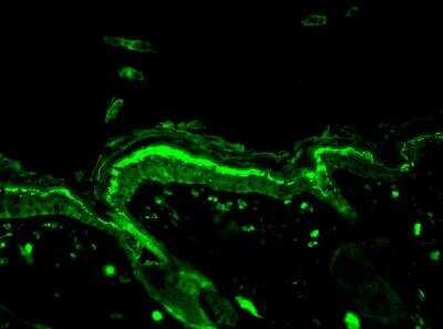
Applications for DNA/RNA Damage Antibody (15A3) - BSA Free
Immunohistochemistry
Immunohistochemistry-Paraffin
Flow Cytometry Panel Builder
Bio-Techne Knows Flow Cytometry
Save time and reduce costly mistakes by quickly finding compatible reagents using the Panel Builder Tool.
Advanced Features
- Spectra Viewer - Custom analysis of spectra from multiple fluorochromes
- Spillover Popups - Visualize the spectra of individual fluorochromes
- Antigen Density Selector - Match fluorochrome brightness with antigen density

Formulation, Preparation, and Storage
Purification
Formulation
Format
Preservative
Concentration
Shipping
Stability & Storage
Background: DNA/RNA Damage
Alternate Names
Additional DNA/RNA Damage Products
Product Documents for DNA/RNA Damage Antibody (15A3) - BSA Free
Certificate of Analysis
To download a Certificate of Analysis, please enter a lot or batch number in the search box below.
Product Specific Notices for DNA/RNA Damage Antibody (15A3) - BSA Free
This product is for research use only and is not approved for use in humans or in clinical diagnosis. Primary Antibodies are guaranteed for 1 year from date of receipt.
Citations for DNA/RNA Damage Antibody (15A3) - BSA Free
 Powered by Bioz
Customer Reviews for DNA/RNA Damage Antibody (15A3) - BSA Free
There are currently no reviews for this product. Be the first to review DNA/RNA Damage Antibody (15A3) - BSA Free and earn rewards!
Have you used DNA/RNA Damage Antibody (15A3) - BSA Free?
Submit a review and receive an Amazon gift card!
$25/€18/£15/$25CAN/¥2500 Yen for a review with an image
$10/€7/£6/$10CAN/¥1110 Yen for a review without an image
Submit a review
Protocols
View specific protocols for DNA/RNA Damage Antibody (15A3) - BSA Free (NB110-96878):
Tissue Preparation
8-OHdG monoclonal antibody reacts on both 50 um frozen tissue sections and paraffin-embedded sections. Tissue should be dissected fresh and fixed in periodate-lysine-paraformaldehyde (PLP) at 4C over night.
PLP
Heat 1 L dH2O to 60C.
Add 60 g paraformaldehyde.
Add 33 g dibasic NaPO4.
Cool to room temperature in a cold water bath.
Add 9 g monobasic NaPO4.
Add 6.45 g Na-m-periodate.
Add 41.1 g lysine (HCl salt).
Filter and dilute to 3 L with dH2O.
Adjust pH to 7.6 with 1.0 N NaOH approx. (20-30 ml).
Tissue prepared for frozen sectioning must be cryoprotected in a 20% glycerol-2% DMSO solution in phosphate buffer for 24-48 hours. Tissue will sink to the bottom of container when fully penetrated. This will eliminate freezing artifact from cutting.
Glycerol-DMSO (for 3 L)
2.4 L 0.1 M phosphate buffer
600 ml glycerol
60 ml DMSO
0.1 M Phosphate Buffer, pH 7.4 (for 1 L)
1 L dH2O
11 g dibasic NaPO4
3 g monobasic NaPO4
After frozen sectioning, tissue should be stored in phosphate buffer with 0.08% sodium azide.
Staining Sections By DAB Procedure
Paraffin-embedded sections must be deparaffinized by sequential immersion in the following for 3 minutes each: xylene (twice), absolute ethanol (twice). Agitate gently in each solution. Proceed with the following procedure.
1. Pretreat sections with a methanol-peroxide solution to eliminate endogenous peroxidases.
Methanol-Peroxide
100 ml absolute methanol
1 ml 33% H2O2
Incubate sections in methanol-peroxide solution for 30 minutes, room temperature.
2. Wash sections 3 times for 10 minutes each in 0.1 M phosphate buffered saline (PBS)
PBS, pH 7.4 (for 1 L)
1 L dH2O
11 g dibasic NaPO4
3 g monobasic NaPO4
8.5 g NaCl
3. Incubate sections for 1 hour in 10% normal goat serum in PBS.
4. Incubate sections in the primary antibody for 18-24 hours at room temperature. Depending on the nature of the sample, a shorter incubation time may be used. It is recommended that a concentration range of 1-10 ug/ml be evaluated in order to determine the optimal concentration for each type of tissue sample. Dilute antibody in PBS containing 0.3% Triton X-100, 0.08% sodium azide and 2% normal goat serum.
NOTE: A humidified chamber is necessary when staining paraffin sections. Slides should be placed flat and primary antibody applied over the section, covering it completely.
5. Rinse sections 3 times for 10 minutes each in PBS.
6. Incubate for 3 hours with peroxidase-conjugated goat anti-mouse IgG (Boehringer-Mannheim, Indianapolis, IN) diluted 1:300 in PBS with 2% normal goat serum.
7. Rinse sections 3 times for 10 minutes each in PBS.
8. Incubate sections for 5-10 minutes in a solution of 0.5 mg/ml 3,3' diamino-benzidine tetrahydrochloride (DAB, Sigma, St. Louis, MO) and 0.005% hydrogen peroxide in 0.05 M tris HCl buffer, pH 7.6 plus imidazole (10 ml/110 ml Tris buffer).
50 mM Tris Buffer, pH 7.6
1 L dH2O
6 g Trizma base
3 ml concentrated HCl (37%)
Sodium Imidazole
100 ml 0.1 M phosphate buffer
0.7 g sodium imidazole
9. Rinse sections 3 times for 10 minutes each in PBS.
10. Mount free-floating sections on subbed slides and air dry.
Subbing Solution
500 ml dH2O
2.5 g gelatin
0.25 g chromium potassium sulfate
Heat to 60C. Filter and proceed to coat slides. Once slides are air dried, sections can be mounted.
11. Dehydrate mounted/paraffin sections by sequential immersion in the following for 3 minutes each: 70% ethanol, 95% ethanol, absolute ethanol, xylene. Agitate gently in each solution.
12. Apply coverslip with Permount in a chemical fume hood
Find general support by application which include: protocols, troubleshooting, illustrated assays, videos and webinars.
- 7-Amino Actinomycin D (7-AAD) Cell Viability Flow Cytometry Protocol
- Antigen Retrieval Protocol (PIER)
- Antigen Retrieval for Frozen Sections Protocol
- Appropriate Fixation of IHC/ICC Samples
- Cellular Response to Hypoxia Protocols
- Chromogenic IHC Staining of Formalin-Fixed Paraffin-Embedded (FFPE) Tissue Protocol
- Chromogenic Immunohistochemistry Staining of Frozen Tissue
- ClariTSA™ Fluorophore Kits
- Detection & Visualization of Antibody Binding
- ELISA Sample Preparation & Collection Guide
- ELISA Troubleshooting Guide
- Extracellular Membrane Flow Cytometry Protocol
- Flow Cytometry Protocol for Cell Surface Markers
- Flow Cytometry Protocol for Staining Membrane Associated Proteins
- Flow Cytometry Staining Protocols
- Flow Cytometry Troubleshooting Guide
- Fluorescent IHC Staining of Frozen Tissue Protocol
- Graphic Protocol for Heat-induced Epitope Retrieval
- Graphic Protocol for the Preparation and Fluorescent IHC Staining of Frozen Tissue Sections
- Graphic Protocol for the Preparation and Fluorescent IHC Staining of Paraffin-embedded Tissue Sections
- Graphic Protocol for the Preparation of Gelatin-coated Slides for Histological Tissue Sections
- How to Run an R&D Systems DuoSet ELISA
- How to Run an R&D Systems Quantikine ELISA
- How to Run an R&D Systems Quantikine™ QuicKit™ ELISA
- ICC Cell Smear Protocol for Suspension Cells
- ICC Immunocytochemistry Protocol Videos
- ICC for Adherent Cells
- IHC Sample Preparation (Frozen sections vs Paraffin)
- Immunocytochemistry (ICC) Protocol
- Immunocytochemistry Troubleshooting
- Immunofluorescence of Organoids Embedded in Cultrex Basement Membrane Extract
- Immunofluorescent IHC Staining of Formalin-Fixed Paraffin-Embedded (FFPE) Tissue Protocol
- Immunohistochemistry (IHC) and Immunocytochemistry (ICC) Protocols
- Immunohistochemistry Frozen Troubleshooting
- Immunohistochemistry Paraffin Troubleshooting
- Immunoprecipitation Protocol
- Intracellular Flow Cytometry Protocol Using Alcohol (Methanol)
- Intracellular Flow Cytometry Protocol Using Detergents
- Intracellular Nuclear Staining Flow Cytometry Protocol Using Detergents
- Intracellular Staining Flow Cytometry Protocol Using Alcohol Permeabilization
- Intracellular Staining Flow Cytometry Protocol Using Detergents to Permeabilize Cells
- Preparing Samples for IHC/ICC Experiments
- Preventing Non-Specific Staining (Non-Specific Binding)
- Primary Antibody Selection & Optimization
- Propidium Iodide Cell Viability Flow Cytometry Protocol
- Protocol for Heat-Induced Epitope Retrieval (HIER)
- Protocol for Liperfluo
- Protocol for Making a 4% Formaldehyde Solution in PBS
- Protocol for VisUCyte™ HRP Polymer Detection Reagent
- Protocol for the Characterization of Human Th22 Cells
- Protocol for the Characterization of Human Th9 Cells
- Protocol for the Fluorescent ICC Staining of Cell Smears - Graphic
- Protocol for the Fluorescent ICC Staining of Cultured Cells on Coverslips - Graphic
- Protocol for the Preparation & Fixation of Cells on Coverslips
- Protocol for the Preparation and Chromogenic IHC Staining of Frozen Tissue Sections
- Protocol for the Preparation and Chromogenic IHC Staining of Frozen Tissue Sections - Graphic
- Protocol for the Preparation and Chromogenic IHC Staining of Paraffin-embedded Tissue Sections
- Protocol for the Preparation and Chromogenic IHC Staining of Paraffin-embedded Tissue Sections - Graphic
- Protocol for the Preparation and Fluorescent ICC Staining of Cells on Coverslips
- Protocol for the Preparation and Fluorescent ICC Staining of Non-adherent Cells
- Protocol for the Preparation and Fluorescent ICC Staining of Stem Cells on Coverslips
- Protocol for the Preparation and Fluorescent IHC Staining of Frozen Tissue Sections
- Protocol for the Preparation and Fluorescent IHC Staining of Paraffin-embedded Tissue Sections
- Protocol for the Preparation of Gelatin-coated Slides for Histological Tissue Sections
- Protocol for the Preparation of a Cell Smear for Non-adherent Cell ICC - Graphic
- Protocol: Annexin V and PI Staining by Flow Cytometry
- Protocol: Annexin V and PI Staining for Apoptosis by Flow Cytometry
- Quantikine HS ELISA Kit Assay Principle, Alkaline Phosphatase
- Quantikine HS ELISA Kit Principle, Streptavidin-HRP Polymer
- Sandwich ELISA (Colorimetric) – Biotin/Streptavidin Detection Protocol
- Sandwich ELISA (Colorimetric) – Direct Detection Protocol
- TUNEL and Active Caspase-3 Detection by IHC/ICC Protocol
- The Importance of IHC/ICC Controls
- Troubleshooting Guide: ELISA
- Troubleshooting Guide: Fluorokine Flow Cytometry Kits
- Troubleshooting Guide: Immunohistochemistry
- View all Protocols, Troubleshooting, Illustrated assays and Webinars
FAQs for DNA/RNA Damage Antibody (15A3) - BSA Free
-
Q: What is the concentration of this antiboby in mol/ml units or give us the molar mass?
A: Unfortunately, we do not calculate the molar concentration or mass of our antibodies. This antibody is provided as 0.1mg at 1.0mg/ml.
-
Q: Would you please help check if the description of Specificity on the product page of NB110-96878 is complete? We would like to ensure the antibody can recognize both 8OHdG and 8OHG
A: Yes, it is reactive to both 8-hydroxy-2’-deoxyguanosine (8OHdG) and 8-hydroxyguanine or 8-hydroxyguanosine (8OHG).
-
Q: What is the concentration of this antiboby in mol/ml units or give us the molar mass?
A: Unfortunately, we do not calculate the molar concentration or mass of our antibodies. This antibody is provided as 0.1mg at 1.0mg/ml.
-
Q: Would you please help check if the description of Specificity on the product page of NB110-96878 is complete? We would like to ensure the antibody can recognize both 8OHdG and 8OHG
A: Yes, it is reactive to both 8-hydroxy-2’-deoxyguanosine (8OHdG) and 8-hydroxyguanine or 8-hydroxyguanosine (8OHG).