![Western Blot: DNCIC1 Antibody (74.1) [NB300-726]](https://resources.rndsystems.com/images/products/DNCIC1-Antibody-74-1-Western-Blot-NB300-726-img0002.jpg "Western Blot: DNCIC1 Antibody (74.1) [NB300-726]")
Key Product Details
Species Reactivity
Applications
Label
Antibody Source
Product Specifications
Immunogen
Reactivity Notes
Localization
Specificity
Clonality
Host
Isotype
Scientific Data Images for DNCIC1 Antibody (74.1)
Western Blot: DNCIC1 Antibody (74.1) [NB300-726]
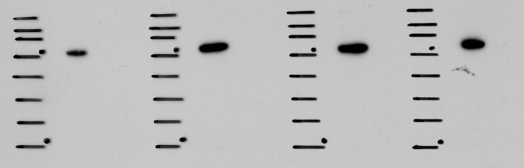
Western Blot: DNCIC1 Antibody (74.1) [NB300-726] - Analysis of cytoplasmic dynein in HeLa cell lysate.![Immunocytochemistry/ Immunofluorescence: DNCIC1 Antibody (74.1) [NB300-726]](https://resources.rndsystems.com/images/products/DNCIC1-Antibody-74-1-Immunocytochemistry-Immunofluorescence-NB300-726-img0006.jpg "Immunocytochemistry/ Immunofluorescence: DNCIC1 Antibody (74.1) [NB300-726]")
Immunocytochemistry/ Immunofluorescence: DNCIC1 Antibody (74.1) [NB300-726]
Immunocytochemistry/Immunofluorescence: DNCIC1 Antibody (74.1) [NB300-726] - Analysis of Dynein in C6 Cells. Cells were grown on chamber slides and fixed with formaldehyde prior to staining. Cells were probed without (control) or with a Dynein monoclonal antibody at a dilution of 1:20 overnight at 4C, washed with PBS and incubated with a DyLight-488 conjugated secondary antibody. Dynein staining (green), F-Actin staining with Phalloidin (red) and nuclei with DAPI (blue) is shown.![Immunohistochemistry-Paraffin: DNCIC1 Antibody (74.1) [NB300-726]](https://resources.rndsystems.com/images/products/DNCIC1-Antibody-74-1-Immunohistochemistry-Paraffin-NB300-726-img0009.jpg "Immunohistochemistry-Paraffin: DNCIC1 Antibody (74.1) [NB300-726]")
Immunohistochemistry-Paraffin: DNCIC1 Antibody (74.1) [NB300-726]
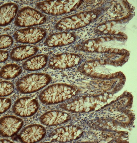
Immunohistochemistry-Paraffin: DNCIC1 Antibody (74.1) [NB300-726] - analysis of DNCIC1 in human colon tissue using anti-DNCIC1 antibody. Image from verified customer review.![Flow Cytometry: DNCIC1 Antibody (74.1) [NB300-726]](https://resources.rndsystems.com/images/products/DNCIC1-Antibody-74-1-Flow-Cytometry-NB300-726-img0012.jpg "Flow Cytometry: DNCIC1 Antibody (74.1) [NB300-726]")
Flow Cytometry: DNCIC1 Antibody (74.1) [NB300-726]
Flow Cytometry: DNCIC1 Antibody (74.1) [NB300-726] - Analysis of Dynein in C6 cells compared to an isotype control (blue).![Western Blot: DNCIC1 Antibody (74.1) [NB300-726]](https://resources.rndsystems.com/images/products/DNCIC1-Antibody-74-1-Western-Blot-NB300-726-img0007.jpg "Western Blot: DNCIC1 Antibody (74.1) [NB300-726]")
Western Blot: DNCIC1 Antibody (74.1) [NB300-726]
Western Blot: DNCIC1 Antibody (74.1) [NB300-726] - analysis of DNCIC1 in human colon cancer cell lysates using anti-DNCIC1 antibody. Image from verified customer review.![Immunocytochemistry/ Immunofluorescence: DNCIC1 Antibody (74.1) [NB300-726]](https://resources.rndsystems.com/images/products/DNCIC1-Antibody-74-1-Immunocytochemistry-Immunofluorescence-NB300-726-img0008.jpg "Immunocytochemistry/ Immunofluorescence: DNCIC1 Antibody (74.1) [NB300-726]")
Immunocytochemistry/ Immunofluorescence: DNCIC1 Antibody (74.1) [NB300-726]
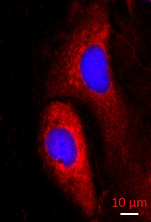
Immunocytochemistry/Immunofluorescence: DNCIC1 Antibody (74.1) [NB300-726] - analysis of DNCIC1 in caco-2 cells using anti-DNCIC1 antibody. Image from verified customer review.![Immunocytochemistry/ Immunofluorescence: DNCIC1 Antibody (74.1) [NB300-726]](https://resources.rndsystems.com/images/products/DNCIC1-Antibody-74-1-Immunocytochemistry-Immunofluorescence-NB300-726-img0004.jpg "Immunocytochemistry/ Immunofluorescence: DNCIC1 Antibody (74.1) [NB300-726]")
Immunocytochemistry/ Immunofluorescence: DNCIC1 Antibody (74.1) [NB300-726]
Immunocytochemistry/Immunofluorescence: DNCIC1 Antibody (74.1) [NB300-726] - Analysis of Dynein in HeLa Cells. Cells were grown on chamber slides and fixed with formaldehyde prior to staining. Cells were probed without (control) or with a Dynein monoclonal antibody at a dilution of 1:20 overnight at 4C, washed with PBS and incubated with a DyLight-488 conjugated secondary antibody. Dynein staining (green), F-Actin staining with Phalloidin (red) and nuclei with DAPI (blue) is shown.![Immunocytochemistry/ Immunofluorescence: DNCIC1 Antibody (74.1) [NB300-726]](https://resources.rndsystems.com/images/products/DNCIC1-Antibody-74-1-Immunocytochemistry-Immunofluorescence-NB300-726-img0005.jpg "Immunocytochemistry/ Immunofluorescence: DNCIC1 Antibody (74.1) [NB300-726]")
Immunocytochemistry/ Immunofluorescence: DNCIC1 Antibody (74.1) [NB300-726]
Immunocytochemistry/Immunofluorescence: DNCIC1 Antibody (74.1) [NB300-726] - Analysis of Dynein in U87-MG Cells. Cells were grown on chamber slides and fixed with formaldehyde prior to staining. Cells were probed without (control) or with a Dynein monoclonal antibody at a dilution of 1:20 overnight at 4C, washed with PBS and incubated with a DyLight-488 conjugated secondary antibody. Dynein staining (green), F-Actin staining with Phalloidin (red) and nuclei with DAPI (blue) is shown.![Flow Cytometry: DNCIC1 Antibody (74.1) [NB300-726]](https://resources.rndsystems.com/images/products/DNCIC1-Antibody-74-1-Flow-Cytometry-NB300-726-img0010.jpg "Flow Cytometry: DNCIC1 Antibody (74.1) [NB300-726]")
Flow Cytometry: DNCIC1 Antibody (74.1) [NB300-726]
Flow Cytometry: DNCIC1 Antibody (74.1) [NB300-726] - Analysis of Dynein in U87-MG cells compared to an isotype control (blue).![Flow Cytometry: DNCIC1 Antibody (74.1) [NB300-726]](https://resources.rndsystems.com/images/products/DNCIC1-Antibody-74-1-Flow-Cytometry-NB300-726-img0011.jpg "Flow Cytometry: DNCIC1 Antibody (74.1) [NB300-726]")
Flow Cytometry: DNCIC1 Antibody (74.1) [NB300-726]
Flow Cytometry: DNCIC1 Antibody (74.1) [NB300-726] - Analysis of Dynein in Hela cells compared to an isotype control (blue).Applications for DNCIC1 Antibody (74.1)
Flow Cytometry
Immunocytochemistry/ Immunofluorescence
Immunohistochemistry-Paraffin
Immunoprecipitation
Western Blot
Reviewed Applications
Read 3 reviews rated 4.7 using NB300-726 in the following applications:
Flow Cytometry Panel Builder
Bio-Techne Knows Flow Cytometry
Save time and reduce costly mistakes by quickly finding compatible reagents using the Panel Builder Tool.
Advanced Features
- Spectra Viewer - Custom analysis of spectra from multiple fluorochromes
- Spillover Popups - Visualize the spectra of individual fluorochromes
- Antigen Density Selector - Match fluorochrome brightness with antigen density

Formulation, Preparation, and Storage
Purification
Formulation
Preservative
Concentration
Shipping
Stability & Storage
Background: DNCIC1
Alternate Names
Entrez Gene IDs
Gene Symbol
UniProt
Additional DNCIC1 Products
Product Documents for DNCIC1 Antibody (74.1)
Certificate of Analysis
To download a Certificate of Analysis, please enter a lot or batch number in the search box below.
Product Specific Notices for DNCIC1 Antibody (74.1)
This product is for research use only and is not approved for use in humans or in clinical diagnosis. Primary Antibodies are guaranteed for 1 year from date of receipt.
Customer Reviews for DNCIC1 Antibody (74.1) (3)
Have you used DNCIC1 Antibody (74.1)?
Submit a review and receive an Amazon gift card!
$25/€18/£15/$25CAN/¥2500 Yen for a review with an image
$10/€7/£6/$10CAN/¥1110 Yen for a review without an image
Submit a review
Customer Images
-
Application: Immunohistochemistry-ParaffinSample Tested: human colon tissueSpecies: HumanVerified Customer | Posted 03/25/2015DNCIC1 in colon epithelial cells

-
Application: ImmunofluorescenceSample Tested: Caco-2 cell lineSpecies: HumanVerified Customer | Posted 03/23/2015DYNC1I1 - IF
-
Application: Western BlotSample Tested: Human colon cancer cell lysatesSpecies: HumanVerified Customer | Posted 03/23/2015DYNC1I1
There are no reviews that match your criteria.
Protocols
Find general support by application which include: protocols, troubleshooting, illustrated assays, videos and webinars.
- 7-Amino Actinomycin D (7-AAD) Cell Viability Flow Cytometry Protocol
- Antigen Retrieval Protocol (PIER)
- Antigen Retrieval for Frozen Sections Protocol
- Appropriate Fixation of IHC/ICC Samples
- Cellular Response to Hypoxia Protocols
- Chromogenic IHC Staining of Formalin-Fixed Paraffin-Embedded (FFPE) Tissue Protocol
- Chromogenic Immunohistochemistry Staining of Frozen Tissue
- ClariTSA™ Fluorophore Kits
- Detection & Visualization of Antibody Binding
- Extracellular Membrane Flow Cytometry Protocol
- Flow Cytometry Protocol for Cell Surface Markers
- Flow Cytometry Protocol for Staining Membrane Associated Proteins
- Flow Cytometry Staining Protocols
- Flow Cytometry Troubleshooting Guide
- Fluorescent IHC Staining of Frozen Tissue Protocol
- Graphic Protocol for Heat-induced Epitope Retrieval
- Graphic Protocol for the Preparation and Fluorescent IHC Staining of Frozen Tissue Sections
- Graphic Protocol for the Preparation and Fluorescent IHC Staining of Paraffin-embedded Tissue Sections
- Graphic Protocol for the Preparation of Gelatin-coated Slides for Histological Tissue Sections
- ICC Cell Smear Protocol for Suspension Cells
- ICC Immunocytochemistry Protocol Videos
- ICC for Adherent Cells
- IHC Sample Preparation (Frozen sections vs Paraffin)
- Immunocytochemistry (ICC) Protocol
- Immunocytochemistry Troubleshooting
- Immunofluorescence of Organoids Embedded in Cultrex Basement Membrane Extract
- Immunofluorescent IHC Staining of Formalin-Fixed Paraffin-Embedded (FFPE) Tissue Protocol
- Immunohistochemistry (IHC) and Immunocytochemistry (ICC) Protocols
- Immunohistochemistry Frozen Troubleshooting
- Immunohistochemistry Paraffin Troubleshooting
- Immunoprecipitation Protocol
- Intracellular Flow Cytometry Protocol Using Alcohol (Methanol)
- Intracellular Flow Cytometry Protocol Using Detergents
- Intracellular Nuclear Staining Flow Cytometry Protocol Using Detergents
- Intracellular Staining Flow Cytometry Protocol Using Alcohol Permeabilization
- Intracellular Staining Flow Cytometry Protocol Using Detergents to Permeabilize Cells
- Preparing Samples for IHC/ICC Experiments
- Preventing Non-Specific Staining (Non-Specific Binding)
- Primary Antibody Selection & Optimization
- Propidium Iodide Cell Viability Flow Cytometry Protocol
- Protocol for Heat-Induced Epitope Retrieval (HIER)
- Protocol for Liperfluo
- Protocol for Making a 4% Formaldehyde Solution in PBS
- Protocol for VisUCyte™ HRP Polymer Detection Reagent
- Protocol for the Characterization of Human Th22 Cells
- Protocol for the Characterization of Human Th9 Cells
- Protocol for the Fluorescent ICC Staining of Cell Smears - Graphic
- Protocol for the Fluorescent ICC Staining of Cultured Cells on Coverslips - Graphic
- Protocol for the Preparation & Fixation of Cells on Coverslips
- Protocol for the Preparation and Chromogenic IHC Staining of Frozen Tissue Sections
- Protocol for the Preparation and Chromogenic IHC Staining of Frozen Tissue Sections - Graphic
- Protocol for the Preparation and Chromogenic IHC Staining of Paraffin-embedded Tissue Sections
- Protocol for the Preparation and Chromogenic IHC Staining of Paraffin-embedded Tissue Sections - Graphic
- Protocol for the Preparation and Fluorescent ICC Staining of Cells on Coverslips
- Protocol for the Preparation and Fluorescent ICC Staining of Non-adherent Cells
- Protocol for the Preparation and Fluorescent ICC Staining of Stem Cells on Coverslips
- Protocol for the Preparation and Fluorescent IHC Staining of Frozen Tissue Sections
- Protocol for the Preparation and Fluorescent IHC Staining of Paraffin-embedded Tissue Sections
- Protocol for the Preparation of Gelatin-coated Slides for Histological Tissue Sections
- Protocol for the Preparation of a Cell Smear for Non-adherent Cell ICC - Graphic
- Protocol: Annexin V and PI Staining by Flow Cytometry
- Protocol: Annexin V and PI Staining for Apoptosis by Flow Cytometry
- R&D Systems Quality Control Western Blot Protocol
- TUNEL and Active Caspase-3 Detection by IHC/ICC Protocol
- The Importance of IHC/ICC Controls
- Troubleshooting Guide: Fluorokine Flow Cytometry Kits
- Troubleshooting Guide: Immunohistochemistry
- Troubleshooting Guide: Western Blot Figures
- Western Blot Conditions
- Western Blot Protocol
- Western Blot Protocol for Cell Lysates
- Western Blot Troubleshooting
- Western Blot Troubleshooting Guide
- View all Protocols, Troubleshooting, Illustrated assays and Webinars
FAQs for DNCIC1 Antibody (74.1)
-
Q: Is the DNCIC1 antibody with the number NB300-726 is only specific for DNCIC1 protein? Does it detect the DNCIC2 protein as well?
A: For our product NB300-726, we have not yet tested this product for cross reactivity against the DNCIC2 protein. These two proteins share quite a few regions of 100% homology, so I think it is highly likely that this product would cross react with the DNCIC2 protein.