DUSP27/DUPD1 Antibody - BSA Free
Novus Biologicals | Catalog # NBP1-84040
![Immunohistochemistry-Paraffin: DUSP27/DUPD1 Antibody [NBP1-84040]](https://resources.rndsystems.com/images/products/DUSP27-DUPD1-Antibody-Immunohistochemistry-Paraffin-NBP1-84040-img0003.jpg "Immunohistochemistry-Paraffin: DUSP27/DUPD1 Antibody [NBP1-84040]")
Loading...
Key Product Details
Species Reactivity
Human
Applications
Immunohistochemistry, Immunohistochemistry-Paraffin, Western Blot
Label
Unconjugated
Antibody Source
Polyclonal Rabbit IgG
Format
BSA Free
Loading...
Product Specifications
Immunogen
This antibody was developed against Recombinant Protein corresponding to amino acids: MTSGEVKTSLKNAYSSAKRLSPKMEEEGEEEDYCTPGAFELERLFWKGSPQYTHVNEVWPKLYIGDEATALDRYRL
Reactivity Notes
Rat reactivity reported from a verified customer review.
Clonality
Polyclonal
Host
Rabbit
Isotype
IgG
Scientific Data Images for DUSP27/DUPD1 Antibody - BSA Free
Immunohistochemistry-Paraffin: DUSP27/DUPD1 Antibody [NBP1-84040]
Immunohistochemistry-Paraffin: DUSP27/DUPD1 Antibody [NBP1-84040] - Staining of human skeletal muscle shows moderate cytoplasmic positivity in myocytes.
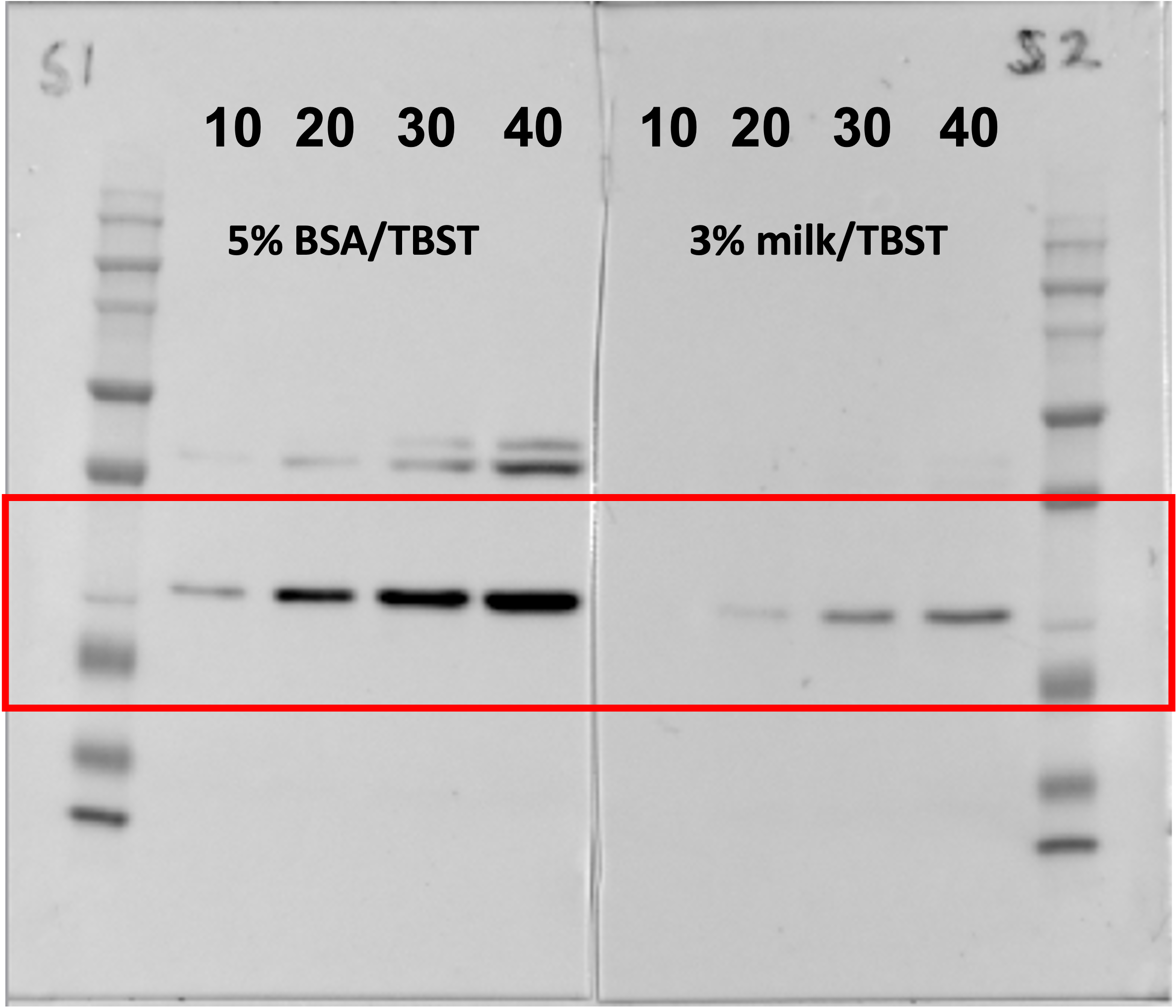
Western Blot: DUSP27/DUPD1 Antibody [NBP1-84040] -
Western Blot: DUSP27/DUPD1 Antibody [NBP1-84040] - Rat soleus muscle loaded at 10, 20, 30 and 40 µg protein in lanes 2-5, and lanes 6-9. DUSP29 ab was diluted in 5% BSA/TBST or 3% milk/TBST and incubated overnight at 4°C. Secondary ab in 5%milk/TBST at 1:20K for 1 hour at RT. Image from verified customer review.![DUSP27/DUPD1 Antibody - BSA Free Western Blot: DUSP27/DUPD1 Antibody - BSA Free [NBP1-84040]](https://resources.rndsystems.com/images/products/nbp1-84040_rabbit-polyclonal-dusp27-dupd1-antibody-84202517195638.jpg "Western Blot: DUSP27/DUPD1 Antibody - BSA Free [NBP1-84040]")
Western Blot: DUSP27/DUPD1 Antibody - BSA Free [NBP1-84040]
Analysis in control (vector only transfected HEK293T lysate) and DUPD1 over-expression lysate (Co-expressed with a C-terminal myc-DDK tag (~3.1 kDa) in mammalian HEK293T cells).Applications for DUSP27/DUPD1 Antibody - BSA Free
Application
Recommended Usage
Immunohistochemistry
1:200 - 1:500
Immunohistochemistry-Paraffin
1:200 - 1:500
Western Blot
0.04-0.4 ug/ml
Application Notes
For IHC-Paraffin, HIER pH 6 retrieval is recommended.
Reviewed Applications
Read 1 review rated 5 using NBP1-84040 in the following applications:
Formulation, Preparation, and Storage
Purification
Affinity purified
Formulation
PBS (pH 7.2) and 40% Glycerol
Format
BSA Free
Preservative
0.02% Sodium Azide
Concentration
Concentrations vary lot to lot. See vial label for concentration. If unlisted please contact technical services.
Shipping
The product is shipped with polar packs. Upon receipt, store it immediately at the temperature recommended below.
Stability & Storage
Store at 4C short term. Aliquot and store at -20C long term. Avoid freeze-thaw cycles.
Background: DUSP27/DUPD1
Alternate Names
atypical dual-specific protein phosphatase, Dual specificity phosphatase 27, dual specificity phosphatase and pro isomerase domain containing 1, DUSP27dual specificity phosphatase DUPD1, EC 3.1.3.16, EC 3.1.3.48, FMDSP
Gene Symbol
DUPD1
Additional DUSP27/DUPD1 Products
Product Documents for DUSP27/DUPD1 Antibody - BSA Free
Certificate of Analysis
To download a Certificate of Analysis, please enter a lot or batch number in the search box below.
Product Specific Notices for DUSP27/DUPD1 Antibody - BSA Free
This product is for research use only and is not approved for use in humans or in clinical diagnosis. Primary Antibodies are guaranteed for 1 year from date of receipt.
Citations for DUSP27/DUPD1 Antibody - BSA Free
 Powered by Bioz
Customer Reviews for DUSP27/DUPD1 Antibody - BSA Free (1)
5 out of 5
1 Customer Rating
Have you used DUSP27/DUPD1 Antibody - BSA Free?
Submit a review and receive an Amazon gift card!
$25/€18/£15/$25CAN/¥2500 Yen for a review with an image
$10/€7/£6/$10CAN/¥1110 Yen for a review without an image
Submit a review
Customer Images
Showing
1
-
1 of
1 review
Showing All
Filter By:
-
Application: Western BlotSample Tested: Rat skeletal muscle tissueSpecies: RatVerified Customer | Posted 03/10/2023Rat soleus muscle loaded at 10, 20, 30 and 40 µg protein in lanes 2-5, and lanes 6-9. DUSP29 ab was diluted in 5% BSA/TBST or 3% milk/TBST and incubated overnight at 4°C. Secondary ab in 5%milk/TBST at 1:20K for 1 hour at RT.
 Bio-Techne ResponseThis review was submitted through the legacy Novus Innovators Program, reflecting a new species or application tested on a primary antibody.
There are no reviews that match your criteria.
Protocols
Find general support by application which include: protocols, troubleshooting, illustrated assays, videos and webinars.
- Antigen Retrieval Protocol (PIER)
- Antigen Retrieval for Frozen Sections Protocol
- Appropriate Fixation of IHC/ICC Samples
- Cellular Response to Hypoxia Protocols
- Chromogenic IHC Staining of Formalin-Fixed Paraffin-Embedded (FFPE) Tissue Protocol
- Chromogenic Immunohistochemistry Staining of Frozen Tissue
- ClariTSA™ Fluorophore Kits
- Detection & Visualization of Antibody Binding
- Fluorescent IHC Staining of Frozen Tissue Protocol
- Graphic Protocol for Heat-induced Epitope Retrieval
- Graphic Protocol for the Preparation and Fluorescent IHC Staining of Frozen Tissue Sections
- Graphic Protocol for the Preparation and Fluorescent IHC Staining of Paraffin-embedded Tissue Sections
- Graphic Protocol for the Preparation of Gelatin-coated Slides for Histological Tissue Sections
- IHC Sample Preparation (Frozen sections vs Paraffin)
- Immunofluorescent IHC Staining of Formalin-Fixed Paraffin-Embedded (FFPE) Tissue Protocol
- Immunohistochemistry (IHC) and Immunocytochemistry (ICC) Protocols
- Immunohistochemistry Frozen Troubleshooting
- Immunohistochemistry Paraffin Troubleshooting
- Preparing Samples for IHC/ICC Experiments
- Preventing Non-Specific Staining (Non-Specific Binding)
- Primary Antibody Selection & Optimization
- Protocol for Heat-Induced Epitope Retrieval (HIER)
- Protocol for Making a 4% Formaldehyde Solution in PBS
- Protocol for VisUCyte™ HRP Polymer Detection Reagent
- Protocol for the Preparation & Fixation of Cells on Coverslips
- Protocol for the Preparation and Chromogenic IHC Staining of Frozen Tissue Sections
- Protocol for the Preparation and Chromogenic IHC Staining of Frozen Tissue Sections - Graphic
- Protocol for the Preparation and Chromogenic IHC Staining of Paraffin-embedded Tissue Sections
- Protocol for the Preparation and Chromogenic IHC Staining of Paraffin-embedded Tissue Sections - Graphic
- Protocol for the Preparation and Fluorescent IHC Staining of Frozen Tissue Sections
- Protocol for the Preparation and Fluorescent IHC Staining of Paraffin-embedded Tissue Sections
- Protocol for the Preparation of Gelatin-coated Slides for Histological Tissue Sections
- R&D Systems Quality Control Western Blot Protocol
- TUNEL and Active Caspase-3 Detection by IHC/ICC Protocol
- The Importance of IHC/ICC Controls
- Troubleshooting Guide: Immunohistochemistry
- Troubleshooting Guide: Western Blot Figures
- Western Blot Conditions
- Western Blot Protocol
- Western Blot Protocol for Cell Lysates
- Western Blot Troubleshooting
- Western Blot Troubleshooting Guide
- View all Protocols, Troubleshooting, Illustrated assays and Webinars
Loading...