EGLN1/PHD2 Antibody - BSA Free
Novus Biologicals | Catalog # NB100-2219
![Immunohistochemistry: EGLN1/PHD2 Antibody - BSA Free [NB100-2219]](https://resources.rndsystems.com/images/products/EGLN1-PHD2-Antibody-Immunohistochemistry-NB100-2219-img0015.jpg "Immunohistochemistry: EGLN1/PHD2 Antibody - BSA Free [NB100-2219]")
Key Product Details
Validated by
Knockout/Knockdown
Species Reactivity
Validated:
Human, Mouse, Rat
Cited:
Human, Mouse, Rat
Applications
Validated:
Knockout Validated, Immunohistochemistry, Immunohistochemistry-Paraffin, Western Blot, Immunocytochemistry/ Immunofluorescence, Simple Western, Immunoprecipitation, Knockdown Validated
Cited:
Knockout Validated, Immunohistochemistry-Paraffin, Western Blot, Simple Western, Immunoprecipitation, IF/IHC
Label
Unconjugated
Antibody Source
Polyclonal Rabbit IgG
Format
BSA Free
Loading...
Product Specifications
Immunogen
This EGLN1/PHD2 antibody was developed against a synthetic peptide made to an internal portion of mouse PHD2/HIF Prolyl Hydroxylase 2 (between residues 300-400). [Uniprot: Q91YE3]
Clonality
Polyclonal
Host
Rabbit
Isotype
IgG
Theoretical MW
43 kDa.
Disclaimer note: The observed molecular weight of the protein may vary from the listed predicted molecular weight due to post translational modifications, post translation cleavages, relative charges, and other experimental factors.
Disclaimer note: The observed molecular weight of the protein may vary from the listed predicted molecular weight due to post translational modifications, post translation cleavages, relative charges, and other experimental factors.
Scientific Data Images for EGLN1/PHD2 Antibody - BSA Free
Immunohistochemistry: EGLN1/PHD2 Antibody - BSA Free [NB100-2219]
EGLN1-PHD2-Antibody-Immunohistochemistry-NB100-2219-img0015.jpg![Knockdown Validated: EGLN1/PHD2 Antibody - BSA Free [NB100-2219]](https://resources.rndsystems.com/images/products/EGLN1-PHD2-Antibody-Knockdown-Validated-NB100-2219-img0016.jpg "Western Blot: EGLN1/PHD2 Antibody - BSA Free [NB100-2219]")
![Immunohistochemistry-Paraffin: EGLN1/PHD2 Antibody - BSA Free [NB100-2219]](https://resources.rndsystems.com/images/products/EGLN1-PHD2-Antibody-Immunohistochemistry-Paraffin-NB100-2219-img0014.jpg "Immunohistochemistry-Paraffin: EGLN1/PHD2 Antibody - BSA Free [NB100-2219]")
Immunohistochemistry-Paraffin: EGLN1/PHD2 Antibody - BSA Free [NB100-2219]
Immunohistochemistry-Paraffin: EGLN1/PHD2 Antibody [NB100-2219] - Analysis of an FFPE mouse lung section using 1:200 dilution of EGLN1/PHD2 antibody. The staining was developed using HRP conjugated anti-rabbit secondary antibody and DAB reagent. The antibody generated a specific staining in the cytoplasm and nuclei of alveolar as well as bronchiolar epithelial cells. Cytoplasmic staining was observed in almost all cells while the nuclear positivity was seen in a subset of cells only.![Western Blot: EGLN1/PHD2 AntibodyBSA Free [NB100-2219]](https://resources.rndsystems.com/images/products/EGLN1-PHD2-Antibody-Western-Blot-NB100-2219-img0013.jpg "Western Blot: EGLN1/PHD2 AntibodyBSA Free [NB100-2219]")
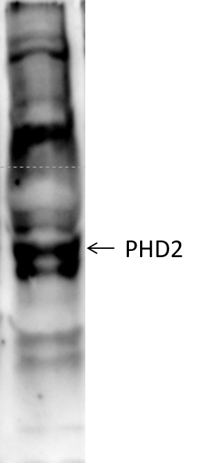
Western Blot: EGLN1/PHD2 AntibodyBSA Free [NB100-2219]
Western Blot: EGLN1/PHD2 Antibody [NB100-2219] - Detection of EGLN1/PHD2 in mouse kidney lysate. ECL exposure, 20 seconds.![Immunocytochemistry/ Immunofluorescence: EGLN1/PHD2 Antibody - BSA Free [NB100-2219]](https://resources.rndsystems.com/images/products/EGLN1-PHD2-Antibody-Immunocytochemistry-Immunofluorescence-NB100-2219-img0012.jpg "Immunocytochemistry/ Immunofluorescence: EGLN1/PHD2 Antibody - BSA Free [NB100-2219]")
Immunocytochemistry/ Immunofluorescence: EGLN1/PHD2 Antibody - BSA Free [NB100-2219]
Immunocytochemistry/Immunofluorescence: EGLN1/PHD2 Antibody [NB100-2219] - EGLN1/PHD2 antibody at 1:500 in HeLa cells with DyLight 488 (green). Nuclei and alpha-tubulin were counterstained with DAPI (blue) and DyLight 550 (red).![Immunocytochemistry/ Immunofluorescence: EGLN1/PHD2 Antibody - BSA Free [NB100-2219]](https://resources.rndsystems.com/images/products/EGLN1-PHD2-Antibody-Immunocytochemistry-Immunofluorescence-NB100-2219-img0009.jpg "Immunocytochemistry/ Immunofluorescence: EGLN1/PHD2 Antibody - BSA Free [NB100-2219]")
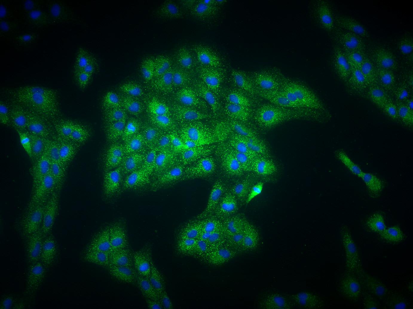
Immunocytochemistry/ Immunofluorescence: EGLN1/PHD2 Antibody - BSA Free [NB100-2219]
Immunocytochemistry/Immunofluorescence: EGLN1/PHD2 Antibody [NB100-2219] - Analysis of EGLN1/PHD2 in ARPE-19 cells using anti-PHD2 antibody. ICC/IF image submitted by a veriifed customer review.![Immunohistochemistry: EGLN1/PHD2 Antibody - BSA Free [NB100-2219]](https://resources.rndsystems.com/images/products/EGLN1-PHD2-Antibody-Immunohistochemistry-NB100-2219-img0010.jpg "Immunohistochemistry: EGLN1/PHD2 Antibody - BSA Free [NB100-2219]")
Immunohistochemistry: EGLN1/PHD2 Antibody - BSA Free [NB100-2219]
Immunohistochemistry: EGLN1/PHD2 Antibody [NB100-2219] - Staining of renal tubular epithelium in mouse using NB100-2219 at 2.5 ug/mL.Applications for EGLN1/PHD2 Antibody - BSA Free
Application
Recommended Usage
Immunocytochemistry/ Immunofluorescence
1:50 - 1:500
Immunohistochemistry
2.5 - 5.0 ug/mL
Immunohistochemistry-Paraffin
2.5 - 5.0 ug/mL
Immunoprecipitation
1:10 - 1:500
Knockout Validated
reported in scientific literature (PMID 24695462)
Simple Western
1:200
Western Blot
2 ug/mL
Application Notes
In Western blot a band is seen ~43 kDa representing HIF Prolyl Hydroxylase 2. There is also a non-specific band of similar intensity at ~75 kDa.
Reviewed Applications
Read 3 reviews rated 4.7 using NB100-2219 in the following applications:
Formulation, Preparation, and Storage
Purification
Immunogen affinity purified
Formulation
PBS
Format
BSA Free
Preservative
0.02% Sodium Azide
Concentration
1 mg/ml
Shipping
The product is shipped with polar packs. Upon receipt, store it immediately at the temperature recommended below.
Stability & Storage
Store at 4C. Do not freeze.
Background: EGLN1/PHD2
EGLN1/PHD2 has been implicated in several critical processes including erythropoiesis, angiogenesis, and metabolism as well as various pathologies such as cancer (2, 5, 6). Studies in mice have found that somatic deletion of PHD2 resulted in higher vascular endothelial growth factor A (VEGF-A) levels, increased blood vessel formation, and more erythropoietin (EPO), leading to severe polycythemia or erythrocytosis (high red blood cell (RBC) volume) (6). Another study revealed that specific point mutations in EGLN1/PHD2 led to elevated EPO and RBC mass associated with hemorrhages and strokes (6). Accordingly, given the known role of PHD2 in inhibition of EPO production, PHD2 inhibitors are being studied as a potential therapeutic for anemia (6). Additionally, dysregulation in EGLN1, and specifically the PHD2-VHL-HIF-1alpha pathway, has been associated with the development of pheochromocytomas (PCC) and sympathetic paragangliomas (PGL), which are rare neuroendocrine tumors (2). Besides pathological features, EGLN1/PHD2 may also be important for high altitude adaptation as two coding sequence variants in PHD2 are prevalent in the Tibetan population but is very rare in people at lower altitudes (2).
Alternate names for EGLN1/PHD2 include HIF Prolyl Hydroxylase 2, PH2, Prolyl hydroxylase domain containing protein 2, HIF2PH2, HIF-Prolyl hydroxylase 2, egl nine homolog 1, and C1orf12.
References
1. Amorim-Pires, D., Peixoto, J., & Lima, J. (2016). Hypoxia Pathway Mutations in Pheochromocytomas and Paragangliomas. Cytogenetic and genome research. https://doi.org/10.1159/000457479
2. Gardie, B., Percy, M. J., Hoogewijs, D., Chowdhury, R., Bento, C., Arsenault, P. R., Richard, S., Almeida, H., Ewing, J., Lambert, F., McMullin, M. F., Schofield, C. J., & Lee, F. S. (2014). The role of PHD2 mutations in the pathogenesis of erythrocytosis. Hypoxia (Auckland, N.Z.). https://doi.org/10.2147/HP.S54455
3. Minervini, G., Quaglia, F., & Tosatto, S. C. (2015). Insights into the proline hydroxylase (PHD) family, molecular evolution and its impact on human health. Biochimie. https://doi.org/10.1016/j.biochi.2015.07.009
4. Semenza G. L. (2007). Hypoxia-inducible factor 1 (HIF-1) pathway. Science's STKE : signal transduction knowledge environment. https://doi.org/10.1126/stke.4072007cm8
5. Chan, D. A., & Giaccia, A. J. (2010). PHD2 in tumour angiogenesis. British journal of cancer. https://doi.org/10.1038/sj.bjc.6605682
6. Meneses, A. M., & Wielockx, B. (2016). PHD2: from hypoxia regulation to disease progression. Hypoxia (Auckland, N.Z.). https://doi.org/10.2147/HP.S53576
Long Name
Egl Nine Homolog 1/Prolyl Hydroxylase Domain-containing Protein 2
Alternate Names
C1orf12, HIFPH2, HPH2, PHD2, SM20, ZMYND6
Gene Symbol
EGLN1
Additional EGLN1/PHD2 Products
Product Documents for EGLN1/PHD2 Antibody - BSA Free
Certificate of Analysis
To download a Certificate of Analysis, please enter a lot or batch number in the search box below.
Product Specific Notices for EGLN1/PHD2 Antibody - BSA Free
This product is for research use only and is not approved for use in humans or in clinical diagnosis. Primary Antibodies are guaranteed for 1 year from date of receipt.
Related Research Areas
Citations for EGLN1/PHD2 Antibody - BSA Free
 Powered by Bioz
Customer Reviews for EGLN1/PHD2 Antibody - BSA Free (3)
4.7 out of 5
3 Customer Ratings
Have you used EGLN1/PHD2 Antibody - BSA Free?
Submit a review and receive an Amazon gift card!
$25/€18/£15/$25CAN/¥2500 Yen for a review with an image
$10/€7/£6/$10CAN/¥1110 Yen for a review without an image
Submit a review
Customer Images
Showing
1
-
3 of
3 reviews
Showing All
Filter By:
-
Application: Western BlotSample Tested: murine bone marrow derived macrophagesSpecies: MouseVerified Customer | Posted 02/16/2017PHD2 in bone marrow derived macrophages

-
Application: ImmunofluorescenceSample Tested: ARPE-19 cellsSpecies: HumanVerified Customer | Posted 05/14/2015ARPE-19 cells
-
Application: Western BlotSample Tested: nuclear extract (brain), Sample Amount: 20ugSpecies: MouseVerified Customer | Posted 01/18/2012
There are no reviews that match your criteria.
Protocols
View specific protocols for EGLN1/PHD2 Antibody - BSA Free (NB100-2219):
Western Blot Protocol
1. Perform SDS-PAGE (4-12%) on samples to be analyzed, loading 40 ug of total protein per lane.
2. Transfer proteins to Nitrocellulose according to the instructions provided by the manufacturer of the transfer apparatus.
3. Rinse membrane with dH2O and then stain the blot using ponceau S for 1-2 minutes to access the transfer of proteins onto the nitrocellulose membrane. Rinse the blot in water to remove excess stain and mark the lane locations and locations of molecular weight markers using a pencil.
4. Rinse the blot in TBS for approximately 5 minutes.
5. Block the membrane using 5% non-fat dry milk + 1% BSA in TBS for 2 hours at room temperature (RT).
6. Rinse the membrane in dH2O and then wash the membrane in wash buffer [TBS + 0.1% Tween] 3 times for 10 minutes each.
7. Dilute the rabbit anti-PHD2 (murine) primary antibody (NB 100-2219) in blocking buffer and incubate 1 hour at RT.
8. Rinse the membrane in dH2O and then wash the membrane in wash buffer [TBS + 0.1% Tween] 3 times for 10 minutes each.
9. Apply the diluted rabbit-IgG HRP-conjugated secondary antibody in blocking buffer (as per manufacturer's instructions) and incubate 1 hour at RT.
10. Wash the blot in wash buffer [TBS + 0.1% Tween] 3 times for 10 minutes each (this step can be repeated as required to reduce background).
11. Apply the detection reagent of choice in accordance with the manufacturer's instructions (we used BioFX Super Plus ECL). Note: Tween-20 can be added to the blocking or antibody dilution buffer at a final concentration of 0.05-0.2%, provided it does not interfere with antibody-antigen binding.
IHC-FFPE sections
I. Deparaffinization:
A. Treat slides with Xylene: 3 changes for 5 minutes each. Drain slides for 10 seconds between changes.
B. Treat slides with 100% Reagent Alcohol: 3 changes for 5 minutes each. Drain slides for 10 seconds between changes.
II. Quench Endogenous Peroxidase:
A. Place slides in peroxidase quenching solution: 15-30 minutes. To Prepare 200 ml of Quenching Solution: Add 3 ml of 30% Hydrogen Peroxide to 200 ml of Methanol.
Use within 4 hours of preparation
B. Place slides in distilled water: 2 changes for 2 minutes each.
III. Retrieve Epitopes:
A. Preheat Citrate Buffer. Place 200 ml of Citrate Buffer Working Solution into container, cover and place into steamer. Heat to 90-96 degrees Celsius.
B. Place rack of slides into hot Citrate Buffer for 20 minutes. Cover.
C. Carefully remove container with slides from steamer and cool on bench, uncovered, for 20 minutes.
D. Slowly add distilled water to further cool for 5 minutes.
E. Rinse slides with distilled water. 2 changes for 2 minutes each.
IV. Immunostaining Procedure:
A. Remove each slide from rack and circle tissue section with a hydrophobic barrier pen (e.g. Liquid Blocker-Super Pap-Pen).
B. Flood slide with Wash Solution. Do not allow tissue sections to dry for the rest of the procedure.
C. Drain wash solution and apply 4 drops of Blocking Reagent to each slide and incubate for 15 minutes.
D. Drain Blocking Reagent (do not wash off the Blocking Reagent), apply 200 ul of Primary Antibody solution to each slide, and incubate for 1 hour.
E. Wash slides with Wash Solution: 3 changes for 5 minutes each.
F. Drain wash solution, apply 4 drops of Secondary antibody to each slide and incubate for 1 hour.
G. Wash slides with Wash Solution: 3 changes for 5 minutes each.
H. Drain wash solution, apply 4 drops of DAB Substrate to each slide and develop for 5-10 minutes. Check development with microscope.
I. Wash slides with Wash Solution: 3 changes for 5 minutes each. Wash slides with Wash Solution: 3 changes for 5 minutes each
J. Drain wash solution, apply 4 drops of Hematoxylin to each slide and stain for 1-3 minutes. Increase time if darker counterstaining is desired.
K. Wash slides with Wash Solution: 2-3 changes for 2 minutes each.
L. Drain wash solution and apply 4 drops of Bluing Solution to each slide for 1-2 minutes.
M. Rinse slides in distilled water.
N. Soak slides in 70% reagent alcohol: 3 minutes with intermittent agitation.
O. Soak slides in 95% reagent alcohol: 2 changes for 3 minutes each with intermittent agitation.
P. Soak slides in 100% reagent alcohol: 3 changes for 3 minutes each with intermittent agitation. Drain slides for 10 seconds between each change.
Q. Soak slides in Xylene: 3 changes for 3 minutes each with intermittent agitation. Drain slides for 10 seconds between each change.
R. Apply 2-3 drops of non-aqueous mounting media to each slide and mount coverslip.
S. Lay slides on a flat surface to dry prior to viewing under microscope.
NOTES:
-Use treated slides (e.g. HistoBond) to assure adherence of FFPE sections to slide.
-Prior to deparaffinization, heat slides overnight in a 60 degrees Celsius oven.
-All steps in which Xylene is used should be performed in a fume hood.
-For Epitope Retrieval, a microwave or pressure cooker may be substituted for the steamer method. Adjust times as necessary depending on conditions.
-For the initial IHC run with a new primary antibody, test tissues with and without Epitope Retrieval. In some instances, Epitope Retrieval may not be necessary.
-200 ul is the recommended maximum volume to apply to a slide for full coverage. Using more than 200 ul may allow solutions to wick off the slide and create drying artifacts. For small tissue sections less than 200 ul may be used.
-5 minutes of development with DAB Substrate should be sufficient. Do not develop for more than 10 minutes. If 5 minutes of development causes background staining, further dilution of the primary antibody may be necessary.
-Hematoxylin should produce a light nuclear counterstain so as not to obscure the DAB staining. Counterstain for 1-1.5 minutes for nuclear antigens. Counterstain for 2-3 minutes for cytoplasmic and membranous antigens. If darker counterstaining is desired increase time (up to 10 minutes).
Find general support by application which include: protocols, troubleshooting, illustrated assays, videos and webinars.
- Antigen Retrieval Protocol (PIER)
- Antigen Retrieval for Frozen Sections Protocol
- Appropriate Fixation of IHC/ICC Samples
- Cellular Response to Hypoxia Protocols
- Chromogenic IHC Staining of Formalin-Fixed Paraffin-Embedded (FFPE) Tissue Protocol
- Chromogenic Immunohistochemistry Staining of Frozen Tissue
- ClariTSA™ Fluorophore Kits
- Detection & Visualization of Antibody Binding
- Fluorescent IHC Staining of Frozen Tissue Protocol
- Graphic Protocol for Heat-induced Epitope Retrieval
- Graphic Protocol for the Preparation and Fluorescent IHC Staining of Frozen Tissue Sections
- Graphic Protocol for the Preparation and Fluorescent IHC Staining of Paraffin-embedded Tissue Sections
- Graphic Protocol for the Preparation of Gelatin-coated Slides for Histological Tissue Sections
- ICC Cell Smear Protocol for Suspension Cells
- ICC Immunocytochemistry Protocol Videos
- ICC for Adherent Cells
- IHC Sample Preparation (Frozen sections vs Paraffin)
- Immunocytochemistry (ICC) Protocol
- Immunocytochemistry Troubleshooting
- Immunofluorescence of Organoids Embedded in Cultrex Basement Membrane Extract
- Immunofluorescent IHC Staining of Formalin-Fixed Paraffin-Embedded (FFPE) Tissue Protocol
- Immunohistochemistry (IHC) and Immunocytochemistry (ICC) Protocols
- Immunohistochemistry Frozen Troubleshooting
- Immunohistochemistry Paraffin Troubleshooting
- Immunoprecipitation Protocol
- Preparing Samples for IHC/ICC Experiments
- Preventing Non-Specific Staining (Non-Specific Binding)
- Primary Antibody Selection & Optimization
- Protocol for Heat-Induced Epitope Retrieval (HIER)
- Protocol for Making a 4% Formaldehyde Solution in PBS
- Protocol for VisUCyte™ HRP Polymer Detection Reagent
- Protocol for the Fluorescent ICC Staining of Cell Smears - Graphic
- Protocol for the Fluorescent ICC Staining of Cultured Cells on Coverslips - Graphic
- Protocol for the Preparation & Fixation of Cells on Coverslips
- Protocol for the Preparation and Chromogenic IHC Staining of Frozen Tissue Sections
- Protocol for the Preparation and Chromogenic IHC Staining of Frozen Tissue Sections - Graphic
- Protocol for the Preparation and Chromogenic IHC Staining of Paraffin-embedded Tissue Sections
- Protocol for the Preparation and Chromogenic IHC Staining of Paraffin-embedded Tissue Sections - Graphic
- Protocol for the Preparation and Fluorescent ICC Staining of Cells on Coverslips
- Protocol for the Preparation and Fluorescent ICC Staining of Non-adherent Cells
- Protocol for the Preparation and Fluorescent ICC Staining of Stem Cells on Coverslips
- Protocol for the Preparation and Fluorescent IHC Staining of Frozen Tissue Sections
- Protocol for the Preparation and Fluorescent IHC Staining of Paraffin-embedded Tissue Sections
- Protocol for the Preparation of Gelatin-coated Slides for Histological Tissue Sections
- Protocol for the Preparation of a Cell Smear for Non-adherent Cell ICC - Graphic
- R&D Systems Quality Control Western Blot Protocol
- TUNEL and Active Caspase-3 Detection by IHC/ICC Protocol
- The Importance of IHC/ICC Controls
- Troubleshooting Guide: Immunohistochemistry
- Troubleshooting Guide: Western Blot Figures
- Western Blot Conditions
- Western Blot Protocol
- Western Blot Protocol for Cell Lysates
- Western Blot Troubleshooting
- Western Blot Troubleshooting Guide
- View all Protocols, Troubleshooting, Illustrated assays and Webinars
FAQs for EGLN1/PHD2 Antibody - BSA Free
Showing
1
-
2 of
2 FAQs
Showing All
-
Q: How do I choose secondary antibodies to label the same cells when I have two primary antibodies from the same host?
A: Use isotype-specific secondary antibodies if the primary antibodies are of different isotypes. You can also make direct conjugates of the primary antibodies by use of antibody labeling kits, dyes, or custom conjugations (please contact Technical Support for custom orders).
-
Q: I would like to use this antibody but it has not been validated in my species of interest. Is there any way I can find out if it will work?
A: We offer risk-free testing of all of our primary antibodies. Please check out our Innovator's Reward Program and test this EGLN1-PHD2 antibody in any unvalidated species or application, without the financial risk of failure.
-
Q: How do I choose secondary antibodies to label the same cells when I have two primary antibodies from the same host?
A: Use isotype-specific secondary antibodies if the primary antibodies are of different isotypes. You can also make direct conjugates of the primary antibodies by use of antibody labeling kits, dyes, or custom conjugations (please contact Technical Support for custom orders).
-
Q: I would like to use this antibody but it has not been validated in my species of interest. Is there any way I can find out if it will work?
A: We offer risk-free testing of all of our primary antibodies. Please check out our Innovator's Reward Program and test this EGLN1-PHD2 antibody in any unvalidated species or application, without the financial risk of failure.
Loading...