![Western Blot: EMG1 Antibody [NBP1-57410]](https://resources.rndsystems.com/images/products/EMG1-Antibody-Western-Blot-NBP1-57410-img0001.jpg "Western Blot: EMG1 Antibody [NBP1-57410]")
Loading...
Key Product Details
Species Reactivity
Human
Applications
Immunohistochemistry, Immunohistochemistry-Paraffin, Western Blot
Label
Unconjugated
Antibody Source
Polyclonal Rabbit IgG
Format
BSA Free
Loading...
Product Specifications
Immunogen
Synthetic peptides corresponding to EMG1 (EMG1 nucleolar protein homolog (S. cerevisiae)). The peptide sequence was selected from the N terminal of EMG1. Peptide sequence SLETVKVGKTYELLNCDKHKSILLKNGRDPGEARPDITHQSLLMLMDSPL The peptide sequence for this immunogen was taken from within the described region.
Clonality
Polyclonal
Host
Rabbit
Isotype
IgG
Description
The addition of 50% glycerol is optional for those storing this antibody at -20C and not aliquoting smaller units. However, please note that glycerol may interrupt some downstream antibody applications and should be added with caution.
Scientific Data Images for EMG1 Antibody - BSA Free
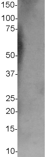
Western Blot: EMG1 Antibody [NBP1-57410]
Western Blot: EMG1 Antibody [NBP1-57410] - HepG2 cell lysate, recommended antibody concentration: 0.2 - 1 ug/mL.![Immunohistochemistry-Paraffin: EMG1 Antibody [NBP1-57410]](https://resources.rndsystems.com/images/products/EMG1-Antibody-Immunohistochemistry-Paraffin-NBP1-57410-img0002.jpg "Immunohistochemistry-Paraffin: EMG1 Antibody [NBP1-57410]")
Immunohistochemistry-Paraffin: EMG1 Antibody [NBP1-57410]
Immunohistochemistry-Paraffin: EMG1 Antibody [NBP1-57410] - Human kidney tissue. Recommended antibody concentration 4 - 8 ug/mL. Cells with positive label: renal corpuscle cells (indicated with arrows) 400X magnifiation.Applications for EMG1 Antibody - BSA Free
Application
Recommended Usage
Immunohistochemistry
1:10 - 1:500
Immunohistochemistry-Paraffin
1:10 - 1:500
Western Blot
1.0 ug/ml
Reviewed Applications
Read 1 review rated 1 using NBP1-57410 in the following applications:
Formulation, Preparation, and Storage
Purification
Affinity purified
Formulation
PBS, 2% Sucrose
Format
BSA Free
Preservative
0.09% Sodium Azide
Concentration
0.5 mg/ml
Shipping
The product is shipped with polar packs. Upon receipt, store it immediately at the temperature recommended below.
Stability & Storage
Store at 4C short term. Aliquot and store at -20C long term. Avoid freeze-thaw cycles.
Background: EMG1
Alternate Names
18S rRNA (pseudouridine-N1-)-methyltransferase NEP1, BWCNS, C2F18S rRNA Psi1248 methyltransferase, EMG1 nucleolar protein homolog (S. cerevisiae), essential for mitotic growth 1, FLJ60792, Grcc2f, NEP1EC 2.1.1.-, Nucleolar protein EMG1 homolog, probable ribosome biogenesis protein NEP1, Protein C2f, Ribosome biogenesis protein NEP1
Gene Symbol
EMG1
UniProt
Additional EMG1 Products
Product Documents for EMG1 Antibody - BSA Free
Certificate of Analysis
To download a Certificate of Analysis, please enter a lot or batch number in the search box below.
Product Specific Notices for EMG1 Antibody - BSA Free
This product is for research use only and is not approved for use in humans or in clinical diagnosis. Primary Antibodies are guaranteed for 1 year from date of receipt.
Customer Reviews for EMG1 Antibody - BSA Free (1)
1 out of 5
1 Customer Rating
Have you used EMG1 Antibody - BSA Free?
Submit a review and receive an Amazon gift card!
$25/€18/£15/$25CAN/¥2500 Yen for a review with an image
$10/€7/£6/$10CAN/¥1110 Yen for a review without an image
Submit a review
Customer Images
Showing
1
-
1 of
1 review
Showing All
Filter By:
-
Application: Western BlotSample Tested: b-cell lymphocyte and B-cell whole cell lysate 40ugSpecies: HumanVerified Customer | Posted 03/25/2019B-cell Lymphocyte Western BlotI used this antibody at a 1:500 dilution (1ug/mL) overnight 4C. I ran 40ug of total cell lysate from B-cell lymphocytes. I also ran the same sample with a second EMG1 antibody (Origene TA501209) which had a very strong signal. The image submitted is a 3 minute exposure. A longer exposure did not reveal any specific bands.

There are no reviews that match your criteria.
Protocols
Find general support by application which include: protocols, troubleshooting, illustrated assays, videos and webinars.
- Antigen Retrieval Protocol (PIER)
- Antigen Retrieval for Frozen Sections Protocol
- Appropriate Fixation of IHC/ICC Samples
- Cellular Response to Hypoxia Protocols
- Chromogenic IHC Staining of Formalin-Fixed Paraffin-Embedded (FFPE) Tissue Protocol
- Chromogenic Immunohistochemistry Staining of Frozen Tissue
- ClariTSA™ Fluorophore Kits
- Detection & Visualization of Antibody Binding
- Fluorescent IHC Staining of Frozen Tissue Protocol
- Graphic Protocol for Heat-induced Epitope Retrieval
- Graphic Protocol for the Preparation and Fluorescent IHC Staining of Frozen Tissue Sections
- Graphic Protocol for the Preparation and Fluorescent IHC Staining of Paraffin-embedded Tissue Sections
- Graphic Protocol for the Preparation of Gelatin-coated Slides for Histological Tissue Sections
- IHC Sample Preparation (Frozen sections vs Paraffin)
- Immunofluorescent IHC Staining of Formalin-Fixed Paraffin-Embedded (FFPE) Tissue Protocol
- Immunohistochemistry (IHC) and Immunocytochemistry (ICC) Protocols
- Immunohistochemistry Frozen Troubleshooting
- Immunohistochemistry Paraffin Troubleshooting
- Preparing Samples for IHC/ICC Experiments
- Preventing Non-Specific Staining (Non-Specific Binding)
- Primary Antibody Selection & Optimization
- Protocol for Heat-Induced Epitope Retrieval (HIER)
- Protocol for Making a 4% Formaldehyde Solution in PBS
- Protocol for VisUCyte™ HRP Polymer Detection Reagent
- Protocol for the Preparation & Fixation of Cells on Coverslips
- Protocol for the Preparation and Chromogenic IHC Staining of Frozen Tissue Sections
- Protocol for the Preparation and Chromogenic IHC Staining of Frozen Tissue Sections - Graphic
- Protocol for the Preparation and Chromogenic IHC Staining of Paraffin-embedded Tissue Sections
- Protocol for the Preparation and Chromogenic IHC Staining of Paraffin-embedded Tissue Sections - Graphic
- Protocol for the Preparation and Fluorescent IHC Staining of Frozen Tissue Sections
- Protocol for the Preparation and Fluorescent IHC Staining of Paraffin-embedded Tissue Sections
- Protocol for the Preparation of Gelatin-coated Slides for Histological Tissue Sections
- R&D Systems Quality Control Western Blot Protocol
- TUNEL and Active Caspase-3 Detection by IHC/ICC Protocol
- The Importance of IHC/ICC Controls
- Troubleshooting Guide: Immunohistochemistry
- Troubleshooting Guide: Western Blot Figures
- Western Blot Conditions
- Western Blot Protocol
- Western Blot Protocol for Cell Lysates
- Western Blot Troubleshooting
- Western Blot Troubleshooting Guide
- View all Protocols, Troubleshooting, Illustrated assays and Webinars
Loading...