F4/80 Antibody (Cl:A3-1) - BSA Free
Novus Biologicals | Catalog # NB600-404
![Immunohistochemistry: F4/80 Antibody (CI-A3-1) - BSA Free [NB600-404]](https://resources.rndsystems.com/images/products/F4-80-Antibody-CI-A3-1-Immunohistochemistry-NB600-404-img0052.jpg "Immunohistochemistry: F4/80 Antibody (CI-A3-1) - BSA Free [NB600-404]")
Key Product Details
Validated by
Biological Validation
Species Reactivity
Validated:
Mouse
Cited:
Human, Mouse, Bacteria - Escherichia coli, Nematode - Caenorhabditis elegans
Applications
Validated:
Immunohistochemistry, Immunohistochemistry-Paraffin, Immunohistochemistry-Frozen, Western Blot, Flow Cytometry, Immunocytochemistry/ Immunofluorescence, Immunoprecipitation, Radioimmunoassay
Cited:
Immunohistochemistry, Immunohistochemistry-Paraffin, Immunohistochemistry-Frozen, Western Blot, Flow Cytometry, Immunocytochemistry, Immunocytochemistry/ Immunofluorescence, IF/IHC
Label
Unconjugated
Antibody Source
Monoclonal Rat IgG2B Clone # Cl:A3-1
Format
BSA Free
Loading...
Product Specifications
Immunogen
Thioglycollate stimulated peritoneal macrophages from C57BL/6 mice
Localization
Cell surface
Marker
Macrophage Marker
Clonality
Monoclonal
Host
Rat
Isotype
IgG2B
Theoretical MW
160 kDa.
Disclaimer note: The observed molecular weight of the protein may vary from the listed predicted molecular weight due to post translational modifications, post translation cleavages, relative charges, and other experimental factors.
Disclaimer note: The observed molecular weight of the protein may vary from the listed predicted molecular weight due to post translational modifications, post translation cleavages, relative charges, and other experimental factors.
Scientific Data Images for F4/80 Antibody (Cl:A3-1) - BSA Free
![Immunocytochemistry/ Immunofluorescence: F4/80 Antibody (CI-A3-1) - BSA Free [NB600-404]](https://resources.rndsystems.com/images/products/F4-80-Antibody-CI-A3-1-Immunocytochemistry-Immunofluorescence-NB600-404-img0045.jpg "Immunocytochemistry/ Immunofluorescence: F4/80 Antibody (CI-A3-1) - BSA Free [NB600-404]")
Immunocytochemistry/ Immunofluorescence: F4/80 Antibody (CI-A3-1) - BSA Free [NB600-404]
Immunocytochemistry/Immunofluorescence: F4/80 Antibody (CI-A3-1) [NB600-404] - Lymph node stained for macrophages, red and B cells CD79b, green; nuclei are stained with DAPI, blue.![Flow Cytometry: F4/80 Antibody (CI-A3-1) - BSA Free [NB600-404]](https://resources.rndsystems.com/images/products/F4-80-Antibody-CI-A3-1-Flow-Cytometry-NB600-404-img0048.jpg "Flow Cytometry: F4/80 Antibody (CI-A3-1) - BSA Free [NB600-404]")
Flow Cytometry: F4/80 Antibody (CI-A3-1) - BSA Free [NB600-404]
Flow Cytometry: F4/80 Antibody (CI-A3-1) [NB600-404] - A surface stain was performed on RAW 246.7 cells with F4/80 antibody (CI-A3-1) NB600-404AF488 (blue) and a matched isotype control (orange). Cells were incubated in an antibody dilution of 10 ug/mL for 20 minutes at room temperature. Both antibodies were conjugated to Alexa Fluor 488.![Immunohistochemistry-Paraffin: F4/80 Antibody (CI-A3-1) - BSA Free [NB600-404]](https://resources.rndsystems.com/images/products/F4-80-Antibody-CI-A3-1-Immunohistochemistry-Paraffin-NB600-404-img0050.jpg "Immunohistochemistry-Paraffin: F4/80 Antibody (CI-A3-1) - BSA Free [NB600-404]")
Immunohistochemistry-Paraffin: F4/80 Antibody (CI-A3-1) - BSA Free [NB600-404]
F4-80-Antibody-CI-A3-1-Immunohistochemistry-Paraffin-NB600-404-img0050.jpg![Immunocytochemistry/ Immunofluorescence: F4/80 Antibody (CI-A3-1) - BSA Free [NB600-404]](https://resources.rndsystems.com/images/products/F4-80-Antibody-CI-A3-1-Immunocytochemistry-Immunofluorescence-NB600-404-img0053.jpg "Immunocytochemistry/ Immunofluorescence: F4/80 Antibody (CI-A3-1) - BSA Free [NB600-404]")
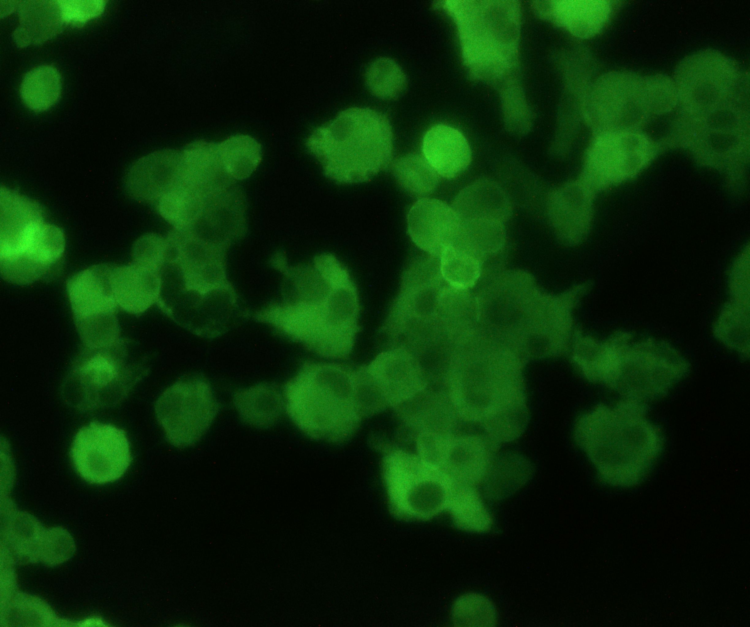
Immunocytochemistry/ Immunofluorescence: F4/80 Antibody (CI-A3-1) - BSA Free [NB600-404]
Immunocytochemistry/Immunofluorescence: F4/80 Antibody (CI-A3-1) [NB600-404] - Analysis of monocytes cultured in M-CSF using F4/80 antibody. Primary antibody dilution: 1:200. Overnight incubation in RT. Image from verified customer review.![Flow Cytometry: F4/80 Antibody (CI-A3-1) - BSA Free [NB600-404]](https://resources.rndsystems.com/images/products/F4-80-Antibody-CI-A3-1-BSA-Free-Flow-Cytometry-NB600-404-img0054.jpg "Flow Cytometry: F4/80 Antibody (CI-A3-1) - BSA Free [NB600-404]")
Flow Cytometry: F4/80 Antibody (CI-A3-1) - BSA Free [NB600-404]
Flow Cytometry: F4/80 Antibody (CI-A3-1) - BSA Free [NB600-404] - Surface staining of F4/80 in CT26 colorectal carcinoma tumor model. Using Allophycocyanin conjugated version of the antibody (NB600-404APC). Image from verified customer review![Immunohistochemistry-Frozen: F4/80 Antibody (CI-A3-1) - BSA Free [NB600-404]](https://resources.rndsystems.com/images/products/F4-80-Antibody-CI-A3-1-Immunohistochemistry-Frozen-NB600-404-img0046.jpg "Immunohistochemistry-Frozen: F4/80 Antibody (CI-A3-1) - BSA Free [NB600-404]")
Immunohistochemistry-Frozen: F4/80 Antibody (CI-A3-1) - BSA Free [NB600-404]
Immunohistochemistry-Frozen: F4/80 Antibody (CI-A3-1) [NB600-404] - Frozen mouse spleen with F4/80 anti-mouse antibody.![Immunohistochemistry-Frozen: F4/80 Antibody (CI-A3-1) - BSA Free [NB600-404]](https://resources.rndsystems.com/images/products/F4-80-Antibody-CI-A3-1-Immunohistochemistry-Frozen-NB600-404-img0047.jpg "Immunohistochemistry-Frozen: F4/80 Antibody (CI-A3-1) - BSA Free [NB600-404]")
Immunohistochemistry-Frozen: F4/80 Antibody (CI-A3-1) - BSA Free [NB600-404]
Immunohistochemistry-Frozen: F4/80 Antibody (CI-A3-1) [NB600-404] - Mouse spleen stained with A: Rat anti Mouse. B: CI-A3-1 clone preincubated with 2 molar excess of human anti Idiotypic.![Flow Cytometry: F4/80 Antibody (CI-A3-1) - BSA Free [NB600-404]](https://resources.rndsystems.com/images/products/F4-80-Antibody-CI-A3-1-Flow-Cytometry-NB600-404-img0037.jpg "Flow Cytometry: F4/80 Antibody (CI-A3-1) - BSA Free [NB600-404]")
Flow Cytometry: F4/80 Antibody (CI-A3-1) - BSA Free [NB600-404]
Flow Cytometry: F4/80 Antibody (CI-A3-1) [NB600-404] - Staining of J774 cells with F4/80 antibody.![Flow Cytometry: F4/80 Antibody (CI-A3-1) - BSA Free [NB600-404]](https://resources.rndsystems.com/images/products/F4-80-Antibody-CI-A3-1-Flow-Cytometry-NB600-404-img0043.jpg "Flow Cytometry: F4/80 Antibody (CI-A3-1) - BSA Free [NB600-404]")
Flow Cytometry: F4/80 Antibody (CI-A3-1) - BSA Free [NB600-404]
Flow Cytometry: F4/80 Antibody (CI-A3-1) [NB600-404] - Staining of mouse peritoneal macrophages with clone CI-A3-1 F4/80 antibody.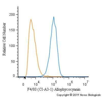
Flow Cytometry: F4/80 Antibody (CI-A3-1) [NB600-404]
 [NB600-404] -")
Immunohistochemistry: F4/80 Antibody (Cl:A3-1) [NB600-404] -
Immunohistochemistry: F4/80 Antibody (Cl:A3-1) [NB600-404] - Lymphocyte interaction inside the DC harboring scaffoldsDC scaffolds (beDC) or scaffolds only, placed in tumor bearing mice or DC-scaffold placed in normal mice were recovered two weeks post-implantation & processed for immunohistochemistry to demonstrate various cell types. (A) Relative size of the biomatrices harvested from various mice groups as depicted in the figure (far left panel). n = 3–5. The dotted line in ‘gross specimen’ differentiates the biomatrix from the reactive host-tissue. Representative images of biomatrix sections stained by the respective antibodies as indicated in the figure. The bar in the ‘gross structure’ is equivalent to 1 mm, whereas for other panels is equal to 100 μm. (B) Snapshots from Supplementary Movie S2 showing lymphocyte (green) movement & interaction with DCs (red) inside the biomatrix. The start point of the observation was set as the zero time point. (C) Lymphocyte division (white arrows) inside the biomatrix in the vicinity of DC (red). Representative snapshot from Supplementary Movie S3. Image collected & cropped by CiteAb from the following publication (https://pubmed.ncbi.nlm.nih.gov/27223090), licensed under a CC-BY license. Not internally tested by Novus Biologicals. - BSA Free [NB600-404] -")
Western Blot: F4/80 Antibody (Cl:A3-1) - BSA Free [NB600-404] -
MFD alters immune signaling in adipose tissue during (a) acute and (b) chronic Mtb infection. Immunoblot analysis of immune cells (CD4, CD8, and F4/80) and inflammatory markers (IFN gamma, TNF alpha, IL-6, and IL-10) in the WAT lysates of RD-fed and MFD-fed uninfected and infected C57BL/6 mice during acute infection (30 DPI) and chronic (3 months) post-infection is shown. The error bars represent standard error of the mean. * p < 0.05, ** p < 0.01, and *** p < 0.001 between indicated groups. Image collected and cropped by CiteAb from the following open publication (https://pubmed.ncbi.nlm.nih.gov/36676177), licensed under a CC-BY license. Not internally tested by Novus Biologicals. - BSA Free [NB600-404] -")
Western Blot: F4/80 Antibody (Cl:A3-1) - BSA Free [NB600-404] -
MFD alters the level of infiltrated immune cells and inflammatory markers in the lungs during chronic Mtb infection. Immunoblot analysis of (a) immune cells (CD4+, CD8+, and F4/80+) and (b) inflammatory markers (IFN gamma, TNF alpha, and IL-6) in the lung lysates of RD-fed and MFD-fed uninfected and infected C57BL/6 mice during chronic (90 DPI) infection. The error bars represent standard error of the mean. * p < 0.05, ** p < 0.01, and *** p < 0.001 between indicated groups. Image collected and cropped by CiteAb from the following open publication (https://pubmed.ncbi.nlm.nih.gov/36676177), licensed under a CC-BY license. Not internally tested by Novus Biologicals. - BSA Free [NB600-404] -")
Western Blot: F4/80 Antibody (Cl:A3-1) - BSA Free [NB600-404] -
MFD alters immune signaling in adipose tissue during (a) acute and (b) chronic Mtb infection. Immunoblot analysis of immune cells (CD4, CD8, and F4/80) and inflammatory markers (IFN gamma, TNF alpha, IL-6, and IL-10) in the WAT lysates of RD-fed and MFD-fed uninfected and infected C57BL/6 mice during acute infection (30 DPI) and chronic (3 months) post-infection is shown. The error bars represent standard error of the mean. * p < 0.05, ** p < 0.01, and *** p < 0.001 between indicated groups. Image collected and cropped by CiteAb from the following open publication (https://pubmed.ncbi.nlm.nih.gov/36676177), licensed under a CC-BY license. Not internally tested by Novus Biologicals.Applications for F4/80 Antibody (Cl:A3-1) - BSA Free
Application
Recommended Usage
Flow Cytometry
1:50-1:100
Immunocytochemistry/ Immunofluorescence
1:10-1:500
Immunohistochemistry
1:10-1:500
Immunohistochemistry-Frozen
1:10-1:500
Immunohistochemistry-Paraffin
1:10-1:500
Immunoprecipitation
1:10-1:500RadioImmunoassay 1:100-1:2000
Radioimmunoassay
1:100-1:2000
Western Blot
1:100-1:2000
Application Notes
This product requires pretreatment of paraffin sections prior to staining. Proteinase K is recommended for tissues fixed for less than 24 hours. Citrate buffer pH 6.0 is recommended for tissues fixed for more than 24 hours. E. coli reactivity reported in scientific literature (PMID:31181736).
Reviewed Applications
Read 2 reviews rated 4 using NB600-404 in the following applications:
Flow Cytometry Panel Builder
Bio-Techne Knows Flow Cytometry
Save time and reduce costly mistakes by quickly finding compatible reagents using the Panel Builder Tool.
Advanced Features
- Spectra Viewer - Custom analysis of spectra from multiple fluorochromes
- Spillover Popups - Visualize the spectra of individual fluorochromes
- Antigen Density Selector - Match fluorochrome brightness with antigen density

Formulation, Preparation, and Storage
Purification
Protein G purified
Formulation
PBS
Format
BSA Free
Preservative
0.09% Sodium Azide
Concentration
1.0 mg/ml
Shipping
The product is shipped with polar packs. Upon receipt, store it immediately at the temperature recommended below.
Stability & Storage
Store at 4C short term. Aliquot and store at -20C long term. Avoid freeze-thaw cycles.
Background: F4/80
Depending on factors such as differentiation, activation, and location, the level of F4/80 expression can vary between cell-types (1,2). For example, peritoneal macrophages are subdivided based on morphology and expression with large peritoneal macrophages having high levels of F4/80 and small peritoneal macrophages having low F4/80 expression (1). Furthermore, splenic macrophages are classified based on location (red pulp, marginal zone, and white pulp) and F4/80 expression level varies within these populations as well; the red pulp macrophages have high levels of F4/80 and CD68 by comparison (1). Originally, the F4/80 monoclonal antibody was generated for a macrophage-specific surface antigen, however F4/80 staining is also observed in other hematopoietic cell populations, thus other immune cell markers should be used in conjunction with F4/80 to distinguish between different populations (1). Though the full extent of F4/80 ligands and biological functions remains elusive, studies have suggested that F4/80 is critical for macrophage-natural killer cell interaction and cytokine release (1,2). Additionally, a study of anterior chamber-associated immune deviation (ACAID) indicated a possible role for F4/80 in the generation of regulatory T cells (1,2).
References
1. Dos Anjos Cassado A. (2017). F4/80 as a Major Macrophage Marker: The Case of the Peritoneum and Spleen. Results and Problems in Cell Differentiation. https://doi.org/10.1007/978-3-319-54090-0_7
2. Hamann, J., Hsiao, C. C., Lee, C. S., Ravichandran, K. S., & Lin, H. H. (2016). Adhesion GPCRs as Modulators of Immune Cell Function. Handbook of Experimental Pharmacology. https://doi.org/10.1007/978-3-319-41523-9_15
Additional F4/80 Products
Product Documents for F4/80 Antibody (Cl:A3-1) - BSA Free
Certificate of Analysis
To download a Certificate of Analysis, please enter a lot or batch number in the search box below.
Product Specific Notices for F4/80 Antibody (Cl:A3-1) - BSA Free
This product is for research use only and is not approved for use in humans or in clinical diagnosis. Primary Antibodies are guaranteed for 1 year from date of receipt.
Related Research Areas
Citations for F4/80 Antibody (Cl:A3-1) - BSA Free
 Powered by Bioz
Customer Reviews for F4/80 Antibody (Cl:A3-1) - BSA Free (2)
4 out of 5
2 Customer Ratings
Have you used F4/80 Antibody (Cl:A3-1) - BSA Free?
Submit a review and receive an Amazon gift card!
$25/€18/£15/$25CAN/¥2500 Yen for a review with an image
$10/€7/£6/$10CAN/¥1110 Yen for a review without an image
Submit a review
Customer Images

Showing
1
-
2 of
2 reviews
Showing All
Filter By:
-
Application: ImmunocytochemistrySample Tested: Bone Marrrow Derived MacrophagesSpecies: MouseVerified Customer | Posted 02/07/2022Monocytes cultured in M-CSF are positive for F4/801:200 Dilution Overnight incubation in RT

-
Application: Immunohistochemistry-ParaffinSample Tested: mouse lymph node FFPESpecies: MouseVerified Customer | Posted 03/26/2015mouse lymph node
There are no reviews that match your criteria.
Protocols
View specific protocols for F4/80 Antibody (Cl:A3-1) - BSA Free (NB600-404):
Immunocytochemistry Protocol
Culture cells to appropriate density in 35 mm culture dishes or 6-well plates.
1. Remove culture medium and add 10% formalin to the dish. Fix at room temperature for 30 minutes.
2. Remove the formalin and add ice cold methanol. Incubate for 5-10 minutes.
3. Remove methanol and add washing solution (i.e. PBS). Be sure to not let the specimen dry out. Wash three times for 10 minutes.
4. To block nonspecific antibody binding incubate in 10% normal goat serum from 1 hour to overnight at room temperature.
5. Add primary antibody at appropriate dilution and incubate at room temperature from 2 hours to overnight at room temperature.
6. Remove primary antibody and replace with washing solution. Wash three times for 10 minutes.
7. Add secondary antibody at appropriate dilution. Incubate for 1 hour at room temperature.
8. Remove antibody and replace with wash solution, then wash for 10 minutes. Add Hoechst 33258 to wash solution at 1:25,0000 and incubate for 10 minutes. Wash a third time for 10 minutes.
9. Cells can be viewed directly after washing. The plates can also be stored in PBS containing Azide covered in Parafilm (TM). Cells can also be cover-slipped using Fluoromount, with appropriate sealing.
*The above information is only intended as a guide. The researcher should determine what protocol best meets their needs. Please follow safe laboratory procedures.
Western Blot Protocol
1. Perform SDS-PAGE on samples to be analyzed, loading 30 ug of total protein per lane.
2. Transfer proteins to membrane according to the instructions provided by the manufacturer of the membrane and transfer apparatus.
3. Stain according to standard Ponceau S procedure (or similar product) to assess transfer success, and mark molecular weight standards where appropriate.
4. Rinse the blot.
5. Block the membrane using standard blocking buffer for at least 1 hour.
6. Wash the membrane in wash buffer three times for 10 minutes each.
7. Dilute primary antibody in blocking buffer and incubate 1 hour at room temperature.
8. Wash the membrane in wash buffer three times for 10 minutes each.
9. Apply the diluted HRP conjugated secondary antibody in blocking buffer (as per manufacturers instructions) and incubate 1 hour at room temperature.
10. Wash the blot in wash buffer three times for 10 minutes each (this step can be repeated as required to reduce background).
11. Apply the detection reagent of choice in accordance with the manufacturers instructions.
**Note: Tween-20 can be added to the blocking or antibody dilution buffer at a final concentration of 0.05-0.2%.
Find general support by application which include: protocols, troubleshooting, illustrated assays, videos and webinars.
- 7-Amino Actinomycin D (7-AAD) Cell Viability Flow Cytometry Protocol
- Antigen Retrieval Protocol (PIER)
- Antigen Retrieval for Frozen Sections Protocol
- Appropriate Fixation of IHC/ICC Samples
- Cellular Response to Hypoxia Protocols
- Chromogenic IHC Staining of Formalin-Fixed Paraffin-Embedded (FFPE) Tissue Protocol
- Chromogenic Immunohistochemistry Staining of Frozen Tissue
- ClariTSA™ Fluorophore Kits
- Detection & Visualization of Antibody Binding
- Extracellular Membrane Flow Cytometry Protocol
- Flow Cytometry Protocol for Cell Surface Markers
- Flow Cytometry Protocol for Staining Membrane Associated Proteins
- Flow Cytometry Staining Protocols
- Flow Cytometry Troubleshooting Guide
- Fluorescent IHC Staining of Frozen Tissue Protocol
- Graphic Protocol for Heat-induced Epitope Retrieval
- Graphic Protocol for the Preparation and Fluorescent IHC Staining of Frozen Tissue Sections
- Graphic Protocol for the Preparation and Fluorescent IHC Staining of Paraffin-embedded Tissue Sections
- Graphic Protocol for the Preparation of Gelatin-coated Slides for Histological Tissue Sections
- ICC Cell Smear Protocol for Suspension Cells
- ICC Immunocytochemistry Protocol Videos
- ICC for Adherent Cells
- IHC Sample Preparation (Frozen sections vs Paraffin)
- Immunocytochemistry (ICC) Protocol
- Immunocytochemistry Troubleshooting
- Immunofluorescence of Organoids Embedded in Cultrex Basement Membrane Extract
- Immunofluorescent IHC Staining of Formalin-Fixed Paraffin-Embedded (FFPE) Tissue Protocol
- Immunohistochemistry (IHC) and Immunocytochemistry (ICC) Protocols
- Immunohistochemistry Frozen Troubleshooting
- Immunohistochemistry Paraffin Troubleshooting
- Immunoprecipitation Protocol
- Intracellular Flow Cytometry Protocol Using Alcohol (Methanol)
- Intracellular Flow Cytometry Protocol Using Detergents
- Intracellular Nuclear Staining Flow Cytometry Protocol Using Detergents
- Intracellular Staining Flow Cytometry Protocol Using Alcohol Permeabilization
- Intracellular Staining Flow Cytometry Protocol Using Detergents to Permeabilize Cells
- Preparing Samples for IHC/ICC Experiments
- Preventing Non-Specific Staining (Non-Specific Binding)
- Primary Antibody Selection & Optimization
- Propidium Iodide Cell Viability Flow Cytometry Protocol
- Protocol for Heat-Induced Epitope Retrieval (HIER)
- Protocol for Liperfluo
- Protocol for Making a 4% Formaldehyde Solution in PBS
- Protocol for VisUCyte™ HRP Polymer Detection Reagent
- Protocol for the Characterization of Human Th22 Cells
- Protocol for the Characterization of Human Th9 Cells
- Protocol for the Fluorescent ICC Staining of Cell Smears - Graphic
- Protocol for the Fluorescent ICC Staining of Cultured Cells on Coverslips - Graphic
- Protocol for the Preparation & Fixation of Cells on Coverslips
- Protocol for the Preparation and Chromogenic IHC Staining of Frozen Tissue Sections
- Protocol for the Preparation and Chromogenic IHC Staining of Frozen Tissue Sections - Graphic
- Protocol for the Preparation and Chromogenic IHC Staining of Paraffin-embedded Tissue Sections
- Protocol for the Preparation and Chromogenic IHC Staining of Paraffin-embedded Tissue Sections - Graphic
- Protocol for the Preparation and Fluorescent ICC Staining of Cells on Coverslips
- Protocol for the Preparation and Fluorescent ICC Staining of Non-adherent Cells
- Protocol for the Preparation and Fluorescent ICC Staining of Stem Cells on Coverslips
- Protocol for the Preparation and Fluorescent IHC Staining of Frozen Tissue Sections
- Protocol for the Preparation and Fluorescent IHC Staining of Paraffin-embedded Tissue Sections
- Protocol for the Preparation of Gelatin-coated Slides for Histological Tissue Sections
- Protocol for the Preparation of a Cell Smear for Non-adherent Cell ICC - Graphic
- Protocol: Annexin V and PI Staining by Flow Cytometry
- Protocol: Annexin V and PI Staining for Apoptosis by Flow Cytometry
- R&D Systems Quality Control Western Blot Protocol
- TUNEL and Active Caspase-3 Detection by IHC/ICC Protocol
- The Importance of IHC/ICC Controls
- Troubleshooting Guide: Fluorokine Flow Cytometry Kits
- Troubleshooting Guide: Immunohistochemistry
- Troubleshooting Guide: Western Blot Figures
- Western Blot Conditions
- Western Blot Protocol
- Western Blot Protocol for Cell Lysates
- Western Blot Troubleshooting
- Western Blot Troubleshooting Guide
- View all Protocols, Troubleshooting, Illustrated assays and Webinars
FAQs for F4/80 Antibody (Cl:A3-1) - BSA Free
Showing
1
-
4 of
4 FAQs
Showing All
-
Q: Are the F4/80/EMR1 antibodies validated in Simple Western?
A: No, F4/80/EMR1 antibodies are not tested in Simple Western at this moment.
-
Q: Does F4/80/EMR1 antibodies come in lyophilized form?
A: Yes, we carry 2 F4/80 antibodies in lyophilized form: MAB5580 and NBP1-06975
-
Q: I'm hoping to use F4/80 as a macrophage marker in skeletal muscle using IHC-Fr but I haven't seen this done successfully (and heard people have tried without much luck). Do you know of any validation that has been done on that tissue?
A: We have not validated NB600-404 on skeletal muscle specifically, and we haven't yet had any customer feedback regarding this antibody being used on skeletal muscle. If you are interested in using this antibody for your testing, and have any trouble getting it to work for you, please let us know. We do offer a guarantee on our products for the species and applications listed, and if we can't get this product working for you, we will compensate you for the purchase.
-
Q: What is the theoretical molecular weight for F4/80/EMR1 antibodies?
A: The TMW of F4/80/EMR1 antibodies is approximately 160 kDa.
-
Q: Are the F4/80/EMR1 antibodies validated in Simple Western?
A: No, F4/80/EMR1 antibodies are not tested in Simple Western at this moment.
-
Q: Does F4/80/EMR1 antibodies come in lyophilized form?
A: Yes, we carry 2 F4/80 antibodies in lyophilized form: MAB5580 and NBP1-06975
-
Q: I'm hoping to use F4/80 as a macrophage marker in skeletal muscle using IHC-Fr but I haven't seen this done successfully (and heard people have tried without much luck). Do you know of any validation that has been done on that tissue?
A: We have not validated NB600-404 on skeletal muscle specifically, and we haven't yet had any customer feedback regarding this antibody being used on skeletal muscle. If you are interested in using this antibody for your testing, and have any trouble getting it to work for you, please let us know. We do offer a guarantee on our products for the species and applications listed, and if we can't get this product working for you, we will compensate you for the purchase.
-
Q: What is the theoretical molecular weight for F4/80/EMR1 antibodies?
A: The TMW of F4/80/EMR1 antibodies is approximately 160 kDa.
Loading...