Fibroblast Antibody (ER-TR7) - BSA Free
Novus Biologicals | Catalog # NB100-64932
Key Product Details
Validated by
Species Reactivity
Validated:
Cited:
Applications
Validated:
Cited:
Label
Antibody Source
Format
Product Specifications
Immunogen
Marker
Specificity
Clonality
Host
Isotype
Scientific Data Images for Fibroblast Antibody (ER-TR7) - BSA Free
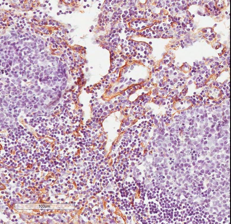
![Immunohistochemistry: Fibroblast Antibody (ER-TR7) - BSA Free [NB100-64932]](https://resources.rndsystems.com/images/products/Fibroblast-Antibody-ER-TR7-Immunohistochemistry-NB100-64932-img0002.jpg "Immunohistochemistry: Fibroblast Antibody (ER-TR7) - BSA Free [NB100-64932]")
![Immunohistochemistry-Frozen: Fibroblast Antibody (ER-TR7) - BSA Free [NB100-64932]](https://resources.rndsystems.com/images/products/Fibroblast-Antibody-ER-TR7-Immunohistochemistry-Frozen-NB100-64932-img0001.jpg "Immunohistochemistry-Frozen: Fibroblast Antibody (ER-TR7) - BSA Free [NB100-64932]")
Immunohistochemistry-Frozen: Fibroblast Antibody (ER-TR7) - BSA Free [NB100-64932]
Immunohistochemistry-Frozen: Fibroblast Antibody (ER-TR7) [NB100-64932] - Fibroblast Antibody (ER-TR7) IHC on Mouse lymph node, frozen section, 20x. Primary antibody diluted 1:500. This image was submitted via customer Review.Applications for Fibroblast Antibody (ER-TR7) - BSA Free
Flow Cytometry
Immunocytochemistry/ Immunofluorescence
Immunohistochemistry
Immunohistochemistry-Frozen
Immunohistochemistry-Paraffin
Reviewed Applications
Read 1 review rated 4 using NB100-64932 in the following applications:
Flow Cytometry Panel Builder
Bio-Techne Knows Flow Cytometry
Save time and reduce costly mistakes by quickly finding compatible reagents using the Panel Builder Tool.
Advanced Features
- Spectra Viewer - Custom analysis of spectra from multiple fluorochromes
- Spillover Popups - Visualize the spectra of individual fluorochromes
- Antigen Density Selector - Match fluorochrome brightness with antigen density

Formulation, Preparation, and Storage
Purification
Formulation
Format
Preservative
Concentration
Shipping
Stability & Storage
Background: Fibroblast
Alternate Names
Additional Fibroblast Products
Product Documents for Fibroblast Antibody (ER-TR7) - BSA Free
Certificate of Analysis
To download a Certificate of Analysis, please enter a lot or batch number in the search box below.
Product Specific Notices for Fibroblast Antibody (ER-TR7) - BSA Free
This product is for research use only and is not approved for use in humans or in clinical diagnosis. Primary Antibodies are guaranteed for 1 year from date of receipt.
Citations for Fibroblast Antibody (ER-TR7) - BSA Free
 Powered by Bioz
Customer Reviews for Fibroblast Antibody (ER-TR7) - BSA Free (1)
Have you used Fibroblast Antibody (ER-TR7) - BSA Free?
Submit a review and receive an Amazon gift card!
$25/€18/£15/$25CAN/¥2500 Yen for a review with an image
$10/€7/£6/$10CAN/¥1110 Yen for a review without an image
Submit a review
Customer Images
-
Application: ImmunohistochemistrySample Tested: PFA Fixed Frosen Section Mouse LymphnodeSpecies: MouseVerified Customer | Posted 06/29/2017Fibroblast Antibody (ER-TR7) IHC on Mouse lymph node, frozen section, 20xWD [1:500]; secondary ab RTU Polymer-HRP

There are no reviews that match your criteria.
Protocols
View specific protocols for Fibroblast Antibody (ER-TR7) - BSA Free (NB100-64932):
Immunohistochemistry-Paraffin Embedded Sections
Antigen Unmasking:
Bring slides to a boil in 10 mM sodium citrate buffer (pH 6.0) then maintain at a sub-boiling temperature for 10 minutes. Cool slides on bench-top for 30 minutes.
Staining:
1. Wash sections in deionized water three times for 5 minutes each.
2. Wash sections in wash buffer for 5 minutes.
3. Block each section with 100-400 ul blocking solution for 1 hour at room temperature.
4. Remove blocking solution and add 100-400 ul diluted primary antibody. Incubate overnight at 4 C.
5. Remove antibody solution and wash sections in wash buffer three times for 5 minutes each.
6. Add 100-400 ul biotinylated diluted secondary antibody. Incubate 30 minutes at room temperature.
7. Remove secondary antibody solution and wash sections three times with wash buffer for 5 minutes each.
8. Add 100-400 ul Streptavidin-HRP reagent to each section and incubate for 30 minutes at room temperature.
9. Wash sections three times in wash buffer for 5 minutes each.
10. Add 100-400 ul DAB substrate to each section and monitor staining closely.
11. As soon as the sections develop, immerse slides in deionized water.
12. Counterstain sections in hematoxylin.
13. Wash sections in deionized water two times for 5 minutes each.
14. Dehydrate sections.
15. Mount coverslips.
Find general support by application which include: protocols, troubleshooting, illustrated assays, videos and webinars.
- 7-Amino Actinomycin D (7-AAD) Cell Viability Flow Cytometry Protocol
- Antigen Retrieval Protocol (PIER)
- Antigen Retrieval for Frozen Sections Protocol
- Appropriate Fixation of IHC/ICC Samples
- Cellular Response to Hypoxia Protocols
- Chromogenic IHC Staining of Formalin-Fixed Paraffin-Embedded (FFPE) Tissue Protocol
- Chromogenic Immunohistochemistry Staining of Frozen Tissue
- ClariTSA™ Fluorophore Kits
- Detection & Visualization of Antibody Binding
- Extracellular Membrane Flow Cytometry Protocol
- Flow Cytometry Protocol for Cell Surface Markers
- Flow Cytometry Protocol for Staining Membrane Associated Proteins
- Flow Cytometry Staining Protocols
- Flow Cytometry Troubleshooting Guide
- Fluorescent IHC Staining of Frozen Tissue Protocol
- Graphic Protocol for Heat-induced Epitope Retrieval
- Graphic Protocol for the Preparation and Fluorescent IHC Staining of Frozen Tissue Sections
- Graphic Protocol for the Preparation and Fluorescent IHC Staining of Paraffin-embedded Tissue Sections
- Graphic Protocol for the Preparation of Gelatin-coated Slides for Histological Tissue Sections
- ICC Cell Smear Protocol for Suspension Cells
- ICC Immunocytochemistry Protocol Videos
- ICC for Adherent Cells
- IHC Sample Preparation (Frozen sections vs Paraffin)
- Immunocytochemistry (ICC) Protocol
- Immunocytochemistry Troubleshooting
- Immunofluorescence of Organoids Embedded in Cultrex Basement Membrane Extract
- Immunofluorescent IHC Staining of Formalin-Fixed Paraffin-Embedded (FFPE) Tissue Protocol
- Immunohistochemistry (IHC) and Immunocytochemistry (ICC) Protocols
- Immunohistochemistry Frozen Troubleshooting
- Immunohistochemistry Paraffin Troubleshooting
- Intracellular Flow Cytometry Protocol Using Alcohol (Methanol)
- Intracellular Flow Cytometry Protocol Using Detergents
- Intracellular Nuclear Staining Flow Cytometry Protocol Using Detergents
- Intracellular Staining Flow Cytometry Protocol Using Alcohol Permeabilization
- Intracellular Staining Flow Cytometry Protocol Using Detergents to Permeabilize Cells
- Preparing Samples for IHC/ICC Experiments
- Preventing Non-Specific Staining (Non-Specific Binding)
- Primary Antibody Selection & Optimization
- Propidium Iodide Cell Viability Flow Cytometry Protocol
- Protocol for Heat-Induced Epitope Retrieval (HIER)
- Protocol for Liperfluo
- Protocol for Making a 4% Formaldehyde Solution in PBS
- Protocol for VisUCyte™ HRP Polymer Detection Reagent
- Protocol for the Characterization of Human Th22 Cells
- Protocol for the Characterization of Human Th9 Cells
- Protocol for the Fluorescent ICC Staining of Cell Smears - Graphic
- Protocol for the Fluorescent ICC Staining of Cultured Cells on Coverslips - Graphic
- Protocol for the Preparation & Fixation of Cells on Coverslips
- Protocol for the Preparation and Chromogenic IHC Staining of Frozen Tissue Sections
- Protocol for the Preparation and Chromogenic IHC Staining of Frozen Tissue Sections - Graphic
- Protocol for the Preparation and Chromogenic IHC Staining of Paraffin-embedded Tissue Sections
- Protocol for the Preparation and Chromogenic IHC Staining of Paraffin-embedded Tissue Sections - Graphic
- Protocol for the Preparation and Fluorescent ICC Staining of Cells on Coverslips
- Protocol for the Preparation and Fluorescent ICC Staining of Non-adherent Cells
- Protocol for the Preparation and Fluorescent ICC Staining of Stem Cells on Coverslips
- Protocol for the Preparation and Fluorescent IHC Staining of Frozen Tissue Sections
- Protocol for the Preparation and Fluorescent IHC Staining of Paraffin-embedded Tissue Sections
- Protocol for the Preparation of Gelatin-coated Slides for Histological Tissue Sections
- Protocol for the Preparation of a Cell Smear for Non-adherent Cell ICC - Graphic
- Protocol: Annexin V and PI Staining by Flow Cytometry
- Protocol: Annexin V and PI Staining for Apoptosis by Flow Cytometry
- TUNEL and Active Caspase-3 Detection by IHC/ICC Protocol
- The Importance of IHC/ICC Controls
- Troubleshooting Guide: Fluorokine Flow Cytometry Kits
- Troubleshooting Guide: Immunohistochemistry
- View all Protocols, Troubleshooting, Illustrated assays and Webinars
FAQs for Fibroblast Antibody (ER-TR7) - BSA Free
-
Q: I am currently in search of an antibody for staining of human dermal fibroblasts. Can you recommend me an antibody for this? Is there an antibody that specifically stains only fibroblasts and not mesenchymal stem cells?
A:
We sell a number of antibodies which are suitable for staining fibroblasts. You can see these at the following links: NB100-64932, NB110-15846 and NB100-1845. NB100-64932 recognizes ER-TR7, an antigen that is located in the cytoplasm of reticular fibroblasts and is a component of the extracellular matrix of lymphoid and non-lymphoid organs. The antigen recognized by clone ER-TR7 has not been identified but studies suggest that it is likely to be distinct from laminin, fibronectin, collagen types I-IV, heparin sulphate proteoglycan, entactin and nidogen. NB110-15846 is specific to beta subunit of prolyl 4-hydroxylase. It reacts with fibroblasts and myoepithelial cells, and does not stain lymphocytes, monocytes, dendritic cells or granulocytes. NB100-1845 recognizes an epitope on primary cultured human fibroblasts, fibroblast cell lines, tissue macrophages and 95% of peripheral blood monocytes. It does not react with vascular smooth muscle cells. Unfortunately, we do not have any data regarding the staining of mesenchymal stem cells with any of these antibodies in our QC lab.