Fibroblasts Antibody (TE-7)
Novus Biologicals | Catalog # NBP2-50082
![Immunocytochemistry/ Immunofluorescence: Fibroblasts Antibody (TE-7) [NBP2-50082]](https://resources.rndsystems.com/images/products/Fibroblasts-Antibody-TE-7-Immunocytochemistry-NBP2-50082-img0005.jpg "Immunocytochemistry/ Immunofluorescence: Fibroblasts Antibody (TE-7) [NBP2-50082]")
Loading...
Key Product Details
Species Reactivity
Validated:
Human
Cited:
Human, Mouse
Applications
Validated:
Immunohistochemistry, Immunohistochemistry-Paraffin, Flow Cytometry, Immunocytochemistry/ Immunofluorescence
Cited:
Immunohistochemistry, Immunocytochemistry/ Immunofluorescence
Label
Unconjugated
Antibody Source
Monoclonal Mouse IgG1 Clone # TE-7
Loading...
Product Specifications
Immunogen
Whole human thymic stoma cells (Haynes, B.F., et al. (1984). J. Exp. Med. 159(4):1149-1168).
Specificity
The antibody reacts with fibroblasts in tissue as well as cultured fibroblasts. On skin sections the antibody reacts with stratum basal epidermal cells. The antigen defined by antibody TE-7 appears to be a marker for mesodermally dervied (mesenchymal) human connective tissue and tumors derived from such tissue, e.g. fibrosarcomas (Haynes, B.F., et al. (1984). J. Exp. Med. 159(4):1149-1168). The antibody is useful for the assessment of thymic epithelial culture contamination with fibroblasts, and the identification of cells of mesodermal origin in the fetus.
Clonality
Monoclonal
Host
Mouse
Isotype
IgG1
Scientific Data Images for Fibroblasts Antibody (TE-7)
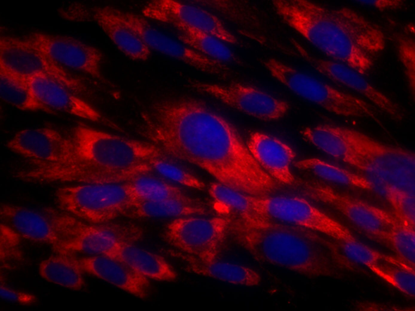
Immunocytochemistry/ Immunofluorescence: Fibroblasts Antibody (TE-7) [NBP2-50082]
Immunocytochemistry/Immunofluorescence: Fibroblasts Antibody (TE-7) [NBP2-50082] - MRC-5 human embryonic lung fibroblast cell line, fixation with PFA 4%, blocking with PBS 1X + 1% BSA + 0.1% Tween, primary antibody used at 1:50 in blocking buffer, O/N. Secondary antibody: goat anti mouse Alexa Fluor 594 for 1 hour at RT. Signal was detected by fluorescence microscopy at 40X. This image was submitted via customer review.![Immunohistochemistry-Paraffin: Fibroblasts Antibody (TE-7) [NBP2-50082]](https://resources.rndsystems.com/images/products/Fibroblasts-Antibody-TE-7-Immunohistochemistry-Paraffin-NBP2-50082-img0004.jpg "Immunohistochemistry-Paraffin: Fibroblasts Antibody (TE-7) [NBP2-50082]")
Immunohistochemistry-Paraffin: Fibroblasts Antibody (TE-7) [NBP2-50082]
Immunohistochemistry-Paraffin: Fibroblasts Antibody (TE-7) [NBP2-50082] - Paraffin-embedded gastrocnemius section from a patient with myopathic condition was performed overnight at 4 degrees C using 10 ug/mL.Applications for Fibroblasts Antibody (TE-7)
Application
Recommended Usage
Flow Cytometry
1:100
Immunocytochemistry/ Immunofluorescence
1:100-1:200
Reviewed Applications
Read 1 review rated 5 using NBP2-50082 in the following applications:
Flow Cytometry Panel Builder
Bio-Techne Knows Flow Cytometry
Save time and reduce costly mistakes by quickly finding compatible reagents using the Panel Builder Tool.
Advanced Features
- Spectra Viewer - Custom analysis of spectra from multiple fluorochromes
- Spillover Popups - Visualize the spectra of individual fluorochromes
- Antigen Density Selector - Match fluorochrome brightness with antigen density

Formulation, Preparation, and Storage
Purification
Protein A or G purified
Formulation
PBS with 1 mg/ml BSA
Preservative
0.07% Sodium Azide
Concentration
0.1 mg/ml
Shipping
The product is shipped with polar packs. Upon receipt, store it immediately at the temperature recommended below.
Stability & Storage
Store at 4C short term. Aliquot and store at -20C long term. Avoid freeze-thaw cycles.
Background: Fibroblasts
Alternate Names
fibro-blasts
Additional Fibroblasts Products
Product Documents for Fibroblasts Antibody (TE-7)
Certificate of Analysis
To download a Certificate of Analysis, please enter a lot or batch number in the search box below.
Product Specific Notices for Fibroblasts Antibody (TE-7)
This product is for research use only and is not approved for use in humans or in clinical diagnosis. Primary Antibodies are guaranteed for 1 year from date of receipt.
Citations for Fibroblasts Antibody (TE-7)
 Powered by Bioz
Customer Reviews for Fibroblasts Antibody (TE-7) (1)
5 out of 5
1 Customer Rating
Have you used Fibroblasts Antibody (TE-7)?
Submit a review and receive an Amazon gift card!
$25/€18/£15/$25CAN/¥2500 Yen for a review with an image
$10/€7/£6/$10CAN/¥1110 Yen for a review without an image
Submit a review
Customer Images
Showing
1
-
1 of
1 review
Showing All
Filter By:
-
Application: ImmunocytochemistrySample Tested: MRC-5 human embryonic lung fibroblast cell lineSpecies: HumanVerified Customer | Posted 03/22/2018MRC-5 human embryonic lung fibroblast cellsMRC-5 human embryonic lung fibroblast cell line, fixation with PFA 4%, blocking with PBS 1X + BSA 1% + 0.1% Tween, primary antibody used at 1:50 in blocking buffer, O/N

There are no reviews that match your criteria.
Protocols
Find general support by application which include: protocols, troubleshooting, illustrated assays, videos and webinars.
- 7-Amino Actinomycin D (7-AAD) Cell Viability Flow Cytometry Protocol
- Antigen Retrieval Protocol (PIER)
- Antigen Retrieval for Frozen Sections Protocol
- Appropriate Fixation of IHC/ICC Samples
- Cellular Response to Hypoxia Protocols
- Chromogenic IHC Staining of Formalin-Fixed Paraffin-Embedded (FFPE) Tissue Protocol
- Chromogenic Immunohistochemistry Staining of Frozen Tissue
- ClariTSA™ Fluorophore Kits
- Detection & Visualization of Antibody Binding
- Extracellular Membrane Flow Cytometry Protocol
- Flow Cytometry Protocol for Cell Surface Markers
- Flow Cytometry Protocol for Staining Membrane Associated Proteins
- Flow Cytometry Staining Protocols
- Flow Cytometry Troubleshooting Guide
- Fluorescent IHC Staining of Frozen Tissue Protocol
- Graphic Protocol for Heat-induced Epitope Retrieval
- Graphic Protocol for the Preparation and Fluorescent IHC Staining of Frozen Tissue Sections
- Graphic Protocol for the Preparation and Fluorescent IHC Staining of Paraffin-embedded Tissue Sections
- Graphic Protocol for the Preparation of Gelatin-coated Slides for Histological Tissue Sections
- ICC Cell Smear Protocol for Suspension Cells
- ICC Immunocytochemistry Protocol Videos
- ICC for Adherent Cells
- IHC Sample Preparation (Frozen sections vs Paraffin)
- Immunocytochemistry (ICC) Protocol
- Immunocytochemistry Troubleshooting
- Immunofluorescence of Organoids Embedded in Cultrex Basement Membrane Extract
- Immunofluorescent IHC Staining of Formalin-Fixed Paraffin-Embedded (FFPE) Tissue Protocol
- Immunohistochemistry (IHC) and Immunocytochemistry (ICC) Protocols
- Immunohistochemistry Frozen Troubleshooting
- Immunohistochemistry Paraffin Troubleshooting
- Intracellular Flow Cytometry Protocol Using Alcohol (Methanol)
- Intracellular Flow Cytometry Protocol Using Detergents
- Intracellular Nuclear Staining Flow Cytometry Protocol Using Detergents
- Intracellular Staining Flow Cytometry Protocol Using Alcohol Permeabilization
- Intracellular Staining Flow Cytometry Protocol Using Detergents to Permeabilize Cells
- Preparing Samples for IHC/ICC Experiments
- Preventing Non-Specific Staining (Non-Specific Binding)
- Primary Antibody Selection & Optimization
- Propidium Iodide Cell Viability Flow Cytometry Protocol
- Protocol for Heat-Induced Epitope Retrieval (HIER)
- Protocol for Liperfluo
- Protocol for Making a 4% Formaldehyde Solution in PBS
- Protocol for VisUCyte™ HRP Polymer Detection Reagent
- Protocol for the Characterization of Human Th22 Cells
- Protocol for the Characterization of Human Th9 Cells
- Protocol for the Fluorescent ICC Staining of Cell Smears - Graphic
- Protocol for the Fluorescent ICC Staining of Cultured Cells on Coverslips - Graphic
- Protocol for the Preparation & Fixation of Cells on Coverslips
- Protocol for the Preparation and Chromogenic IHC Staining of Frozen Tissue Sections
- Protocol for the Preparation and Chromogenic IHC Staining of Frozen Tissue Sections - Graphic
- Protocol for the Preparation and Chromogenic IHC Staining of Paraffin-embedded Tissue Sections
- Protocol for the Preparation and Chromogenic IHC Staining of Paraffin-embedded Tissue Sections - Graphic
- Protocol for the Preparation and Fluorescent ICC Staining of Cells on Coverslips
- Protocol for the Preparation and Fluorescent ICC Staining of Non-adherent Cells
- Protocol for the Preparation and Fluorescent ICC Staining of Stem Cells on Coverslips
- Protocol for the Preparation and Fluorescent IHC Staining of Frozen Tissue Sections
- Protocol for the Preparation and Fluorescent IHC Staining of Paraffin-embedded Tissue Sections
- Protocol for the Preparation of Gelatin-coated Slides for Histological Tissue Sections
- Protocol for the Preparation of a Cell Smear for Non-adherent Cell ICC - Graphic
- Protocol: Annexin V and PI Staining by Flow Cytometry
- Protocol: Annexin V and PI Staining for Apoptosis by Flow Cytometry
- TUNEL and Active Caspase-3 Detection by IHC/ICC Protocol
- The Importance of IHC/ICC Controls
- Troubleshooting Guide: Fluorokine Flow Cytometry Kits
- Troubleshooting Guide: Immunohistochemistry
- View all Protocols, Troubleshooting, Illustrated assays and Webinars
Loading...