![Western Blot: FLIP Antibody [NB100-56141]](https://resources.rndsystems.com/images/products/FLIP-Antibody-Western-Blot-NB100-56141-img0003.jpg "Western Blot: FLIP Antibody [NB100-56141]")
Loading...
Key Product Details
Species Reactivity
Human
Applications
Immunohistochemistry, Immunohistochemistry-Paraffin, Immunohistochemistry-Frozen, Western Blot, Immunoprecipitation
Label
Unconjugated
Antibody Source
Polyclonal Rabbit IgG
Format
BSA Free
Loading...
Product Specifications
Immunogen
A synthetic peptide corresponding to amino acids 75-92 (DRKAVETHLLRNPHLVS) of human FLIP was used as immunogen, GenBank no. gi|12643547. This amino acid sequnce is 100% conserved in FLIP long and short, and various FLIP isoforms as denoted in the NCBI BLAST data base. http://www.ncbi.nlm.nih.gov/blast/.
Specificity
The gene encoding FLIP produces, by alternative splicing, 24 different transcripts, all with introns, putatively encoding 22 different protein isoforms.
Clonality
Polyclonal
Host
Rabbit
Isotype
IgG
Scientific Data Images for FLIP Antibody - BSA Free
Western Blot: FLIP Antibody [NB100-56141]
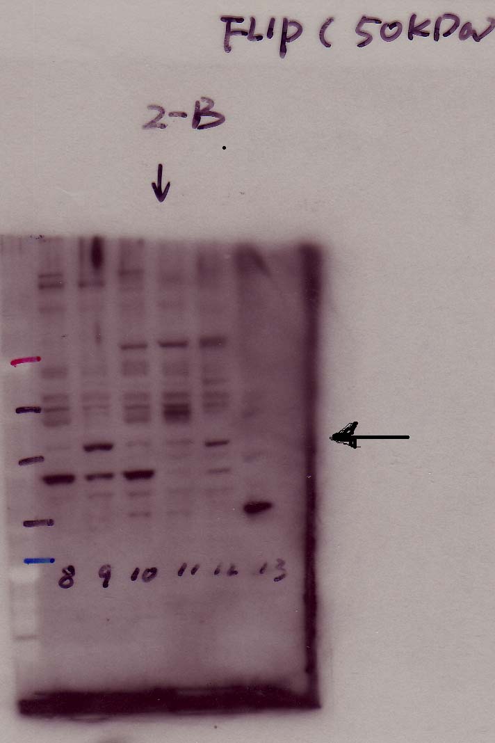
Western Blot: FLIP Antibody [NB100-56141] - analysis of FLIP in human ovarian cancer cell lines using anti-FLIP antibody. The primary antibody was used at a dilution of 1:200 and incubated for overnight at 4C in 5% milk in PBST. Image from verified customer review.![Immunohistochemistry: FLIP Antibody [NB100-56141]](https://resources.rndsystems.com/images/products/FLIP-Antibody-Immunohistochemistry-NB100-56141-img0002.jpg "Immunohistochemistry: FLIP Antibody [NB100-56141]")
Immunohistochemistry: FLIP Antibody [NB100-56141]
Immunohistochemistry: FLIP Antibody [NB100-56141] - Immunhohistochemical analysis of FLIP in tissues cores from a formalin-fixed, paraffin-embedded human brain tumor microarray using NB100-56141 at 1:2000. A-C, tissue cores from a patient with gemistocytoma. A, normal adjacent brain tissue. Astrocytes are positive for Flip expression. B, gemistocytoma (Grade II). Gemistocytes are positive for Flip expression. C, peritumoural oedma brain tissue. Reactive astrocytocytes are positive for FLIP expression. C1, a higher magnification of C. Collectively, the data shows that astrocytes (normal), reactive astrocytes and gemistocytes are positive for Flip expression. Hematoxylin-eosin counterstain.![Immunohistochemistry: FLIP Antibody [NB100-56141]](https://resources.rndsystems.com/images/products/FLIP-Antibody-Immunohistochemistry-NB100-56141-img0001.jpg "Immunohistochemistry: FLIP Antibody [NB100-56141]")
Immunohistochemistry: FLIP Antibody [NB100-56141]
Immunohistochemistry: FLIP Antibody [NB100-56141] - Immunhohistochemical analysis of FLIP in two tissues cores from a formalin-fixed, paraffin-embedded human brain tumor microarray using NB100-56141 at 1:2000. A, gemistocytoma (Grade II) positive for FLIP expression. B, gemistocytoma (Grade II) negative for FLIP expression. A1 and A2 are higher magnifications from A. A, A1, and A2 show expression of FLIP in the gemistocytes of the tumor. Hematoxylin-eosin counterstain.Applications for FLIP Antibody - BSA Free
Application
Recommended Usage
Immunohistochemistry-Paraffin
1:1000-1:5000
Immunoprecipitation
1:50-1:200
Western Blot
1:1000-1:2000
Application Notes
IHC-Frozen: Users should optimize according to model and immunodetection system used (secondary reagents). The molecular weights of these isoforms may vary. Therefore, the molecular weight of FLIP observed by western blot may vary according to the isoforms present in the sample.
Reviewed Applications
Read 1 review rated 4 using NB100-56141 in the following applications:
Formulation, Preparation, and Storage
Purification
Unpurified
Formulation
Whole antisera
Format
BSA Free
Preservative
0.05% Sodium Azide
Concentration
This product is unpurified. The exact concentration of antibody is not quantifiable.
Shipping
The product is shipped with polar packs. Upon receipt, store it immediately at the temperature recommended below.
Stability & Storage
Store at 4C short term. Aliquot and store at -20C long term. Avoid freeze-thaw cycles.
Background: FLIP
Long Name
FLICE Inhibitory Proteins
Alternate Names
CASPER, CFLAR, FLAME-1, I-FLICE
Gene Symbol
CFLAR
Additional FLIP Products
Product Documents for FLIP Antibody - BSA Free
Certificate of Analysis
To download a Certificate of Analysis, please enter a lot or batch number in the search box below.
Product Specific Notices for FLIP Antibody - BSA Free
This product is for research use only and is not approved for use in humans or in clinical diagnosis. Primary Antibodies are guaranteed for 1 year from date of receipt.
Related Research Areas
Customer Reviews for FLIP Antibody - BSA Free (1)
4 out of 5
1 Customer Rating
Have you used FLIP Antibody - BSA Free?
Submit a review and receive an Amazon gift card!
$25/€18/£15/$25CAN/¥2500 Yen for a review with an image
$10/€7/£6/$10CAN/¥1110 Yen for a review without an image
Submit a review
Customer Images
Showing
1
-
1 of
1 review
Showing All
Filter By:
-
Application: Western BlotSample Tested: Human ovarian cancer celll linesSpecies: HumanVerified Customer | Posted 12/05/2014Human ovarian cancer celll lines

There are no reviews that match your criteria.
Protocols
Find general support by application which include: protocols, troubleshooting, illustrated assays, videos and webinars.
- Antigen Retrieval Protocol (PIER)
- Antigen Retrieval for Frozen Sections Protocol
- Appropriate Fixation of IHC/ICC Samples
- Cellular Response to Hypoxia Protocols
- Chromogenic IHC Staining of Formalin-Fixed Paraffin-Embedded (FFPE) Tissue Protocol
- Chromogenic Immunohistochemistry Staining of Frozen Tissue
- ClariTSA™ Fluorophore Kits
- Detection & Visualization of Antibody Binding
- Fluorescent IHC Staining of Frozen Tissue Protocol
- Graphic Protocol for Heat-induced Epitope Retrieval
- Graphic Protocol for the Preparation and Fluorescent IHC Staining of Frozen Tissue Sections
- Graphic Protocol for the Preparation and Fluorescent IHC Staining of Paraffin-embedded Tissue Sections
- Graphic Protocol for the Preparation of Gelatin-coated Slides for Histological Tissue Sections
- IHC Sample Preparation (Frozen sections vs Paraffin)
- Immunofluorescent IHC Staining of Formalin-Fixed Paraffin-Embedded (FFPE) Tissue Protocol
- Immunohistochemistry (IHC) and Immunocytochemistry (ICC) Protocols
- Immunohistochemistry Frozen Troubleshooting
- Immunohistochemistry Paraffin Troubleshooting
- Immunoprecipitation Protocol
- Preparing Samples for IHC/ICC Experiments
- Preventing Non-Specific Staining (Non-Specific Binding)
- Primary Antibody Selection & Optimization
- Protocol for Heat-Induced Epitope Retrieval (HIER)
- Protocol for Making a 4% Formaldehyde Solution in PBS
- Protocol for VisUCyte™ HRP Polymer Detection Reagent
- Protocol for the Preparation & Fixation of Cells on Coverslips
- Protocol for the Preparation and Chromogenic IHC Staining of Frozen Tissue Sections
- Protocol for the Preparation and Chromogenic IHC Staining of Frozen Tissue Sections - Graphic
- Protocol for the Preparation and Chromogenic IHC Staining of Paraffin-embedded Tissue Sections
- Protocol for the Preparation and Chromogenic IHC Staining of Paraffin-embedded Tissue Sections - Graphic
- Protocol for the Preparation and Fluorescent IHC Staining of Frozen Tissue Sections
- Protocol for the Preparation and Fluorescent IHC Staining of Paraffin-embedded Tissue Sections
- Protocol for the Preparation of Gelatin-coated Slides for Histological Tissue Sections
- R&D Systems Quality Control Western Blot Protocol
- TUNEL and Active Caspase-3 Detection by IHC/ICC Protocol
- The Importance of IHC/ICC Controls
- Troubleshooting Guide: Immunohistochemistry
- Troubleshooting Guide: Western Blot Figures
- Western Blot Conditions
- Western Blot Protocol
- Western Blot Protocol for Cell Lysates
- Western Blot Troubleshooting
- Western Blot Troubleshooting Guide
- View all Protocols, Troubleshooting, Illustrated assays and Webinars
Loading...