FXR/NR1H4 Antibody - BSA Free
Novus Biologicals | Catalog # NB400-153
![Western Blot: FXR/NR1H4 Antibody [NB400-153]](https://resources.rndsystems.com/images/products/FXR-NR1H4-Antibody-Western-Blot-NB400-153-img0006.jpg "Western Blot: FXR/NR1H4 Antibody [NB400-153]")
Key Product Details
Species Reactivity
Validated:
Human, Mouse
Cited:
Human, Mouse
Applications
Validated:
Western Blot, Immunocytochemistry/ Immunofluorescence
Cited:
Western Blot
Label
Unconjugated
Antibody Source
Polyclonal Rabbit IgG
Format
BSA Free
Loading...
Product Specifications
Immunogen
A synthetic peptide made to the C-terminus of the human FXR protein. [UniProt# Q96RI1]
Reactivity Notes
Mouse reactivity reported in scientific literature (PMID: 29491146).
Localization
Cytoplasmic, nuclear
Clonality
Polyclonal
Host
Rabbit
Isotype
IgG
Theoretical MW
66 kDa.
Disclaimer note: The observed molecular weight of the protein may vary from the listed predicted molecular weight due to post translational modifications, post translation cleavages, relative charges, and other experimental factors.
Disclaimer note: The observed molecular weight of the protein may vary from the listed predicted molecular weight due to post translational modifications, post translation cleavages, relative charges, and other experimental factors.
Scientific Data Images for FXR/NR1H4 Antibody - BSA Free
Western Blot: FXR/NR1H4 Antibody [NB400-153]
Western Blot: FXR/NR1H4 Antibody [NB400-153] - Total protein from human A549, U2OS cell and mouse 3T3 cells was separated on a 7.5% gel by SDS-PAGE, transferred to PVDF membrane and blocked in 5% non-fat milk in TBST. The membrane was probed with 2.0 ug/ml anti-FXR (NB400-153) in block buffer and detected with an anti-rabbit HRP secondary antibody using NovaLume chemiluminescence detection reagent (NPB2-61915).![Immunocytochemistry/ Immunofluorescence: FXR/NR1H4 Antibody [NB400-153]](https://resources.rndsystems.com/images/products/FXR-NR1H4-Antibody-Immunocytochemistry-Immunofluorescence-NB400-153-img0005.jpg "Immunocytochemistry/ Immunofluorescence: FXR/NR1H4 Antibody [NB400-153]")
Immunocytochemistry/ Immunofluorescence: FXR/NR1H4 Antibody [NB400-153]
Immunocytochemistry/Immunofluorescence: FXR/NR1H4 Antibody [NB400-153] - A549 cells were fixed for 10 minutes using 10% formalin and then permeabilized for 5 minutes using 1X PBS + 0.5% Triton-X100. The cells were incubated with anti-FXR/NR1H4 at 2 ug/ml overnight at 4C and detected with an anti-rabbit Dylight 488 (Green) at a 1:500 dilution. Alpha tubulin (DM1A) NB100-690 was used as a co-stain at a 1:1000 dilution and detected with an anti-mouse Dylight 550 (Red) at a 1:500 dilution. Nuclei were counterstained with DAPI (Blue). Cells were imaged using a 40X objective.![Western Blot: FXR/NR1H4 Antibody [NB400-153]](https://resources.rndsystems.com/images/products/FXR-Antibody-Western-Blot-NB400-153-img0002.jpg "Western Blot: FXR/NR1H4 Antibody [NB400-153]")
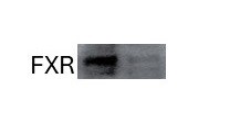
Western Blot: FXR/NR1H4 Antibody [NB400-153]
Western Blot: FXR Antibody [NB400-153] - Detection of FXR in HeLa nuclear extract using NB400-153. ECL 30 minute detection.![Immunocytochemistry/ Immunofluorescence: FXR/NR1H4 Antibody [NB400-153]](https://resources.rndsystems.com/images/products/FXR-NR1H4-Antibody-Immunocytochemistry-Immunofluorescence-NB400-153-img0004.jpg "Immunocytochemistry/ Immunofluorescence: FXR/NR1H4 Antibody [NB400-153]")
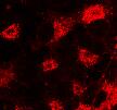
Immunocytochemistry/ Immunofluorescence: FXR/NR1H4 Antibody [NB400-153]
Immunocytochemistry/Immunofluorescence: FXR/NR1H4 Antibody [NB400-153] - NIH3T3 cells were fixed for 10 minutes using 10% formalin and then permeabilized for 5 minutes using 1X PBS + 0.5% Triton-X100. The cells were incubated with anti-FXR/NR1H4 at 2 ug/ml overnight at 4C and detected with an anti-rabbit Dylight 488 (Green) at a 1:500 dilution. Alpha tubulin (DM1A) NB100-690 was used as a co-stain at a 1:1000 dilution and detected with an anti-mouse Dylight 550 (Red) at a 1:500 dilution. Nuclei were counterstained with DAPI (Blue). Cells were imaged using a 40X objective.Applications for FXR/NR1H4 Antibody - BSA Free
Application
Recommended Usage
Immunocytochemistry/ Immunofluorescence
1:100
Western Blot
1 - 4 ug/mL
Application Notes
The observed molecular weight of the protein may vary from the listed predicted molecular weight due to post translational modifications, post translation cleavages, relative charges, and other experimental factors.
Reviewed Applications
Read 4 reviews rated 4.5 using NB400-153 in the following applications:
Formulation, Preparation, and Storage
Purification
Immunogen affinity purified
Formulation
Tris-Glycine, 0.15M NaCl
Format
BSA Free
Preservative
0.05% Sodium Azide
Concentration
1 mg/ml
Shipping
The product is shipped with polar packs. Upon receipt, store it immediately at the temperature recommended below.
Stability & Storage
Store at 4C short term. Aliquot and store at -20C long term. Avoid freeze-thaw cycles.
Background: FXR/NR1H4
Long Name
Farnesoid X-activated receptor
Alternate Names
BAR, HRR-1, NR1H4, RIP14
Gene Symbol
NR1H4
Additional FXR/NR1H4 Products
Product Documents for FXR/NR1H4 Antibody - BSA Free
Certificate of Analysis
To download a Certificate of Analysis, please enter a lot or batch number in the search box below.
Product Specific Notices for FXR/NR1H4 Antibody - BSA Free
This product is for research use only and is not approved for use in humans or in clinical diagnosis. Primary Antibodies are guaranteed for 1 year from date of receipt.
Related Research Areas
Citations for FXR/NR1H4 Antibody - BSA Free
 Powered by Bioz
Customer Reviews for FXR/NR1H4 Antibody - BSA Free (4)
4.5 out of 5
4 Customer Ratings
Have you used FXR/NR1H4 Antibody - BSA Free?
Submit a review and receive an Amazon gift card!
$25/€18/£15/$25CAN/¥2500 Yen for a review with an image
$10/€7/£6/$10CAN/¥1110 Yen for a review without an image
Submit a review
Customer Images


Showing
1
-
4 of
4 reviews
Showing All
Filter By:
-
Application: Western BlotSample Tested: HepG2 liver cells and Huh-7 human hepatoma cell lineSpecies: HumanVerified Customer | Posted 12/18/2023It works well for the WB analysis

-
Application: ImmunocytochemistrySample Tested: hela cellSpecies: HumanVerified Customer | Posted 04/04/2020
-
Application: Western BlotSample Tested: Hela whole cell lysateSpecies: HumanVerified Customer | Posted 12/27/2019
-
Application: Western BlotSample Tested: HepG2 and Human liver cancer cell line (HuH7)Species: HumanVerified Customer | Posted 02/19/2019
There are no reviews that match your criteria.
Protocols
View specific protocols for FXR/NR1H4 Antibody - BSA Free (NB400-153):
FXR/NR1H4 Antibody:
Immunocytochemistry Protocol
Culture cells to appropriate density in 35 mm culture dishes or 6-well plates.
1. Remove culture medium and add 10% formalin to the dish. Fix at room temperature for 30 minutes.
2. Remove the formalin and add ice cold methanol. Incubate for 5-10 minutes.
3. Remove methanol and add washing solution (i.e. PBS). Be sure to not let the specimen dry out. Wash three times for 10 minutes.
4. To block nonspecific antibody binding incubate in 10% normal goat serum from 1 hour to overnight at room temperature.
5. Add primary antibody at appropriate dilution and incubate at room temperature from 2 hours to overnight at room temperature.
6. Remove primary antibody and replace with washing solution. Wash three times for 10 minutes.
7. Add secondary antibody at appropriate dilution. Incubate for 1 hour at room temperature.
8. Remove antibody and replace with wash solution, then wash for 10 minutes. Add Hoechst 33258 to wash solution at 1:25,0000 and incubate for 10 minutes. Wash a third time for 10 minutes.
9. Cells can be viewed directly after washing. The plates can also be stored in PBS containing Azide covered in Parafilm (TM). Cells can also be cover-slipped using Fluoromount, with appropriate sealing.
*The above information is only intended as a guide. The researcher should determine what protocol best meets their needs. Please follow safe laboratory procedures.
FXR/NR1H4 Antibody:
Western Blot Protocol
1. Perform SDS-PAGE (4-12%) on samples to be analyzed, loading 20 ug of total transfected protein per lane.
2. Transfer proteins to Nitrocellulose according to the instructions provided by the manufacturer of the transfer
apparatus.
3. Stain the blot using ponceau S for 1-2 minutes to access the transfer of proteins onto the nitrocellulose membrane. Rinse the blot in water to remove excess stain and mark the lane locations and locations of molecular weight markers using a pencil.
4. Rinse the blot in TBS for approximately 5 minutes.
5. Block the membrane using 5% non-fat dry milk in TBS + 0.5% BSA for 1 hour.
6. Dilute the rabbit anti-FXR primary antibody (NB 400-153) in blocking buffer and incubate 2 hours at room temperature.
7. Wash the membrane in water for 5 minutes and apply the diluted rabbit-IgG HRP-conjugated secondary antibody in blocking buffer (as per manufacturers instructions) and incubate 1 hour at room temperature.
8. Wash the blot in TBS containing 0.05-0.1% Tween-20 for 10-20 minutes.
9. Wash the blot in type I water for an additional 10-20 minutes (this step can be repeated as required to reduce background).
10. Apply the detection reagent of choice in accordance with the manufacturers instructions (Amersham's ECL is the standard reagent used at Novus Biologicals).
Note: Tween-20 can be added to the blocking buffer at a final concentration of 0.05-0.2%, provided it does not interfere with antibody-antigen binding.
Find general support by application which include: protocols, troubleshooting, illustrated assays, videos and webinars.
- Appropriate Fixation of IHC/ICC Samples
- Cellular Response to Hypoxia Protocols
- ClariTSA™ Fluorophore Kits
- Detection & Visualization of Antibody Binding
- ICC Cell Smear Protocol for Suspension Cells
- ICC Immunocytochemistry Protocol Videos
- ICC for Adherent Cells
- Immunocytochemistry (ICC) Protocol
- Immunocytochemistry Troubleshooting
- Immunofluorescence of Organoids Embedded in Cultrex Basement Membrane Extract
- Immunohistochemistry (IHC) and Immunocytochemistry (ICC) Protocols
- Preparing Samples for IHC/ICC Experiments
- Preventing Non-Specific Staining (Non-Specific Binding)
- Primary Antibody Selection & Optimization
- Protocol for VisUCyte™ HRP Polymer Detection Reagent
- Protocol for the Fluorescent ICC Staining of Cell Smears - Graphic
- Protocol for the Fluorescent ICC Staining of Cultured Cells on Coverslips - Graphic
- Protocol for the Preparation and Fluorescent ICC Staining of Cells on Coverslips
- Protocol for the Preparation and Fluorescent ICC Staining of Non-adherent Cells
- Protocol for the Preparation and Fluorescent ICC Staining of Stem Cells on Coverslips
- Protocol for the Preparation of a Cell Smear for Non-adherent Cell ICC - Graphic
- R&D Systems Quality Control Western Blot Protocol
- TUNEL and Active Caspase-3 Detection by IHC/ICC Protocol
- The Importance of IHC/ICC Controls
- Troubleshooting Guide: Western Blot Figures
- Western Blot Conditions
- Western Blot Protocol
- Western Blot Protocol for Cell Lysates
- Western Blot Troubleshooting
- Western Blot Troubleshooting Guide
- View all Protocols, Troubleshooting, Illustrated assays and Webinars
Loading...