GADD153/CHOP Antibody - BSA Free
Novus Biologicals | Catalog # NBP2-13172
![Western Blot: GADD153/CHOP AntibodyBSA Free [NBP2-13172]](https://resources.rndsystems.com/images/products/GADD153-CHOP-Antibody-Western-Blot-NBP2-13172-img0006.jpg "Western Blot: GADD153/CHOP AntibodyBSA Free [NBP2-13172]")
Key Product Details
Validated by
Biological Validation
Species Reactivity
Validated:
Human, Mouse, Rat, Primate
Cited:
Human, Mouse, Rat
Applications
Validated:
Immunohistochemistry, Immunohistochemistry-Paraffin, Western Blot, Flow Cytometry, Flow (Intracellular), Immunocytochemistry/ Immunofluorescence
Cited:
Western Blot, Immunocytochemistry/ Immunofluorescence
Label
Unconjugated
Antibody Source
Polyclonal Rabbit IgG
Format
BSA Free
Loading...
Product Specifications
Immunogen
A synthetic peptide made to an internal portion of the human CHOP/GADD153 protein (between residues 100-150) [UniProt P35638]
Reactivity Notes
Mouse reactivity reported in scientific literature (PMID:32814096).
Localization
Cytoplasm when unactivated, translocation to nucleus upon activation.
Marker
ER Stress Marker
Clonality
Polyclonal
Host
Rabbit
Isotype
IgG
Scientific Data Images for GADD153/CHOP Antibody - BSA Free
![Immunocytochemistry/ Immunofluorescence: GADD153/CHOP Antibody - BSA Free [NBP2-13172]](https://resources.rndsystems.com/images/products/GADD153-CHOP-Antibody-Immunocytochemistry-Immunofluorescence-NBP2-13172-img0008.jpg "Immunocytochemistry/ Immunofluorescence: GADD153/CHOP Antibody - BSA Free [NBP2-13172]")
Immunocytochemistry/ Immunofluorescence: GADD153/CHOP Antibody - BSA Free [NBP2-13172]
Immunocytochemistry/Immunofluorescence: GADD153/CHOP Antibody [NBP2-13172] - HeLa cells were fixed in 4% paraformaldehyde for 10 minutes and permeabilized in 0.5% Triton X-100 in PBS for 5 minutes. The cells were incubated with anti-GADD153/CHOP Antibody NBP2-13172 at 1 ug/ml overnight at 4C and detected with an anti-rabbit Dylight 488 (Green) at a 1:1000 dilution for 60 minutes. Nuclei were counterstained with DAPI (Blue). Cells were imaged using a 100X objective and digitally deconvolved.![Western Blot: GADD153/CHOP AntibodyBSA Free [NBP2-13172]](https://resources.rndsystems.com/images/products/GADD153-CHOP-Antibody-Western-Blot-NBP2-13172-img0007.jpg "Western Blot: GADD153/CHOP AntibodyBSA Free [NBP2-13172]")
![Immunohistochemistry: GADD153/CHOP Antibody - BSA Free [NBP2-13172]](https://resources.rndsystems.com/images/products/GADD153-CHOP-Antibody-Immunohistochemistry-NBP2-13172-img0005.jpg "Immunohistochemistry: GADD153/CHOP Antibody - BSA Free [NBP2-13172]")
Immunohistochemistry: GADD153/CHOP Antibody - BSA Free [NBP2-13172]
Immunohistochemistry: GADD153/CHOP Antibody [NBP2-13172] - Mouse liver tissue. Antigen retreival: Citrate buffer pH=6.0,Blocking: 10% BSA in 0.1%Tween in TBS, Primary antibody: 1:100 in 1% BSA in 0.1%Tween in TBS,Incubation: Overnight at 4C, Secondary antibody: 1:200 incubation for 1hr at room temperature, AB complex: 1:100 for 1hr at room temprature Develpment DAB. IHC-P image submitted by a verified customer review.![Flow Cytometry: GADD153/CHOP Antibody - BSA Free [NBP2-13172]](https://resources.rndsystems.com/images/products/GADD153-CHOP-Antibody-Flow-Cytometry-NBP2-13172-img0004.jpg "Flow Cytometry: GADD153/CHOP Antibody - BSA Free [NBP2-13172]")
Flow Cytometry: GADD153/CHOP Antibody - BSA Free [NBP2-13172]
Flow Cytometry: GADD153/CHOP Antibody [NBP2-13172] - An intracellular stain was performed on SK-MEL-28 cells with GADD153/CHOP Antibody NBP2-13172F (blue) and a matched isotype control (orange). Cells were fixed with 4% PFA and then permeabilized with 0.1% saponin. Cells were incubated in an antibody dilution of 5 ug/mL for 30 minutes at room temperature. Both antibodies were conjugated to FITC.![Western Blot: GADD153/CHOP AntibodyBSA Free [NBP2-13172]](https://resources.rndsystems.com/images/products/GADD153-CHOP-Antibody-Western-Blot-NBP2-13172-img0003.jpg "Western Blot: GADD153/CHOP AntibodyBSA Free [NBP2-13172]")
![Immunohistochemistry: GADD153/CHOP Antibody - BSA Free [NBP2-13172]](https://resources.rndsystems.com/images/products/GADD153-CHOP-Antibody-Immunohistochemistry-NBP2-13172-img0002.jpg "Immunohistochemistry: GADD153/CHOP Antibody - BSA Free [NBP2-13172]")
Immunohistochemistry: GADD153/CHOP Antibody - BSA Free [NBP2-13172]
Immunohistochemistry: GADD153/CHOP Antibody [NBP2-13172] - IHC staining of CHOP/GADD153 in mouse colon using DAB with hematoxylin counterstain.
Western Blot: GADD153/CHOP Antibody - BSA Free [NBP2-13172] -
Western Blot: GADD153/CHOP Antibody - BSA Free [NBP2-13172] - Effects of psoralen on ER-stress related protein expression in SMMC7721. a Western blot assays showing the effects of psoralen & thapsigargin on ER-stress related protein expression. b Western blot assays showing the effects of TUDC on the expression of ER-stress related protein induced by PSO. c The mRNA levels of GRP78 & DDIT3 with TUDC under PSO & TG treatment. Values are mean ± SD (n = 3) & * is means compared to the Con group, **P < 0.01 Image collected & cropped by CiteAb from the following publication (https://pubmed.ncbi.nlm.nih.gov/31277690), licensed under a CC-BY license. Not internally tested by Novus Biologicals.
Western Blot: GADD153/CHOP Antibody - BSA Free [NBP2-13172] -
Western Blot: GADD153/CHOP Antibody - BSA Free [NBP2-13172] - Reduction of fibronectin prevents Dex-induced ER stress in human GTM-3 cells. (A) GTM3 cells were transfected with a plasmid expressing CRISPR-Cas9 targeting fibronectin (CR-FN) & then treated with Veh or Dex for 48 hours. Fibronectin knockdown partially reduced ER stress as evident from reduced GRP78 & CHOP levels after Dex treatment (n = 2). (B) Human GTM3 cells were treated with Dex (100 nM) with or without CR-FN for 48 hours. Fixed cells were stained for fibronectin & KDEL to examine Dex-induced fibronectin & ER stress. Fibronectin knockdown reduced fibronectin & ER stress in Dex-treated TM cells (n = 2). Image collected & cropped by CiteAb from the following publication (https://www.nature.com/articles/s41598-017-14938-0), licensed under a CC-BY license. Not internally tested by Novus Biologicals.
Western Blot: GADD153/CHOP Antibody - BSA Free [NBP2-13172] -
Western Blot: GADD153/CHOP Antibody - BSA Free [NBP2-13172] - TM cells are sensitive to ER stress from Dex-induced abnormal ECM compared to other ocular cells: Primary human TM cells (A), RPE (B) & corneal fibroblasts (C) were treated with Veh or Dex for 3 days & immunostained for fibronectin & KDEL. Although fibronectin was increased in all of these cell types, ER stress was only observed in TM cells. (D) Human GTM3 or ARPE-19 cells were treated with Veh or Dex for 3 days. Fibronectin & chronic ER stress markers ATF4 & CHOP were examined in cellular lysates (n = 2). Although Dex increased fibronectin in both GTM3 & ARPE-19 cells, chronic ER stress was only observed in GTM3 cells indicating that TM cells are more prone to ER stress from Dex-induced ECM accumulation. (E) GTM-3 or ARPE-19 cells were treated with cellular fibronectin (cFn) for 2 days & ER stress was examined by Western blot analysis. Image collected & cropped by CiteAb from the following publication (https://www.nature.com/articles/s41598-017-14938-0), licensed under a CC-BY license. Not internally tested by Novus Biologicals.
Western Blot: GADD153/CHOP Antibody - BSA Free [NBP2-13172] -
Western Blot: GADD153/CHOP Antibody - BSA Free [NBP2-13172] - Overexpression of extracellular fibronectin induces ER stress in primary human TM cells. (A) Primary human TM cells (n = 2 strains) were treated with cellular fibronectin (10 ug/ml) for 48 hours & cellular lysates were analyzed for ER stress markers. (B) Primary human TM cells (n = 2 strains) were treated with cellular fibronectin (10 ug/ml) for 48 hours & fixed cells were stained with fibronectin & KDEL antibodies. Cellular fibronectin increased extracellular fibronectin & ER stress in GTM3 cells (n = 2). Scale bar = 50 μm. (C) GTM3 cells were transduced with fibronectin activation lentiviral particles for 48 hours & stained with fibronectin & KDEL antibodies. Increased fibronectin expression alone is sufficient to induce ER stress in TM cells (n = 2). Scale bar = 50 μm. Image collected & cropped by CiteAb from the following publication (https://www.nature.com/articles/s41598-017-14938-0), licensed under a CC-BY license. Not internally tested by Novus Biologicals.
Western Blot: GADD153/CHOP Antibody - BSA Free [NBP2-13172] -
Western Blot: GADD153/CHOP Antibody - BSA Free [NBP2-13172] - NASTRp induces ER stress & eventually leads to cell death with Bim up-regulation.(A) qRT-PCR analysis of biomarkers of ER stress/UPR related genes in NCI-H441 treated with 20 μM NASTRp in time course manner. (B) NASTRp induced ER stress & activated UPR led to apoptosis with Bim induction in NSCLC cell lines. Cells were treated with the indicated concentrations of NASTRp for 24 hours & cell lysates were subjected to SDS-PAGE/Western blot analysis using the indicated antibodies. (C) Upregulation of CHOP by NASTRp. NCI-H441 cells were treated with 20 NASTRp for 0–24 hours. At the indicated time points, cells were fixed & subjected to immunofluorescence assay with anti-CHOP antibody. Cell images were microphotographed using fluorescence microscopy at 60X magnification. Scale bars: white; 10 μm. (D) TUNEL-positive cells in NASTRp-treated (for 48 hours) A549 cells were counted & analyzed as relative % of TUNEL-positive cells. *P < 0.05 versus vehicle. Image collected & cropped by CiteAb from the following publication (https://dx.plos.org/10.1371/journal.pone.0122628), licensed under a CC-BY license. Not internally tested by Novus Biologicals.
Immunocytochemistry/ Immunofluorescence: GADD153/CHOP Antibody - BSA Free [NBP2-13172] -
Immunocytochemistry/ Immunofluorescence: GADD153/CHOP Antibody - BSA Free [NBP2-13172] - NASTRp induces ER stress & eventually leads to cell death with Bim up-regulation.(A) qRT-PCR analysis of biomarkers of ER stress/UPR related genes in NCI-H441 treated with 20 μM NASTRp in time course manner. (B) NASTRp induced ER stress & activated UPR led to apoptosis with Bim induction in NSCLC cell lines. Cells were treated with the indicated concentrations of NASTRp for 24 hours & cell lysates were subjected to SDS-PAGE/Western blot analysis using the indicated antibodies. (C) Upregulation of CHOP by NASTRp. NCI-H441 cells were treated with 20 NASTRp for 0–24 hours. At the indicated time points, cells were fixed & subjected to immunofluorescence assay with anti-CHOP antibody. Cell images were microphotographed using fluorescence microscopy at 60X magnification. Scale bars: white; 10 μm. (D) TUNEL-positive cells in NASTRp-treated (for 48 hours) A549 cells were counted & analyzed as relative % of TUNEL-positive cells. *P < 0.05 versus vehicle. Image collected & cropped by CiteAb from the following publication (https://dx.plos.org/10.1371/journal.pone.0122628), licensed under a CC-BY license. Not internally tested by Novus Biologicals.Applications for GADD153/CHOP Antibody - BSA Free
Application
Recommended Usage
Flow (Intracellular)
2-5 ug/million cells
Flow Cytometry
2-5 ug/million cells
Immunocytochemistry/ Immunofluorescence
1-5 ug/ml
Immunohistochemistry
1:400
Immunohistochemistry-Paraffin
1:400
Western Blot
1:1000
Reviewed Applications
Read 1 review rated 5 using NBP2-13172 in the following applications:
Flow Cytometry Panel Builder
Bio-Techne Knows Flow Cytometry
Save time and reduce costly mistakes by quickly finding compatible reagents using the Panel Builder Tool.
Advanced Features
- Spectra Viewer - Custom analysis of spectra from multiple fluorochromes
- Spillover Popups - Visualize the spectra of individual fluorochromes
- Antigen Density Selector - Match fluorochrome brightness with antigen density

Formulation, Preparation, and Storage
Purification
Immunogen affinity purified
Formulation
PBS
Format
BSA Free
Preservative
0.02% Sodium Azide
Concentration
1 mg/ml
Shipping
The product is shipped with polar packs. Upon receipt, store it immediately at the temperature recommended below.
Stability & Storage
Store at 4C short term. Aliquot and store at -20C long term. Avoid freeze-thaw cycles.
Background: GADD153
Long Name
Growth Arrest and DNA Damage-inducible Protein GADD153
Alternate Names
CEBP zeta, CHOP, CHOP10, DDIT3
Gene Symbol
DDIT3
UniProt
Additional GADD153 Products
Product Documents for GADD153/CHOP Antibody - BSA Free
Certificate of Analysis
To download a Certificate of Analysis, please enter a lot or batch number in the search box below.
Product Specific Notices for GADD153/CHOP Antibody - BSA Free
This product is for research use only and is not approved for use in humans or in clinical diagnosis. Primary Antibodies are guaranteed for 1 year from date of receipt.
Related Research Areas
Citations for GADD153/CHOP Antibody - BSA Free
 Powered by Bioz
Customer Reviews for GADD153/CHOP Antibody - BSA Free (1)
5 out of 5
1 Customer Rating
Have you used GADD153/CHOP Antibody - BSA Free?
Submit a review and receive an Amazon gift card!
$25/€18/£15/$25CAN/¥2500 Yen for a review with an image
$10/€7/£6/$10CAN/¥1110 Yen for a review without an image
Submit a review
Customer Images
Showing
1
-
1 of
1 review
Showing All
Filter By:
-
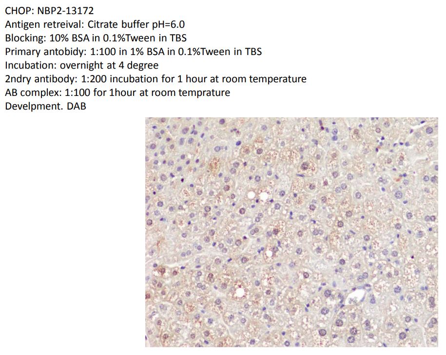
Application: Immunohistochemistry-ParaffinSample Tested: Liver tissueSpecies: MouseVerified Customer | Posted 01/23/2020CHOP: NBP2-13172 Antigen retreival: Citrate buffer pH=6.0 Blocking: 10% BSA in 0.1%Tween in TBS Primary antobidy: 1:100 in 1% BSA in 0.1%Tween in TBS Incubation: overnight at 4 degree 2ndry antibody: 1:200 incubation for 1 hour at room temperature AB complex: 1:100 for 1hour at room temprature Develpment. DAB

There are no reviews that match your criteria.
Protocols
View specific protocols for GADD153/CHOP Antibody - BSA Free (NBP2-13172):
Protocol for Flow Cytometry Intracellular Staining
Sample Preparation.
1. Grow cells to 60-85% confluency. Flow cytometry requires between 2 x 10⁵ and 1 x 10⁶ cells for optimal performance.
2. If cells are adherent, harvest gently by washing once with staining buffer and then scraping. Avoid using trypsin as this can disrupt certain epitopes of interest. If enzymatic harvest is required, use Accutase, Collagenase, or TrypLE Express for a less damaging option.
3. Reserve 100 uL for counting, then transfer cell volume into a 50 mL conical tube and centrifuge for 8 minutes at 400 RCF.
a. Count cells using a hemocytometer and a 1:1 trypan blue exclusion stain to determine cell viability before starting the flow protocol. If cells appear blue, do not proceed.
4. Re-suspend cells to a concentration of 1 x 10⁶ cells/mL in staining buffer (NBP2-26247).
5. Aliquot out 100 uL samples in accordance with your experimental samples.
Tip: When cell surface and intracellular staining are required in the same sample, it is advisable that the cell surface staining be performed first since the fixation and permeabilization steps might reduce the availability of surface antigens.
Intracellular Staining.
Tip: When performing intracellular staining, it is important to use appropriate fixation and permeabilization reagents based upon the target and its subcellular location. Generally, our Intracellular Flow Assay Kit (NBP2-29450) is a good place to start as it contains an optimized combination of reagents for intracellular staining as well as an inhibitor of intracellular protein transport (necessary if staining secreted proteins). Certain targets may require more gentle or transient permeabilization protocols such as the commonly employed methanol or saponin-based methods.
Protocol for Cytoplasmic Targets:
1. Fix the cells by adding 100 uL fixation solution (such as 4% PFA) to each sample for 10-15 minutes.
2. Permeabilize cells by adding 100 uL of a permeabilization buffer to every 1 x 10⁶ cells present in the sample. Mix well and incubate at room temperature for 15 minutes.
a. For cytoplasmic targets, use a gentle permeabilization solution such as 1X PBS + 0.5% Saponin or 1X PBS + 0.5% Tween-20.
b. To maintain the permeabilized state throughout your experiment, use staining buffer + 0.1% of the permeabilization reagent (i.e. 0.1% Tween-20 or 0.1% Saponin).
3. Following the 15 minute incubation, add 2 mL of the staining buffer + 0.1% permeabilizer to each sample.
4. Centrifuge for 1 minute at 400 RCF.
5. Discard supernatant and re-suspend in 100 uL of staining buffer + 0.1% permeabilizer.
6. Add appropriate amount of each antibody (eg. 1 test or 1 ug per sample, as experimentally determined).
7. Mix well and incubate at room temperature for 30 minutes- 1 hour. Gently mix samples every 10-15 minutes.
8. Following the primary/conjugate incubation, add 1-2 mL/sample of staining buffer +0.1% permeabilizer and centrifuge for 1 minute at 400 RCF.
9. Wash twice by re-suspending cells in staining buffer (2 mL for tubes or 200 uL for wells) and centrifuging at 400 RCF for 5 minutes. Discard supernatant.
10. Add appropriate amount of secondary antibody (as experimentally determined) to each sample.
11. Incubate at room temperature in dark for 20 minutes.
12. Add 1-2 mL of staining buffer and centrifuge at 400 RCF for 1 minute and discard supernatant.
13. Wash twice by re-suspending cells in staining buffer (2 mL for tubes or 200 uL for wells) and centrifuging at 400 RCF for 5 minutes. Discard supernatant.
14. Resuspend in an appropriate volume of staining buffer (usually 500 uL per sample) and proceed with analysis on your flow cytometer.
Immunocytochemistry Protocol
Culture cells to appropriate density in 35 mm culture dishes or 6-well plates.
1. Remove culture medium and wash the cells briefly in PBS. Add 10% formalin to the dish and fix at room temperature for 10 minutes.
2. Remove the formalin and wash the cells in PBS.
3. Permeablize the cells with 0.1% Triton X100 or other suitable detergent for 10 min.
4. Remove the permeablization buffer and wash three times for 10 minutes each in PBS. Be sure to not let the specimen dry out.
5. To block nonspecific antibody binding, incubate in 10% normal goat serum from 1 hour to overnight at room temperature.
6. Add primary antibody at appropriate dilution and incubate overnight at 4C.
7. Remove primary antibody and replace with PBS. Wash three times for 10 minutes each.
8. Add secondary antibody at appropriate dilution. Incubate for 1 hour at room temperature.
9. Remove secondary antibody and replace with PBS. Wash three times for 10 minutes each.
10. Counter stain DNA with DAPi if required.
Immunohistochemistry-Paraffin Embedded Sections Protocol
Antigen Unmasking:
Bring slides to a boil in 10 mM sodium citrate buffer (pH 6.0) then maintain at a sub-boiling temperature for 10 minutes. Cool slides on bench-top for 30 minutes.
Staining:
1. Wash sections in deionized water three times for 5 minutes each.
2. Wash sections in wash buffer for 5 minutes.
3. Block each section with 100-400 ul blocking solution for 1 hour at room temperature.
4. Remove blocking solution and add 100-400 ul diluted primary antibody. Incubate overnight at 4 C.
5. Remove antibody solution and wash sections in wash buffer three times for 5 minutes each.
6. Add 100-400 ul biotinylated diluted secondary antibody. Incubate 30 minutes at room temperature.
7. Remove secondary antibody solution and wash sections three times with wash buffer for 5 minutes each.
8. Add 100-400 ul Streptavidin-HRP reagent to each section and incubate for 30 minutes at room temperature.
9. Wash sections three times in wash buffer for 5 minutes each.
10. Add 100-400 ul DAB substrate to each section and monitor staining closely.
11. As soon as the sections develop, immerse slides in deionized water.
12. Counterstain sections in hematoxylin.
13. Wash sections in deionized water two times for 5 minutes each.
14. Dehydrate sections.
15. Mount coverslips.
*The above information is only intended as a guide. The researcher should determine what protocol best meets their needs. Please follow safe laboratory procedures.
Western Blot Protocol
1. Perform SDS-PAGE on samples to be analyzed, loading 40 ug of total protein per lane.
2. Transfer proteins to membrane according to the instructions provided by the manufacturer of the membrane and transfer apparatus.
3. Stain according to standard Ponceau S procedure (or similar product) to assess transfer success, and mark molecular weight standards where appropriate.
4. Rinse the blot.
5. Block the membrane using standard blocking buffer for at least 1 hour.
6. Wash the membrane in wash buffer three times for 10 minutes each.
7. Dilute primary antibody in blocking buffer and incubate 1 hour at room temperature.
8. Wash the membrane in wash buffer three times for 10 minutes each.
9. Apply the diluted HRP conjugated secondary antibody in blocking buffer (as per manufacturers instructions) and incubate 1 hour at room temperature.
10. Wash the blot in wash buffer three times for 10 minutes each (this step can be repeated as required to reduce background).
11. Apply the detection reagent of choice in accordance with the manufacturers instructions.
Note: Tween-20 can be added to the blocking or antibody dilution buffer at a final concentration of 0.05-0.2%.
*The above information is only intended as a guide. The researcher should determine what protocol best meets their needs. Please follow safe laboratory procedures.
Find general support by application which include: protocols, troubleshooting, illustrated assays, videos and webinars.
- 7-Amino Actinomycin D (7-AAD) Cell Viability Flow Cytometry Protocol
- Antigen Retrieval Protocol (PIER)
- Antigen Retrieval for Frozen Sections Protocol
- Appropriate Fixation of IHC/ICC Samples
- Cellular Response to Hypoxia Protocols
- Chromogenic IHC Staining of Formalin-Fixed Paraffin-Embedded (FFPE) Tissue Protocol
- Chromogenic Immunohistochemistry Staining of Frozen Tissue
- ClariTSA™ Fluorophore Kits
- Detection & Visualization of Antibody Binding
- Extracellular Membrane Flow Cytometry Protocol
- Flow Cytometry Protocol for Cell Surface Markers
- Flow Cytometry Protocol for Staining Membrane Associated Proteins
- Flow Cytometry Staining Protocols
- Flow Cytometry Troubleshooting Guide
- Fluorescent IHC Staining of Frozen Tissue Protocol
- Graphic Protocol for Heat-induced Epitope Retrieval
- Graphic Protocol for the Preparation and Fluorescent IHC Staining of Frozen Tissue Sections
- Graphic Protocol for the Preparation and Fluorescent IHC Staining of Paraffin-embedded Tissue Sections
- Graphic Protocol for the Preparation of Gelatin-coated Slides for Histological Tissue Sections
- ICC Cell Smear Protocol for Suspension Cells
- ICC Immunocytochemistry Protocol Videos
- ICC for Adherent Cells
- IHC Sample Preparation (Frozen sections vs Paraffin)
- Immunocytochemistry (ICC) Protocol
- Immunocytochemistry Troubleshooting
- Immunofluorescence of Organoids Embedded in Cultrex Basement Membrane Extract
- Immunofluorescent IHC Staining of Formalin-Fixed Paraffin-Embedded (FFPE) Tissue Protocol
- Immunohistochemistry (IHC) and Immunocytochemistry (ICC) Protocols
- Immunohistochemistry Frozen Troubleshooting
- Immunohistochemistry Paraffin Troubleshooting
- Intracellular Flow Cytometry Protocol Using Alcohol (Methanol)
- Intracellular Flow Cytometry Protocol Using Detergents
- Intracellular Nuclear Staining Flow Cytometry Protocol Using Detergents
- Intracellular Staining Flow Cytometry Protocol Using Alcohol Permeabilization
- Intracellular Staining Flow Cytometry Protocol Using Detergents to Permeabilize Cells
- Preparing Samples for IHC/ICC Experiments
- Preventing Non-Specific Staining (Non-Specific Binding)
- Primary Antibody Selection & Optimization
- Propidium Iodide Cell Viability Flow Cytometry Protocol
- Protocol for Heat-Induced Epitope Retrieval (HIER)
- Protocol for Liperfluo
- Protocol for Making a 4% Formaldehyde Solution in PBS
- Protocol for VisUCyte™ HRP Polymer Detection Reagent
- Protocol for the Characterization of Human Th22 Cells
- Protocol for the Characterization of Human Th9 Cells
- Protocol for the Fluorescent ICC Staining of Cell Smears - Graphic
- Protocol for the Fluorescent ICC Staining of Cultured Cells on Coverslips - Graphic
- Protocol for the Preparation & Fixation of Cells on Coverslips
- Protocol for the Preparation and Chromogenic IHC Staining of Frozen Tissue Sections
- Protocol for the Preparation and Chromogenic IHC Staining of Frozen Tissue Sections - Graphic
- Protocol for the Preparation and Chromogenic IHC Staining of Paraffin-embedded Tissue Sections
- Protocol for the Preparation and Chromogenic IHC Staining of Paraffin-embedded Tissue Sections - Graphic
- Protocol for the Preparation and Fluorescent ICC Staining of Cells on Coverslips
- Protocol for the Preparation and Fluorescent ICC Staining of Non-adherent Cells
- Protocol for the Preparation and Fluorescent ICC Staining of Stem Cells on Coverslips
- Protocol for the Preparation and Fluorescent IHC Staining of Frozen Tissue Sections
- Protocol for the Preparation and Fluorescent IHC Staining of Paraffin-embedded Tissue Sections
- Protocol for the Preparation of Gelatin-coated Slides for Histological Tissue Sections
- Protocol for the Preparation of a Cell Smear for Non-adherent Cell ICC - Graphic
- Protocol: Annexin V and PI Staining by Flow Cytometry
- Protocol: Annexin V and PI Staining for Apoptosis by Flow Cytometry
- R&D Systems Quality Control Western Blot Protocol
- TUNEL and Active Caspase-3 Detection by IHC/ICC Protocol
- The Importance of IHC/ICC Controls
- Troubleshooting Guide: Fluorokine Flow Cytometry Kits
- Troubleshooting Guide: Immunohistochemistry
- Troubleshooting Guide: Western Blot Figures
- Western Blot Conditions
- Western Blot Protocol
- Western Blot Protocol for Cell Lysates
- Western Blot Troubleshooting
- Western Blot Troubleshooting Guide
- View all Protocols, Troubleshooting, Illustrated assays and Webinars
Loading...