GAPDH Antibody - BSA Free
Novus Biologicals | Catalog # NB100-56875
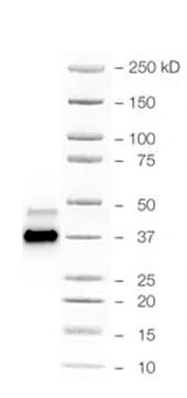
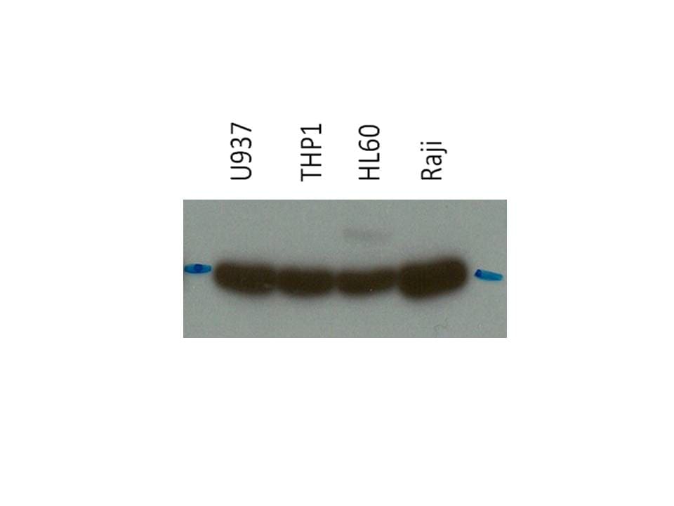
![Western Blot: GAPDH Antibody [NB100-56875]](https://resources.rndsystems.com/images/products/GAPDH-Antibody-Western-Blot-NB100-56875-img0004.jpg "Western Blot: GAPDH Antibody [NB100-56875]")
Loading...
Key Product Details
Validated by
Biological Validation
Species Reactivity
Validated:
Human, Mouse, Rat, Porcine, Canine, Drosophila, Feline, Hamster, Primate
Cited:
Human, Mouse, Rat, Porcine, Canine, Feline, Hamster, Insect - Drosophila, Primate
Predicted:
Gerbil (93%). Backed by our 100% Guarantee.
Applications
Validated:
Immunohistochemistry, Western Blot, Immunoblotting, Immunocytochemistry/ Immunofluorescence, Simple Western
Cited:
Western Blot, Immunoblotting, Immunocytochemistry/ Immunofluorescence, IF/IHC
Label
Unconjugated
Antibody Source
Polyclonal Rabbit IgG
Format
BSA Free
Loading...
Product Specifications
Immunogen
Amino acids 73-87 PITIFQERDPSKIKW of glyceraldehyde 3-phosphate dehydrogenase protein were used as the immunogen of this GAPDH antibody.
Reactivity Notes
Porcine reactivity reported in scientific literature (PMID:32764569). Immunogen displays the following percentage of sequence identity for non-tested species: 100% homologous in baboon, chimp and macaque; salamander(86%)..
Marker
Cytosolic Marker
Clonality
Polyclonal
Host
Rabbit
Isotype
IgG
Theoretical MW
36 kDa.
Disclaimer note: The observed molecular weight of the protein may vary from the listed predicted molecular weight due to post translational modifications, post translation cleavages, relative charges, and other experimental factors.
Disclaimer note: The observed molecular weight of the protein may vary from the listed predicted molecular weight due to post translational modifications, post translation cleavages, relative charges, and other experimental factors.
Scientific Data Images for GAPDH Antibody - BSA Free
Western Blot: GAPDH Antibody [NB100-56875]
Western Blot: GAPDH Antibody [NB100-56875] - Analysis of GAPDH using this antibody at 1:500 in nuclear and cytoplasmic fractions made from Drosophila head extracts. Data courtesy of Dr. Jerry Lin, University of Wisconsin-Madison.![Western Blot: GAPDH Antibody [NB100-56875]](https://resources.rndsystems.com/images/products/GAPDH-Antibody-Western-Blot-NB100-56875-img0006.jpg "Western Blot: GAPDH Antibody [NB100-56875]")
Western Blot: GAPDH Antibody [NB100-56875]
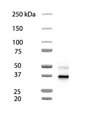
Western Blot: GAPDH Antibody [NB100-56875] - Analysis of GAPDH in the multiple human tumor cell line lysate INSTA-Blot using this antibody. 25 ug/ml. Theoretical molecular weight: 36 kDa.![Western Blot: GAPDH Antibody [NB100-56875]](https://resources.rndsystems.com/images/products/GAPDH-Antibody-Western-Blot-NB100-56875-img0008.jpg "Western Blot: GAPDH Antibody [NB100-56875]")
Western Blot: GAPDH Antibody [NB100-56875]
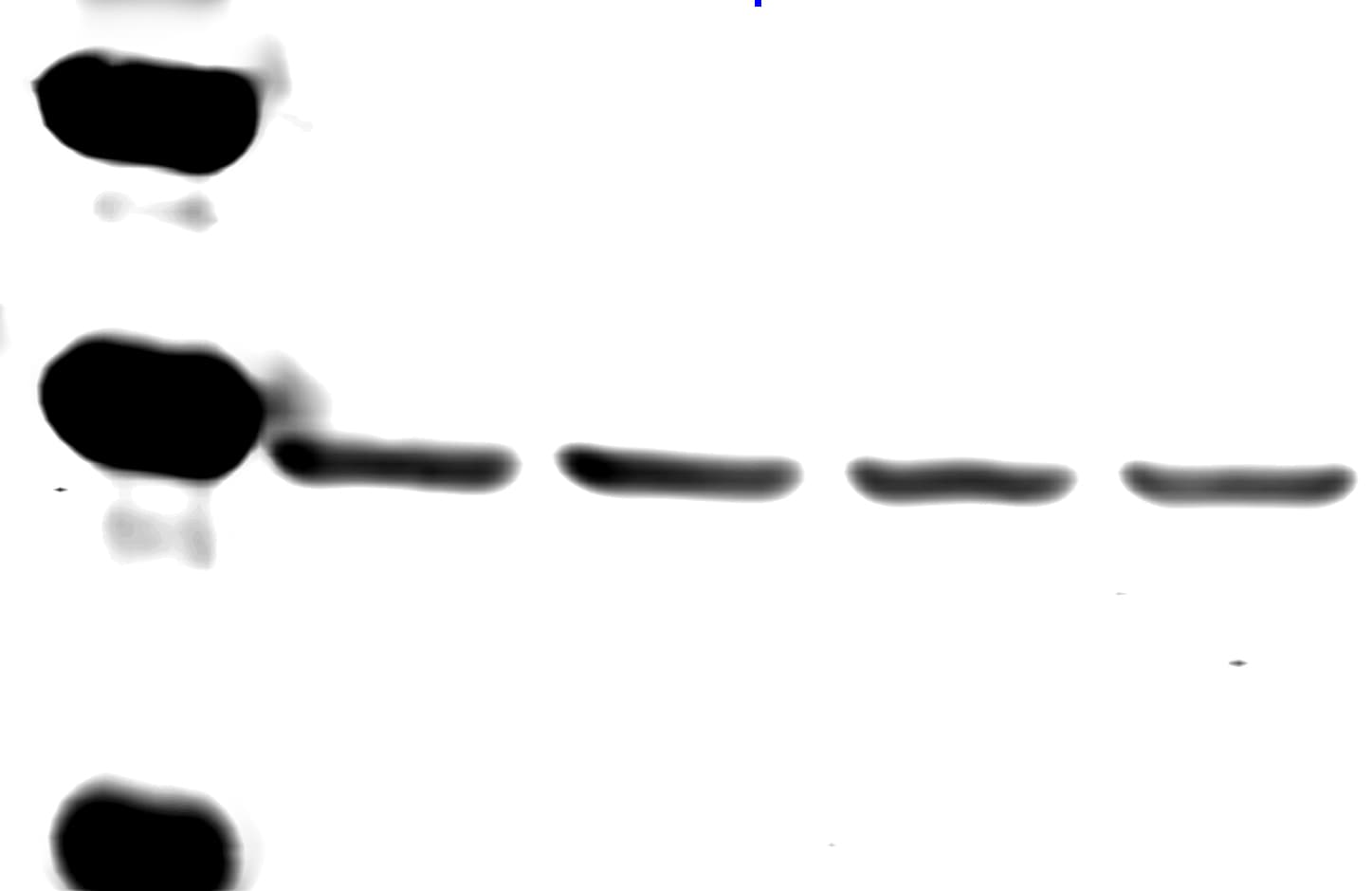
Western Blot: GAPDH Antibody [NB100-56875] - Analysis using HeLa cells. Theoretical molecular weight: 36 kDa.![Western Blot: GAPDH Antibody [NB100-56875]](https://resources.rndsystems.com/images/products/GAPDH-Antibody-Western-Blot-NB100-56875-img0009.jpg "Western Blot: GAPDH Antibody [NB100-56875]")
Western Blot: GAPDH Antibody [NB100-56875]
GAPDH-Antibody-Western-Blot-NB100-56875-img0009.jpg![Western Blot: GAPDH Antibody [NB100-56875]](https://resources.rndsystems.com/images/products/GAPDH-Antibody-Western-Blot-NB100-56875-img0010.jpg "Western Blot: GAPDH Antibody [NB100-56875]")
![Simple Western: GAPDH Antibody [NB100-56875]](https://resources.rndsystems.com/images/products/GAPDH-Antibody-Simple-Western-NB100-56875-img0005.jpg "Simple Western: GAPDH Antibody [NB100-56875]")
Simple Western: GAPDH Antibody [NB100-56875]
Simple Western: GAPDH Antibody [NB100-56875] - Lane view shows a specific band for GAPDH in 0.5 mg/ml of HeLa lysate. This experiment was performed under reducing A246conditions using the 12-230 kDa separation system. Note: band observed higher than predicted 36 kDa molecular weight.
Western Blot: GAPDH Antibody [NB100-56875] -
Western Blot: GAPDH Antibody [NB100-56875] - Vps13 co-fractionates with Rab7 & Rab5.(A) Western blot analysis of control fly head samples fractionated into a cytosolic & membrane fraction from postnuclear supernatant (PNS). EGFR was used as a membrane marker & GAPDH as a cytosolic marker. (B) Membrane fractions from control fly heads treated with 1 M KCl, Na2CO3 pH 11 or 6 M urea were centrifuged to separate the soluble & insoluble (membrane containing) fractions. The level of Vps13 was determined in these fractions. Markers for peripheral membrane proteins (GM130), integral membrane proteins (EGFR) & the cytosolic proteins (GAPDH) were used. The “Vps13 lysate” lane contains a lysate derived from Vps13 homozygous mutant fly heads, as expected no Vps13 is detected, demonstrating the specificity of the antibody against Vps13. (C) Membranes from control fly heads were fractionated on a sucrose gradient. Western blot analysis was performed to analyze the distribution of Vps13 in relation to markers associated with membranes of various organelles: Rab7 (late endosomes), Rab5 (early endosomes), GM130 (golgi), Lamp1 (lysosomes) & ATP5A (mitochondria). (D) Immunoisolation of membranes from fraction 14 of the sucrose gradient using Vps13 NT, Rab7 & Rab5 antibodies. (E) Quantification of the sucrose gradient fractionation of Fig 2C. Image collected & cropped by CiteAb from the following publication (https://pubmed.ncbi.nlm.nih.gov/28107480), licensed under a CC-BY license. Not internally tested by Novus Biologicals.
Western Blot: GAPDH Antibody [NB100-56875] -
Western Blot: GAPDH Antibody [NB100-56875] - Overexpression of HsVps13A rescues phenotypes of Vps13 mutants.(A) Samples from fly heads of Actin-GAL4 / + (as a control) & Actin-GAL4 / UAS-HsVps13A (HsVps13A expressing) flies were separated into a membrane & cytosol fraction & analyzed by Western blot for HsVps13A levels. EGFR & GAPDH used as controls for membrane & cytosolic proteins, respectively. (B) Eclosion rate of Vps13 mutant flies a Actin-GAL4/+ (control) or Actin-GAL4/UAS-HsVp13A (HsVps13A expressing) background at 25°C. (C) Ubiquitylated proteins from samples of 1 day old fly head extracts of Vps13/CyO; Actin-GAL4/+ (as a control), Vps13/ Vps13; Actin-GAL4/+ (representing homozygous mutants) & Vps13/ Vps13; Actin-GAL4/UAS-HsVps13A (representing homozygous mutants expressing human VPS13A). (D) Representative picture of ubiquitylated protein staining of the third instar larval ventral nerve cord of Vps13/CyO; Actin-GAL4/+ (as a control), Vps13/ Vps13; Actin-GAL4/+ & Vps13/ Vps13; Actin-GAL4/UAS-HsVps13A. Arrows indicate accumulations of ubiquitylated positive structures. The scale bar indicates 50 μm & 12,5 μm in the enlargement. (E) Quantification of the number of puncta in third instar larval ventral nerve cord of the experiment presented in Fig 6D. (F) Life span curve of Vps13/ Vps13; Actin-GAL4/+ & Vps13/ Vps13; Actin-GAL4/UAS-HsVps13A. All quantifications show the mean & SEM of at least three independent experiments per condition. For statistical analysis a two-tailed students T-test was used in combination with a Welch’s correction if necessary. P<0.05 is *, P<0.01 is ** & P<0.001 is ***. Image collected & cropped by CiteAb from the following publication (https://pubmed.ncbi.nlm.nih.gov/28107480), licensed under a CC-BY license. Not internally tested by Novus Biologicals.
Western Blot: GAPDH Antibody [NB100-56875] -
Western Blot: GAPDH Antibody [NB100-56875] - No dystrophin expression was detected in cardiac & skeletal muscles of Dmdmdx rats.(A) Male 7 month-old rats of line 61, wild-type littermate controls (WT) & Dmdmdx were sacrificed & biopsies from tibialis cranialis muscles (TC) & hearts (H) were harvested. Western-blot of total proteins (50 µg) was incubated with NCL-DYS2 & Manex1011C monoclonal antibodies (C-terminal & exons 10/11 epitopes, respectively). This revealed undetectable levels of the 427 kDa dystrophin band in line 61 Dmdmdx rats. Muscle from a GRMD dog (GD) was used as negative control & samples from WT rats were used as positive controls. Staining with an anti-GAPDH polyclonal antibody validated equal protein loadings. (B–E) Heart & biceps femoris muscles from the same wild-type (B & C) & Dmdmdx rats (D & E) were assessed for dystrophin expression using immunohistochemistry with Mandys110 monoclonal antibody (against exons 38–39 epitope). Compared to the subsarcolemmal expression of dystrophin in wild-type muscles, no dystrophin was detected in Dmdmdx rats except for the presence in skeletal muscle of only rare scattered revertant positive fibers (arrowheads). Immunolabelling of dystrophin (B–E) Bar = 100 µm. Image collected & cropped by CiteAb from the following publication (https://dx.plos.org/10.1371/journal.pone.0110371), licensed under a CC-BY license. Not internally tested by Novus Biologicals.
Western Blot: GAPDH Antibody [NB100-56875] -
Western Blot: GAPDH Antibody [NB100-56875] - ABCA1, ABCG1 & SR-BI protein expressions. a & b Hepatic protein expressions of ABCA1, ABCG1 & SR-BI were significantly decreased in COMT−/− mice at GD 18.5, compared to C57BL/6 J mice. Decreased hepatic ABCA1 expression was also observed at 10 days postpartum. ATI-5261 increased ABCA1 & ABCG1 expression in the liver at 10 days postpartum. c Placental protein expressions of ABCA1 & ABCG1 was reduced in COMT−/− mice, compared to C57BL/6 J mice. ATI-5261 treatment significantly increased ABCA1 levels in the placenta of COMT−/− mice. d Representative immunoblots of the corresponding proteins in the placenta with mouse RAW264.7 cell lysate included as positive control. Similar results were obtained when the experiment was repeated with lysates prepared from three batches of tissues. Data are presented as mean ± SEM. Groups (n = 8 in all groups) were compared using one-way ANOVA with post-hoc analysis (Tukey’s procedure). *, p < 0.05 Image collected & cropped by CiteAb from the following publication (https://pubmed.ncbi.nlm.nih.gov/30237900), licensed under a CC-BY license. Not internally tested by Novus Biologicals.
Western Blot: GAPDH Antibody [NB100-56875] -
Western Blot: GAPDH Antibody [NB100-56875] - Vps13 co-fractionates with Rab7 & Rab5.(A) Western blot analysis of control fly head samples fractionated into a cytosolic & membrane fraction from postnuclear supernatant (PNS). EGFR was used as a membrane marker & GAPDH as a cytosolic marker. (B) Membrane fractions from control fly heads treated with 1 M KCl, Na2CO3 pH 11 or 6 M urea were centrifuged to separate the soluble & insoluble (membrane containing) fractions. The level of Vps13 was determined in these fractions. Markers for peripheral membrane proteins (GM130), integral membrane proteins (EGFR) & the cytosolic proteins (GAPDH) were used. The “Vps13 lysate” lane contains a lysate derived from Vps13 homozygous mutant fly heads, as expected no Vps13 is detected, demonstrating the specificity of the antibody against Vps13. (C) Membranes from control fly heads were fractionated on a sucrose gradient. Western blot analysis was performed to analyze the distribution of Vps13 in relation to markers associated with membranes of various organelles: Rab7 (late endosomes), Rab5 (early endosomes), GM130 (golgi), Lamp1 (lysosomes) & ATP5A (mitochondria). (D) Immunoisolation of membranes from fraction 14 of the sucrose gradient using Vps13 NT, Rab7 & Rab5 antibodies. (E) Quantification of the sucrose gradient fractionation of Fig 2C. Image collected & cropped by CiteAb from the following publication (https://pubmed.ncbi.nlm.nih.gov/28107480), licensed under a CC-BY license. Not internally tested by Novus Biologicals.
Simple Western: GAPDH Antibody [NB100-56875] -
Simple Western: GAPDH Antibody [NB100-56875] - Analysis of non-canonical inflammasome pathway.A LDH cytotoxicity assay in the presence of LPS or SCGB3A2 alone, or the two together (SCGB3A2 + LPS). B LDH cytotoxicity assay in the presence of various amount of nigericin. Average ± SD from more than three independent experiments. *P < 0.01, **P < 0.0001 by Tukey’s multiple comparison. C Morphology of cells cultured in the presence of LPS or SCGB3A2 alone, SCGB3A2 + LPS, or nigericin for 3 h. For nigericin-treated group, cells were primed with LPS before addition of nigericin. Black arrow indicates ballooned cells, characteristic feature of pyroptosis. D Western blotting for the cleaved forms of CASP1 (p20), CASP4 (p20), & GSDMD (N-terminal). S: SCGB3A2, N: nigericin. Experiments were repeated more than twice & same results were obtained. Image collected & cropped by CiteAb from the following publication (https://pubmed.ncbi.nlm.nih.gov/33452234), licensed under a CC-BY license. Not internally tested by Novus Biologicals.Applications for GAPDH Antibody - BSA Free
Application
Recommended Usage
Simple Western
1:500
Western Blot
1:500-1:2000
Application Notes
In Simple Western only 10 - 15 uL of the recommended dilution is used per data point.
See Simple Western Antibody Database for Simple Western validation: Tested in HeLa lysate 0.5 mg/mL, separated by Size, antibody dilution of 1:500, apparent MW was 43 kDa. Use in immunoblotting reported in scientific literature (PMID: 28545464). Use in ICC/IF was reported in scientific literature (PMID: 31312260). Use in Immunohistochemistry reported in scientific literature (PMID:32123074).
See Simple Western Antibody Database for Simple Western validation: Tested in HeLa lysate 0.5 mg/mL, separated by Size, antibody dilution of 1:500, apparent MW was 43 kDa. Use in immunoblotting reported in scientific literature (PMID: 28545464). Use in ICC/IF was reported in scientific literature (PMID: 31312260). Use in Immunohistochemistry reported in scientific literature (PMID:32123074).
Reviewed Applications
Read 7 reviews rated 4.9 using NB100-56875 in the following applications:
Formulation, Preparation, and Storage
Purification
Protein G purified
Formulation
PBS
Format
BSA Free
Preservative
0.05% Sodium Azide
Concentration
1.0 mg/ml
Shipping
The product is shipped with polar packs. Upon receipt, store it immediately at the temperature recommended below.
Stability & Storage
Store at 4C short term. Aliquot and store at -20C long term. Avoid freeze-thaw cycles.
Background: GAPDH
References
1) Barber RD, Harmer DW, Coleman RA, Clark BJ. (2005) GAPDH as a housekeeping gene: analysis of GAPDH mRNA expression in a panel of 72 human tissues. Physiol Genomics. 21(3):389-95. PMID: 15769908
2) Jia Y, Takimoto K. (2006) Mitogen-activated protein kinases control cardiac KChIP2 gene expression. Circ Res. 98(3):386-93. PMID: 16385079
3) Godsel LM, Hsieh SN, Amargo EV, Bass AE, Pascoe-McGillicuddy LT, Huen AC, Thorne ME, Gaudry CA, Park JK, Myung K, Goldman RD, Chew TL, Green KJ. (2005) Desmoplakin assembly dynamics in four dimensions: multiple phases differentially regulated by intermediate filaments and actin. J Cell Biol. 171(6):1045-59. PMID: 16365169
4) Sirover MA1. (1999) New insights into an old protein: the functional diversity of mammalian glyceraldehyde-3-phosphate dehydrogenase. Biochim Biophys Acta. 1432(2): 159-84. PMID: 10407139
5) Tristan C, Shahani N, Sedlak TW, Sawa A. (2011) The diverse functions of GAPDH: views from different subcellular compartments. Cell Signal. 23(2):317-23. PMID: 20727968
Long Name
Glyceraldehyde-3-phosphate Dehydrogenase
Alternate Names
G3PDH
Gene Symbol
GAPDH
UniProt
Additional GAPDH Products
Product Documents for GAPDH Antibody - BSA Free
Certificate of Analysis
To download a Certificate of Analysis, please enter a lot or batch number in the search box below.
Product Specific Notices for GAPDH Antibody - BSA Free
This product is for research use only and is not approved for use in humans or in clinical diagnosis. Primary Antibodies are guaranteed for 1 year from date of receipt.
Related Research Areas
Citations for GAPDH Antibody - BSA Free
 Powered by Bioz
Customer Reviews for GAPDH Antibody - BSA Free (7)
4.9 out of 5
7 Customer Ratings
Have you used GAPDH Antibody - BSA Free?
Submit a review and receive an Amazon gift card!
$25/€18/£15/$25CAN/¥2500 Yen for a review with an image
$10/€7/£6/$10CAN/¥1110 Yen for a review without an image
Submit a review
Customer Images

Showing
1
-
5 of
7 reviews
Showing All
Filter By:
-
Application: Western BlotSample Tested: HCT-116 human colorectal carcinoma cell lineSpecies: HumanVerified Customer | Posted 02/03/2026GAPDH band in HCT116Diluted to 1:1000, strong band, would be better to reduce concentration

-
Application: Western BlotSample Tested: MDA-MB-231 human breast cancer cell lineSpecies: HumanVerified Customer | Posted 02/03/2026GAPDH band in MDA-231Very clear band with 1:1000 dilution
-
Application: Western BlotSample Tested: 3 human pancreas cancer cell linesSpecies: HumanVerified Customer | Posted 01/08/2019Very good antibody. Specific. Highly recommended.
-
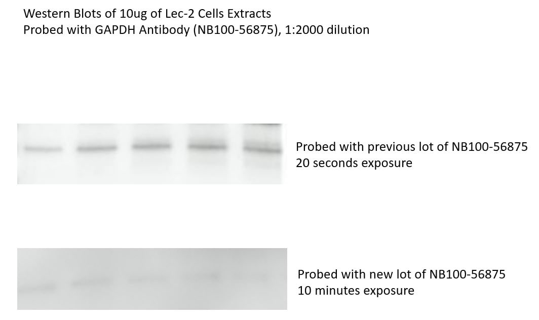
Application: Western BlotSample Tested: protein extracts from Lec-2 cellsSpecies: Chinese HamsterVerified Customer | Posted 10/13/2017Western Blots of 10ug of Lec-2 Cells Extracts Probed with GAPDH Antibody (NB100-56875), 1:2000 dilutionI used GAPDH antibody NB100-56875 for Western blot on Lec-2 cells extracts before. It worked really well. Last lot, 04231895B-10, does not recognize hamster GAPDH. I cannot blame Novus since it is polyclonal antibody and they also did not claim that it recognizes hamster GAPDH.
-
Application: Western BlotSample Tested: Heart lysatesSpecies: FelineVerified Customer | Posted 12/09/2016Licor Odessey Fc 800CW IRdye
-
Application: Western BlotSample Tested:Species: HumanVerified Customer | Posted 09/01/2015GAPDH bands for 4 cell lines
-
Application: Western BlotVerified Customer | Posted 05/20/2014
There are no reviews that match your criteria.
Protocols
Find general support by application which include: protocols, troubleshooting, illustrated assays, videos and webinars.
- Antigen Retrieval Protocol (PIER)
- Antigen Retrieval for Frozen Sections Protocol
- Appropriate Fixation of IHC/ICC Samples
- Cellular Response to Hypoxia Protocols
- Chromogenic IHC Staining of Formalin-Fixed Paraffin-Embedded (FFPE) Tissue Protocol
- Chromogenic Immunohistochemistry Staining of Frozen Tissue
- ClariTSA™ Fluorophore Kits
- Detection & Visualization of Antibody Binding
- Fluorescent IHC Staining of Frozen Tissue Protocol
- Graphic Protocol for Heat-induced Epitope Retrieval
- Graphic Protocol for the Preparation and Fluorescent IHC Staining of Frozen Tissue Sections
- Graphic Protocol for the Preparation and Fluorescent IHC Staining of Paraffin-embedded Tissue Sections
- Graphic Protocol for the Preparation of Gelatin-coated Slides for Histological Tissue Sections
- ICC Cell Smear Protocol for Suspension Cells
- ICC Immunocytochemistry Protocol Videos
- ICC for Adherent Cells
- IHC Sample Preparation (Frozen sections vs Paraffin)
- Immunocytochemistry (ICC) Protocol
- Immunocytochemistry Troubleshooting
- Immunofluorescence of Organoids Embedded in Cultrex Basement Membrane Extract
- Immunofluorescent IHC Staining of Formalin-Fixed Paraffin-Embedded (FFPE) Tissue Protocol
- Immunohistochemistry (IHC) and Immunocytochemistry (ICC) Protocols
- Immunohistochemistry Frozen Troubleshooting
- Immunohistochemistry Paraffin Troubleshooting
- Preparing Samples for IHC/ICC Experiments
- Preventing Non-Specific Staining (Non-Specific Binding)
- Primary Antibody Selection & Optimization
- Protocol for Heat-Induced Epitope Retrieval (HIER)
- Protocol for Making a 4% Formaldehyde Solution in PBS
- Protocol for VisUCyte™ HRP Polymer Detection Reagent
- Protocol for the Fluorescent ICC Staining of Cell Smears - Graphic
- Protocol for the Fluorescent ICC Staining of Cultured Cells on Coverslips - Graphic
- Protocol for the Preparation & Fixation of Cells on Coverslips
- Protocol for the Preparation and Chromogenic IHC Staining of Frozen Tissue Sections
- Protocol for the Preparation and Chromogenic IHC Staining of Frozen Tissue Sections - Graphic
- Protocol for the Preparation and Chromogenic IHC Staining of Paraffin-embedded Tissue Sections
- Protocol for the Preparation and Chromogenic IHC Staining of Paraffin-embedded Tissue Sections - Graphic
- Protocol for the Preparation and Fluorescent ICC Staining of Cells on Coverslips
- Protocol for the Preparation and Fluorescent ICC Staining of Non-adherent Cells
- Protocol for the Preparation and Fluorescent ICC Staining of Stem Cells on Coverslips
- Protocol for the Preparation and Fluorescent IHC Staining of Frozen Tissue Sections
- Protocol for the Preparation and Fluorescent IHC Staining of Paraffin-embedded Tissue Sections
- Protocol for the Preparation of Gelatin-coated Slides for Histological Tissue Sections
- Protocol for the Preparation of a Cell Smear for Non-adherent Cell ICC - Graphic
- R&D Systems Quality Control Western Blot Protocol
- TUNEL and Active Caspase-3 Detection by IHC/ICC Protocol
- The Importance of IHC/ICC Controls
- Troubleshooting Guide: Immunohistochemistry
- Troubleshooting Guide: Western Blot Figures
- Western Blot Conditions
- Western Blot Protocol
- Western Blot Protocol for Cell Lysates
- Western Blot Troubleshooting
- Western Blot Troubleshooting Guide
- View all Protocols, Troubleshooting, Illustrated assays and Webinars
FAQs for GAPDH Antibody - BSA Free
Showing
1
-
5 of
8 FAQs
Showing All
-
Q: Do you offer a smaller size for the GAPDH antibodies?
A: We offer sample sizes for many of our GAPDH loading control antibodies including NB300-221.
-
Q: I need a suitable loading control antibody that I can use in western blot of rat nerve extracts, which do you recommend?
A: This GAPDH antibody (NB300-221) is a good choice for a loading control antibody and has been cited in over 180 publications and used on rat lysates with good results.
-
Q: I want to know why GAPDH is used as a loading control antibody in WB techniques.
A: Our GAPDH antibody makes a good loading control because GAPDH is a housekeeping gene essential to metabolism that is globally expressed in most tissue types and cell lines.
-
Q: I wanted to know which of the two housekeeping genes B-actin or GAPDH can be used for best results. I intend to do an experiment to determine expression of IL-6 and TNF-alpha in liver and kidney tissues.
A: For homogenized tissue, beta-actin and GAPDH are both fine.
-
Q: I'm looking for a loading control between 24kDa - 65kDa for use in simple western, we tried an actin antibody we already had but it didn't work. What can you recommend?
A: Our GAPDH antibody NB300-221 has been validated in simple western and detects around 35 kDa so I think it would be a good choice for a simple western loading control antibody based on your size requirements.
-
Q: We need a loading control antibody for WB working with HeLa cell line, will this GAPDH antibody work for that?
A: GAPDH is expressed in HeLa cell lines but whether or not it is the best option for a loading control antibody depends on the size of the protein you are interested in. GAPDH antibody is a good choice for low to mid MW targets, as GAPDH detects around 35 kDa.
-
Q: We received GAPDH antibody NB600-502 and there was only about 15µl of antibody in the tube. I was under the impression this should have been 100µl.
A: The unit size of this product is 0.1 mg with a concentration of 6.7 mg/ml. Hence the volume should be no less than 14.9 ul.
-
Q: What secondary antibody should I use for visualization of the GAPDH antibody?
A: This GAPDH antibody is raised in mouse so you would want to use an anti-mouse secondary with the dye of your choice.
-
Q: Do you offer a smaller size for the GAPDH antibodies?
A: We offer sample sizes for many of our GAPDH loading control antibodies including NB300-221.
-
Q: I need a suitable loading control antibody that I can use in western blot of rat nerve extracts, which do you recommend?
A: This GAPDH antibody (NB300-221) is a good choice for a loading control antibody and has been cited in over 180 publications and used on rat lysates with good results.
-
Q: I want to know why GAPDH is used as a loading control antibody in WB techniques.
A: Our GAPDH antibody makes a good loading control because GAPDH is a housekeeping gene essential to metabolism that is globally expressed in most tissue types and cell lines.
-
Q: I wanted to know which of the two housekeeping genes B-actin or GAPDH can be used for best results. I intend to do an experiment to determine expression of IL-6 and TNF-alpha in liver and kidney tissues.
A: For homogenized tissue, beta-actin and GAPDH are both fine.
-
Q: I'm looking for a loading control between 24kDa - 65kDa for use in simple western, we tried an actin antibody we already had but it didn't work. What can you recommend?
A: Our GAPDH antibody NB300-221 has been validated in simple western and detects around 35 kDa so I think it would be a good choice for a simple western loading control antibody based on your size requirements.
-
Q: We need a loading control antibody for WB working with HeLa cell line, will this GAPDH antibody work for that?
A: GAPDH is expressed in HeLa cell lines but whether or not it is the best option for a loading control antibody depends on the size of the protein you are interested in. GAPDH antibody is a good choice for low to mid MW targets, as GAPDH detects around 35 kDa.
-
Q: We received GAPDH antibody NB600-502 and there was only about 15µl of antibody in the tube. I was under the impression this should have been 100µl.
A: The unit size of this product is 0.1 mg with a concentration of 6.7 mg/ml. Hence the volume should be no less than 14.9 ul.
-
Q: What secondary antibody should I use for visualization of the GAPDH antibody?
A: This GAPDH antibody is raised in mouse so you would want to use an anti-mouse secondary with the dye of your choice.
Loading...