GFP Antibody - BSA Free
Novus Biologicals | Catalog # NB100-1614
![Immunocytochemistry/ Immunofluorescence: GFP Antibody [NB100-1614]](https://resources.rndsystems.com/images/products/GFP-Antibody-Immunocytochemistry-Immunofluorescence-NB100-1614-img0003.jpg "Immunocytochemistry/ Immunofluorescence: GFP Antibody [NB100-1614]")
Loading...
Key Product Details
Validated by
Biological Validation
Species Reactivity
Validated:
Non-species specific
Cited:
Human, Mouse, Rat, Avian - Chicken, Firefly, Fish - Danio rerio (Zebrafish), Insect - Drosophila, Jellyfish, Primate, Yeast
Applications
Validated:
Immunohistochemistry, Immunohistochemistry-Paraffin, Immunohistochemistry-Frozen, Immunohistochemistry Free-Floating, Western Blot, ELISA, Flow Cytometry, Immunocytochemistry/ Immunofluorescence, In vivo assay
Cited:
Immunohistochemistry, Immunohistochemistry-Paraffin, Immunohistochemistry-Frozen, Immunohistochemistry Free-Floating, Western Blot, Immunocytochemistry/ Immunofluorescence, Chromatin Immunoprecipitation, In vivo assay, IF/IHC, IHC/IF
Label
Unconjugated
Antibody Source
Polyclonal Chicken IgY
Format
BSA Free
Loading...
Product Specifications
Immunogen
This GFP antibody was developed by immunizing chickens with purified recombinant green fluorescent protein (GFP) emulsified in Freund's adjuvant.
Reactivity Notes
NB100-1614 has been tested on transgenic mice expressing recombinant GFP.
Clonality
Polyclonal
Host
Chicken
Isotype
IgY
Scientific Data Images for GFP Antibody - BSA Free
Immunocytochemistry/ Immunofluorescence: GFP Antibody [NB100-1614]
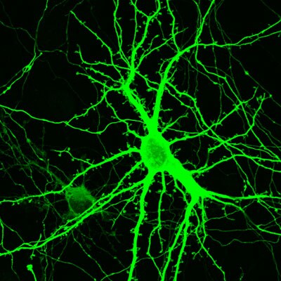
Immunocytochemistry/Immunofluorescence: GFP Antibody [NB100-1614] - Staining of cultured hippocampal neuron from rat. ICC/IF image submitted by a verified customer review.![Immunocytochemistry/ Immunofluorescence: GFP Antibody [NB100-1614]](https://resources.rndsystems.com/images/products/GFP-Antibody-Chromatin-Immunoprecipitation-NB100-1614-img0007.jpg "Immunocytochemistry/ Immunofluorescence: GFP Antibody [NB100-1614]")
Immunocytochemistry/ Immunofluorescence: GFP Antibody [NB100-1614]
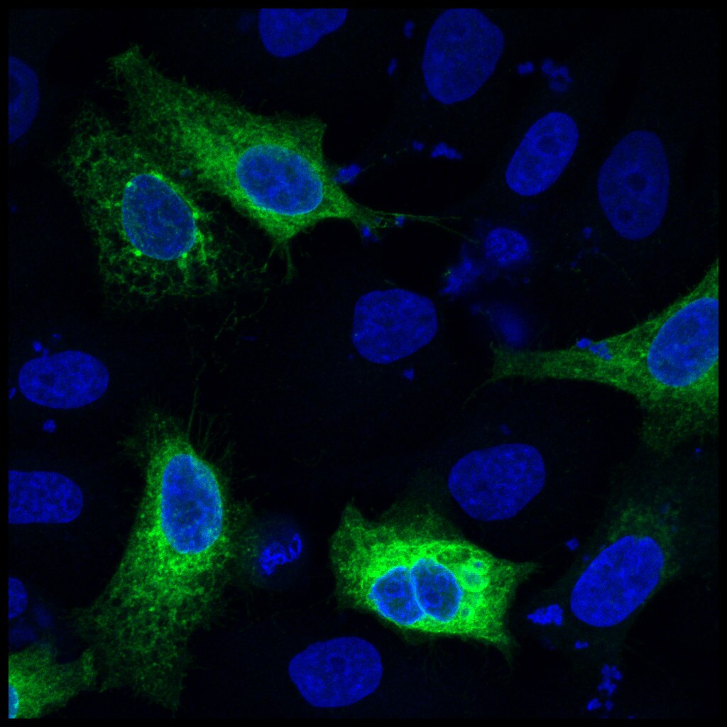
Immunocytochemistry/Immunofluorescence: GFP Antibody [NB100-1614] - HeLa cells transiently expressing GFP were visualized using GFP antibody (green). Nuclei were counterstained with DAPI. ICC/IF image submitted by a verified customer review.![Immunocytochemistry/ Immunofluorescence: GFP Antibody [NB100-1614]](https://resources.rndsystems.com/images/products/GFP-Antibody-Immunocytochemistry-Immunofluorescence-NB100-1614-img0001.jpg "Immunocytochemistry/ Immunofluorescence: GFP Antibody [NB100-1614]")
Immunocytochemistry/ Immunofluorescence: GFP Antibody [NB100-1614]
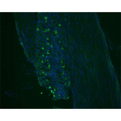
Immunocytochemistry/Immunofluorescence: GFP Antibody [NB100-1614] - IF analysis of GFP in mouse trigeminal ganglia, cornea. Image courtesy of product review by Jessica Newsom.![Immunohistochemistry: GFP Antibody [NB100-1614]](https://resources.rndsystems.com/images/products/GFP-Antibody-Immunohistochemistry-NB100-1614-img0018.jpg "Immunohistochemistry: GFP Antibody [NB100-1614]")
![Immunocytochemistry/ Immunofluorescence: GFP Antibody [NB100-1614]](https://resources.rndsystems.com/images/products/GFP-Antibody-Chromatin-Immunoprecipitation-NB100-1614-img0008.jpg "Immunocytochemistry/ Immunofluorescence: GFP Antibody [NB100-1614]")
Immunocytochemistry/ Immunofluorescence: GFP Antibody [NB100-1614]
Immunocytochemistry/Immunofluorescence: GFP Antibody [NB100-1614] - IF analysis of GFP in rat cortical culture (2 weeks old). ICC/IF image submitted by a verified customer review.![Immunocytochemistry/ Immunofluorescence: GFP Antibody [NB100-1614]](https://resources.rndsystems.com/images/products/GFP-Antibody-Immunocytochemistry-NB100-1614-img0009.jpg "Immunocytochemistry/ Immunofluorescence: GFP Antibody [NB100-1614]")
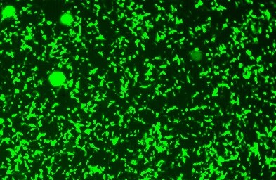
Immunocytochemistry/ Immunofluorescence: GFP Antibody [NB100-1614]
Immunocytochemistry/Immunofluorescence: GFP Antibody [NB100-1614] - LNCaP human prostate cancer cell line imaged after transfection with GFP expressing plasmid. ICC/IF image submitted by a verified customer review.![Immunocytochemistry/ Immunofluorescence: GFP Antibody [NB100-1614]](https://resources.rndsystems.com/images/products/GFP-Antibody-Immunocytochemistry-Immunofluorescence-NB100-1614-img0010.jpg "Immunocytochemistry/ Immunofluorescence: GFP Antibody [NB100-1614]")
Immunocytochemistry/ Immunofluorescence: GFP Antibody [NB100-1614]
Immunocytochemistry/Immunofluorescence: GFP Antibody [NB100-1614] - Staining of a tissue section through the midbrain region of an adult mouse for GFP (green) and Tryptophan Hydroxylase-positive neurons (RED).![Immunohistochemistry-Paraffin: GFP Antibody [NB100-1614]](https://resources.rndsystems.com/images/products/GFP-Antibody-Immunohistochemistry-Paraffin-NB100-1614-img0011.jpg "Immunohistochemistry-Paraffin: GFP Antibody [NB100-1614]")
Immunohistochemistry-Paraffin: GFP Antibody [NB100-1614]
Immunohistochemistry-Paraffin: GFP Antibody [NB100-1614] - Pyramidal neurons in the hippocampal formation of a neonatal mouse brain. Tissue was paraformaldehyde-fixed (4%) and paraffin-embedded. GFP staining is in green in left panel.![Immunohistochemistry-Paraffin: GFP Antibody [NB100-1614]](https://resources.rndsystems.com/images/products/GFP-Antibody-Immunohistochemistry-Paraffin-NB100-1614-img0016.jpg "Immunohistochemistry-Paraffin: GFP Antibody [NB100-1614]")
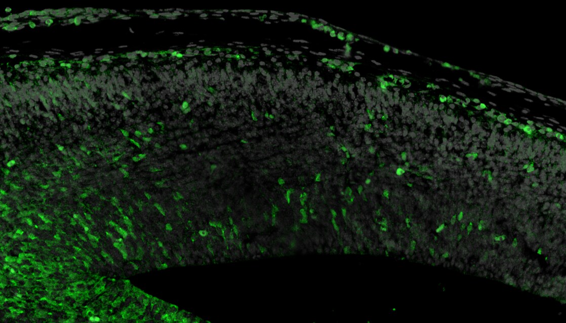
Immunohistochemistry-Paraffin: GFP Antibody [NB100-1614]
Immunohistochemistry-Paraffin: GFP Antibody [NB100-1614] - Embyonic mouse brain section. GFP in green, DAPI in grey. IHC-P image submitted by a verified customer review.![Immunohistochemistry: GFP Antibody [NB100-1614]](https://resources.rndsystems.com/images/products/GFP-Antibody-Immunohistochemistry-NB100-1614-img0017.jpg "Immunohistochemistry: GFP Antibody [NB100-1614]")
Immunohistochemistry: GFP Antibody [NB100-1614]
GFP-Antibody-Immunohistochemistry-NB100-1614-img0017.jpg![GFP Antibody Immunocytochemistry/Immunofluorescence: GFP Antibody [NB100-1614]](https://resources.rndsystems.com/images/products/antibody/nb100-1614_chicken-polyclonal-gfp-antibody-immunocytochemistry-immunofluorescence-245202393523..jpg "Immunocytochemistry/Immunofluorescence: GFP Antibody [NB100-1614]")
Immunocytochemistry/Immunofluorescence: GFP Antibody [NB100-1614]
Comparison between GFP-immunoreactivity using NB100-1614 (left panel in red) and autofluorescence (right panel in green). In this case, the cortical neuron in this unfixed thick section was first photographed for GFP autofluorescence (left), and then the section was fixed (4% paraformaldehyde) and immunostained for GFP-immunoreactivity (1:1000 dilution) using Texas Red-goat anti-chicken IgY antibodies as a secondary. The same cell (left) was then identified. Far right: Western blot showing specific immunolabeling of the GFP protein.
Immunohistochemistry: Chicken Polyclonal GFP Antibody [NB100-1614] -
Immunohistochemistry: Chicken Polyclonal GFP Antibody [NB100-1614] - Chicken Polyclonal GFP Antibody on mouse cancer tissue. DAPI (Red) and Cytopastmic GFP stain (Green). Primary antibody dilution: 1:200 in a 200 um slice. Image from a verified customer review.
Immunohistochemistry-Frozen: Chicken Polyclonal GFP Antibody [NB100-1614] -
Immunohistochemistry-Frozen: Chicken Polyclonal GFP Antibody [NB100-1614] - Image depicting GFP in magenta and CD31 (Catalog # AF3628) in red. Tissue is a fixed frozen section of sciatic nerve obtained from a S100A4 GFP mouse. NB100-1614 was diluted 1 in 10000 and was left on tissue sections overnight at 4 degrees Celsius. Secondary antibody was donkey anti-chicken conjugated to Alexa 647. Image from a verified customer review.
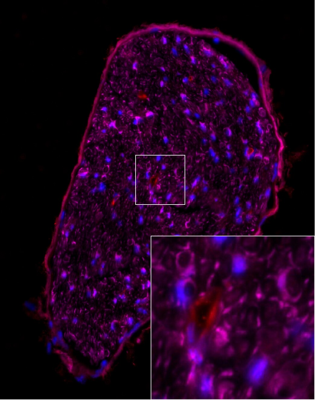
Immunocytochemistry/ Immunofluorescence: GFP Antibody [NB100-1614] -
Immunocytochemistry/ Immunofluorescence: GFP Antibody [NB100-1614] - Macrophages regulate dormancy in tumor cells.a Representative image of triple immunofluorescently stained in E0771-GFP primary tumor tissue for tumor cells, macrophages, & NR2F1. Green = GFP; Red = NR2F1; White = IBA-1; Blue = DAPI. White arrow shows a macrophage. The yellow arrow contact between an NR2F1-positive tumor cell & a macrophage. Mϕ=Macrophage. Scale bar=20 μm. b Quantification showing frequency of distances between NR2F1+ tumor cells to nearest macrophage in primary tumor. Data is normalized to frequency of distances between all DAPI+ nuclei to nearest TMEM. Bar = mean. Error bars = ±SEM. n = 34 fields of view (551 × 316 µm2) in 4 animals. For comparison between 0 & 200 µm bins a two-tailed Mann-Whitney test used (p < 0.0001). ****p < 0.0001. c Representative immunofluorescence images of NR2F1 expression in E0771-GFP tumor cells cultured alone, in direct contact w/ BAC1.2F5 macrophages,/in direct contact w/ HUVEC endothelial cells. White arrows show macrophages/endothelial cells in direct contact w/ a tumor cell. Green = GFP; Red = NR2F1; Blue = DAPI. TC = Tumor Cell. Mϕ = Macrophage. EC = Endothelial Cell. Scale bar = 15 μm. d Percentage of NR2F1-positive tumor cells from each group in C. TC alone: n = 777 cells in 9 independent experiments; TC+Mϕ; n = 226 cells in 6 independent experiments, TC+EC = n = 359 cells in 4 independent experiments. Bar = mean. Error bars = ±SEM. For TC vs. TC+Mϕ (p = 0.0039), & for TC vs. TC+EC (p = 1), a two-tailed Kruskal-Wallis test w/ Dunn’s multiple comparisons adjustment used. For TC+Mϕ vs. TC+EC (0.012), a two-tailed one-way ANOVA w/ Sidak’s multiple comparison adjustment used. *p < 0.05. **p < 0.01; ns = not significant. Source data are provided as a Source Data file. Image collected & cropped by CiteAb from following publication (https://pubmed.ncbi.nlm.nih.gov/35110548), licensed under a CC-BY license. Not internally tested by Novus Biologicals.
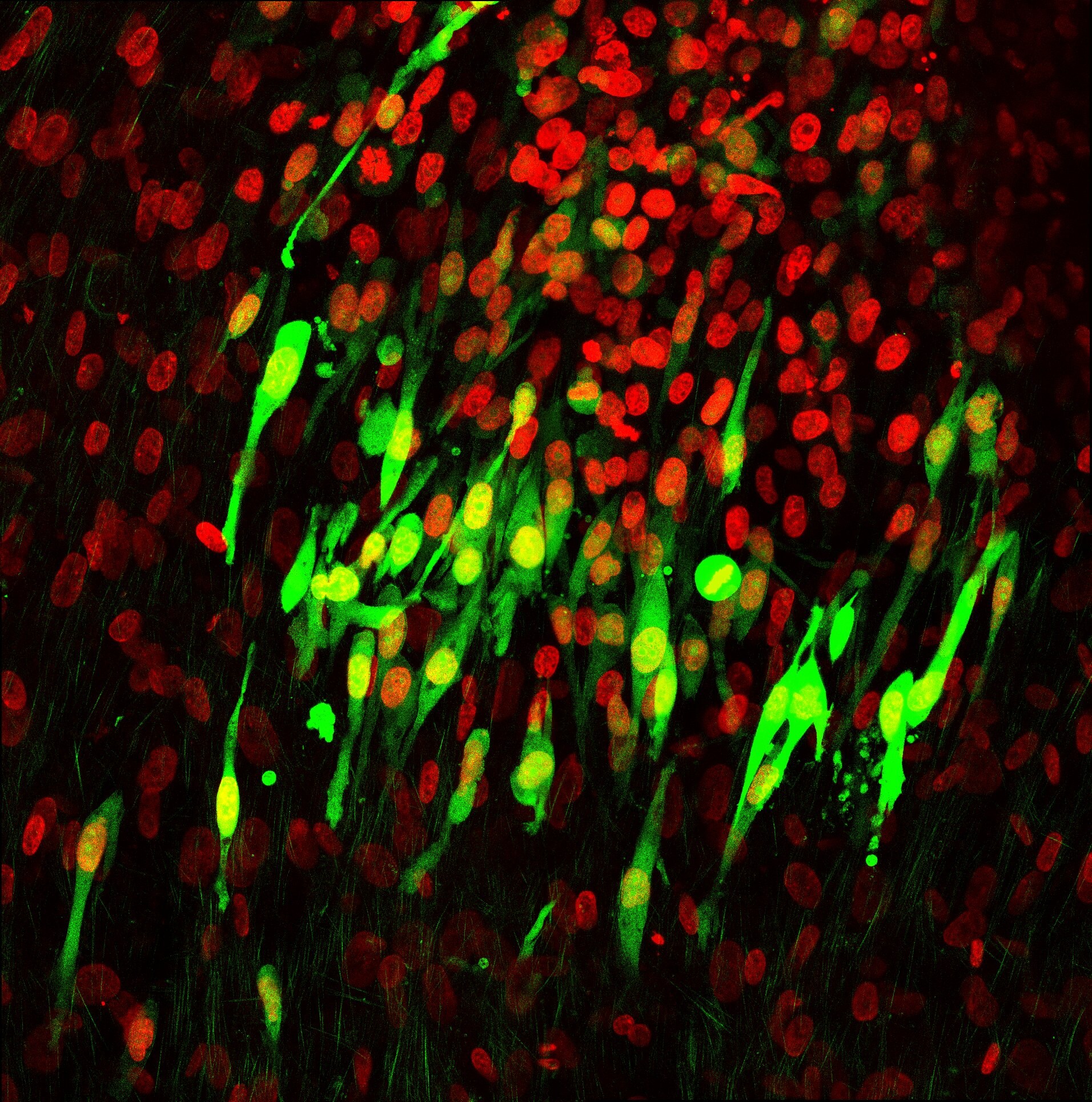
Immunocytochemistry/ Immunofluorescence: GFP Antibody [NB100-1614] -
Immunocytochemistry/ Immunofluorescence: GFP Antibody [NB100-1614] - Spontaneously metastasizing tumor cells survive significantly longer at the secondary site compared to intravenously injected tumor cells.a Representative intravital microscopy images showing the possible fates of extravascular disseminated tumor cells in the lung parenchyma. Top: Images of disseminated tumor cells just after extravasation. Bottom left: Example of an extravascular tumor cell, which has died, as evidenced by small extravascular apoptotic bodies (yellow arrow). Bottom middle: Example of an extravascular tumor cell that survived as a single & solitary tumor cell over time. Bottom right: Example of an extravascular tumor cell that began to divide & grow into a micro-metastasis. Red = tdTomato labeled endothelial cells & 155 kDa Tetramethylrhodamine dextran labeled blood serum, Green = GFP labeled tumor cells. Yellow dashed lines delineate blood vessel boundaries. Scale bar = 15 μm. b Percentage of extravascular E0771-GFP disseminated tumor cells that died, survived, or grew after extravasation in EM & SM models 64 hrs after arrival to the lung vasculature. EM: n = 27 tumor cells in 4 mice. SM: n = 31 tumor cells in 4 mice. Bar = mean. Error bars = ±SEM. For Died & Survived columns, a two-tailed unpaired t-test was used (p = 0.0003 & 0.0005, respectively). For Grew columns, a two-tailed Mann-Whitney test was used (p = 0.14). ***p < 0.001. ns = not significant. Source data are provided as a Source Data file. Image collected & cropped by CiteAb from the following publication (https://pubmed.ncbi.nlm.nih.gov/35110548), licensed under a CC-BY license. Not internally tested by Novus Biologicals.
Immunocytochemistry/ Immunofluorescence: GFP Antibody [NB100-1614] -
Immunocytochemistry/ Immunofluorescence: GFP Antibody [NB100-1614] - Qx protects against ototoxin-induced HC death & promotes supporting cell proliferation. (A–I) TUNEL assay (red) performed in zebrafish incubated w/ vehicle or 300 µM of Qx alone (controls) or w/ corresponding ototoxin w/ or w/out Qx 300 µM. Animals counterstained w/ phalloidin (green). (A) Neomycin (Neo) 200 µM incubation for 30 min. (B) Gentamicin (GM) 50 µM incubation for 1 hour. (C) Cisplatin (CP) 400 µM incubation for 2 hrs. (D) GM 50 µM incubation for 1 hour followed by recovery for 5 hrs. (E) Incubation w/ Qx 300 µM for 8 hrs & Neo 200 µM for 30 min. (F) Incubation w/ Qx 300 µM for 8 hrs & GM 50 µM for 1 hour. (G) Qx 300 µM for 8 hrs + CP 400 µM for 2 hrs. (H) Qx 300 µM incubation for 8 hrs + GM 50 µM for 1 hour followed by 5 hrs recovery. Asterisks denote TUNEL-positive HCs. (I) The % of TUNEL-positive neuromasts calculated for each treatment & represented as mean +/− SEM. (J–R) Proliferation assays performed in 5dpf Tg(brn3c:GFP) in presence/absence of Qx & corresponding ototoxin, by BrdU-labelling method (red). Animals immunostained for GFP (green). (J) control. (K) Neo 200 µM 30 min. (L) GM 50 µM 1 hour. (M) CP: 400 µM 2 hrs. (N) Qx 300 µM 8 hrs. (O) Qx 300 µM 8 hrs + Neo 200 µM 30 min. (P) Qx 300 µM 8 hrs + GM 50 µM 1 hour. (Q) Qx 300 µM 8 hrs + CP 400 µM 2 hrs. Asterisks denote neuromast supporting cells positive for BrdU. (R) The % of BrdU-positive supporting cells per neuromast calculated for each treatment & represented as mean +/− SEM. One-way ANOVA, *p < 0.05, **p < 0.01. Black asterisks compared versus corresponding control. Red asterisk compared versus corresponding ototoxin-only treatment. Scale bar: (A–H) 10 µm, (J–O) 7 μm. Data taken from at least 15 animals & 3 experiments runs. Image collected & cropped by CiteAb from following publication (https://pubmed.ncbi.nlm.nih.gov/30310154), licensed under a CC-BY license. Not internally tested by Novus Biologicals.
Immunocytochemistry/ Immunofluorescence: GFP Antibody [NB100-1614] -
Immunocytochemistry/ Immunofluorescence: GFP Antibody [NB100-1614] - Spontaneously metastasizing tumor cells are more frequently doubly positive for dormancy & stem-like markers compared to intravenously injected tumor cells.a Representative images of triple immunofluorescence staining for GFP, NR2F1, & SOX9 expression in primary tumors, circulating tumor cells (CTCs), & disseminated tumor cells (Lung) from an E0771-GFP SM model (Left) & in disseminated tumor cells (Lung) from an EM model (Right). Green = GFP; Red = NR2F1; Orange = SOX9; Blue = DAPI. Scale bar for Primary Tumor = 50 μm. Scale bar for CTCs & Lung = 15 μm. b Percentage of double-positive tumor cells NR2F1-positive SOX9High from each group in Fig. 5a. Primary Tumor: n = 2383 in 97 fields of view (65 × 65 µm2) in 7 animals; CTCs: n = 379 cells in 8 animals; SM Lung: n = 104 cells in 9 animals; In vitro: n = 413 cells in 3 independent experiments. EM Lung: n = 75 cells in 7 animals. Bar = mean. Error bars = ±SEM. For EM Lung vs. SM Lung (p = 0.0001) & EM Lung vs. in vitro (p = 0.69), a two-tailed Kruskal-Wallis test with Dunn’s multiple comparisons adjustment was used. For PT vs. CTC: (p = 0.0041), PT vs. Lung SM (p = 0.0030), & CTC vs. Lung SM (p = 1.00) a two-tailed ANOVA test with Sidak’s multiple comparisons adjustment was used. **p < 0.01. ***p < 0.001. ns = not significant. Source data are provided as a Source Data file. Image collected & cropped by CiteAb from the following publication (https://pubmed.ncbi.nlm.nih.gov/35110548), licensed under a CC-BY license. Not internally tested by Novus Biologicals.
Immunocytochemistry/ Immunofluorescence: GFP Antibody [NB100-1614] -
Immunocytochemistry/ Immunofluorescence: GFP Antibody [NB100-1614] - Dose protection curve against aminoglycosides. 5dpf larvae were incubated with vehicle (E3, A) or with 50 μM (D–F), 150 μM (G–I) or 300 μM (J–L) of Qx for a total of 8 hours. Gentamicin (GM, 50 μM, B,E,H,K) or neomycin (Neo, 200 µM,C,F,I,L) were added during the last 60 min or 30 min of incubation, respectively. Animals were fixed & stained for otoferlin (red) & GFP (green). (M) Quantification of the number of hair cells per neuromast after the different treatments represented as mean +/− SEM. Note that since no significant differences were found in the number of hair cells per neuromast when animals were incubated with the different Qx concentrations (0–300 µM, A,D,G,J), the control value represents the average of all these treatments. One-way ANOVA, Dunnett post test. *p < 0.05, **p < 0.01, ***p < 0.001. Black asterisks compared versus control. Red asterisks compared versus the corresponding aminoglycoside-only treatment. (N) Scores for neuromast morphology (see Materials & Methods). Scale bar: 7 μm. Data were taken from at least 20 animals & 3 experiments runs. Image collected & cropped by CiteAb from the following publication (https://pubmed.ncbi.nlm.nih.gov/30310154), licensed under a CC-BY license. Not internally tested by Novus Biologicals.
Immunocytochemistry/ Immunofluorescence: GFP Antibody [NB100-1614] -
Immunocytochemistry/ Immunofluorescence: GFP Antibody [NB100-1614] - Qx protects against cisplatin ototoxicity. 5dpf Tg(brn3c:GFP) larvae were incubated with vehicle alone (DMSO, A), 400 μM of cisplatin (CP, B), pre-treated with Qx for 2 hours & then incubated with CP for 6 hours (Qx, CP, C,E,G) or pre-treated with Qx for 2 hours & then co-treated with Qx & CP for 6 more hours (Qx + CP, D,F,H). Animals were fixed & immunostained for GFP (green) & otoferlin (red). (I) Quantification of the number of hair cells per neuromast after the different treatments represented as mean +/− SEM. One-way ANOVA, Dunnett post test. *p < 0.05, ***p < 0.001. Black asterisks compared versus control. Red asterisks compared versus CP 400 µM. (J) Scores for neuromast morphology (see Materials & Methods). Scale bar: 6 μm. Data were taken from at least 20 animals & 3 experiments runs. Image collected & cropped by CiteAb from the following publication (https://pubmed.ncbi.nlm.nih.gov/30310154), licensed under a CC-BY license. Not internally tested by Novus Biologicals.
Immunocytochemistry/ Immunofluorescence: GFP Antibody [NB100-1614] -
Immunocytochemistry/ Immunofluorescence: GFP Antibody [NB100-1614] - Dose protection curve against CP. 5dpf Tg(brn3c:GFP) larvae were incubated with 50 µM to 800 µM of CP (A,C,E,G) for 6 hours or pre-treated with 300 µM of Qx for 2 hours & then co-treated with Qx & CP (50µM-800µM) for 6 hours (B,D,F,H). Animals were fixed & immunostained for GFP (green) & otoferlin (red). Control animals were exposed to vehicle alone (DMSO). (I) Quantification of the number of hair cells per neuromast after the different treatments represented as mean +/− SEM. One-way ANOVA, Dunnett post test. ***p < 0.001. Black asterisks compared versus DMSO-treated animals. Red asterisks compared versus the corresponding CP concentration. (J) Scores for neuromast morphology (see Materials & Methods). Scale bar: 6 μm. Data were taken from at least 20 animals & 3 experiments runs. Image collected & cropped by CiteAb from the following publication (https://pubmed.ncbi.nlm.nih.gov/30310154), licensed under a CC-BY license. Not internally tested by Novus Biologicals.Applications for GFP Antibody - BSA Free
Application
Recommended Usage
ELISA
1:100 - 1:2000
Immunocytochemistry/ Immunofluorescence
1:2000-1:5000
Immunohistochemistry
1:2000 - 1:5000
Immunohistochemistry-Frozen
1:10 - 1:500
Immunohistochemistry-Paraffin
1:10 - 1:500
Western Blot
1:5000 - 1:10000
Application Notes
Use in In vivo reported in scientific literature (PMID:34911937).Use in IHC-FrFl reported in scientific literature (PMID:33558651). Use in IHC-F was reported in the scientific literature (PMID: 23799397).
Reviewed Applications
Read 8 reviews rated 4.5 using NB100-1614 in the following applications:
Flow Cytometry Panel Builder
Bio-Techne Knows Flow Cytometry
Save time and reduce costly mistakes by quickly finding compatible reagents using the Panel Builder Tool.
Advanced Features
- Spectra Viewer - Custom analysis of spectra from multiple fluorochromes
- Spillover Popups - Visualize the spectra of individual fluorochromes
- Antigen Density Selector - Match fluorochrome brightness with antigen density

Formulation, Preparation, and Storage
Purification
Immunogen affinity purified
Formulation
10 mM PBS (0.9% isotonic, w/v, pH 7.2) and 50% (v/v) Glycerol
Format
BSA Free
Preservative
0.02% Sodium Azide
Concentration
10.0 mg/ml
Shipping
The product is shipped with polar packs. Upon receipt, store it immediately at the temperature recommended below.
Stability & Storage
Store at -20C in the dark. Avoid freeze-thaw cycles.
Background: GFP
References
1. Shi, C., Pan, F. C., Kim, J. N., Washington, M. K., Padmanabhan, C., Meyer, C. T.,... Means, A. L. (2019). Differential Cell Susceptibilities to Kras(G12D) in the Setting of Obstructive Chronic Pancreatitis. Cell Mol Gastroenterol Hepatol. doi:10.1016/j.jcmgh.2019.07.001
2. Zhao, S., Fortier, T. M., & Baehrecke, E. H. (2018). Autophagy Promotes Tumor-like Stem Cell Niche Occupancy. Curr Biol, 28(19), 3056-3064.e3053. doi:10.1016/j.cub.2018.07.075
3. Zusso, M., Lunardi, V., Franceschini, D., Pagetta, A., Lo, R., Stifani, S.,... Moro, S. (2019). Ciprofloxacin and levofloxacin attenuate microglia inflammatory response via TLR4/NF-kB pathway. J Neuroinflammation, 16(1), 148. doi:10.1186/s12974-019-1538-9
Long Name
Green Fluorescent Protein
Alternate Names
eGFP, GFPuv
Additional GFP Products
Product Documents for GFP Antibody - BSA Free
Certificate of Analysis
To download a Certificate of Analysis, please enter a lot or batch number in the search box below.
Product Specific Notices for GFP Antibody - BSA Free
Chicken products cannot be exported to Canada.
Purification Notes
Chickens were immunized with purified recombinant green fluorescent protein (GFP) emulsified in Freund's adjuvant. After multiple injections, eggs were collected from the hens, and IgY fractions were prepared from the yolks and then affinity-purified antibodies were prepared using GFP conjugated to an agarose matrix. The final product is a filter-sterilized mixture of both affinity-purified antibodies (30 ug/ml) and purified IgY (10 mg/mL).
Storage Notes
Store at -20C in the dark. Under these conditions, the antibodies should have a shelf life of at least 12 months (provided they remain sterile). Since 50% glycerol is present in the vial, this antibody preparation should remain a liquid at -20C. For longer storage periods, store at -80C, but be aware that freezing this preparation may reduce its activity.
This product is for research use only and is not approved for use in humans or in clinical diagnosis. Primary Antibodies are guaranteed for 1 year from date of receipt.
Citations for GFP Antibody - BSA Free
 Powered by Bioz
Customer Reviews for GFP Antibody - BSA Free (8)
4.5 out of 5
8 Customer Ratings
Have you used GFP Antibody - BSA Free?
Submit a review and receive an Amazon gift card!
$25/€18/£15/$25CAN/¥2500 Yen for a review with an image
$10/€7/£6/$10CAN/¥1110 Yen for a review without an image
Submit a review
Customer Images
Showing
1
-
5 of
8 reviews
Showing All
Filter By:
-
Application: Immunohistochemistry-FrozenSample Tested: Sciatic NerveSpecies: MouseVerified Customer | Posted 04/05/2024Image depicting GFP NB100-1614 in magenta and CD31 AF3628 in red. Tissue is a fixed frozen section of sciatic nerve obtained from a S100A4 GFP mouse.NB100-1614 was diluted 1 in 10000 and was left on tissue sections overnight at 4 degrees Celsius. Secondary antibody was donkey anti chicken conjugated to Alexa 647.

-
Application: ImmunohistochemistrySample Tested: Cancer CellsSpecies: MouseVerified Customer | Posted 10/04/2023DAPI (Red) Cytopastmic GFP stain (Green) Primary antibody dilution: 1:200 in a 200 um slice
-
Application: Immunohistochemistry-ParaffinSample Tested: embryonic mouse brainSpecies: MouseVerified Customer | Posted 02/17/2020GFP in green DAPI in grey
-
Application: ImmunocytochemistrySample Tested: LNCaP human prostate cancer cell lineSpecies: HumanVerified Customer | Posted 05/09/2018LNCap cells imaged after transfection with GFP expressing plasmid.Human LnCap cells were grown in RPMI1640 + 10% FCS in 5% CO2 at 37C. The cells were electroporated with GFP expressing plasmid using Amaxa Nucleofection kit.
-
Application: ImmunocytochemistrySample Tested: hela cellSpecies: HumanVerified Customer | Posted 12/25/2016confocal analysis of HeLa cells transiently expressing GFP. Nuclei were counterstained with DAPIGFP expressing cells were fixed with PFA 4% for 30 min, washed several times with PBS and then permeabilized for 5 min with PBSTx-100 1%. After several washes cells were incubated with the chicken anti-GFP 1:500 in fish blocking solution (PBS + 2.5% fish gelatin + 2.5% FCS) overnight at 4C. The next day after several washes with PBS, cells were incubated with anti-chicken Alexa 488 1:400 in fish gelatin for 1hour at room temperature.
-
Application: ImmunofluorescenceSample Tested: rat cortical culture (2 weeks old)Species: RatVerified Customer | Posted 02/17/2016
-
Application: ImmunofluorescenceSample Tested: cultured hippocampal neuron from ratSpecies: RatVerified Customer | Posted 08/23/2013
-
Application: ImmunohistochemistrySample Tested: Mouse Trigeminal Ganglia, CorneaSpecies: MouseVerified Customer | Posted 05/15/2012
There are no reviews that match your criteria.
Protocols
View specific protocols for GFP Antibody - BSA Free (NB100-1614):
Citrate Buffer Antigen Retrieval Protocol
Background: Formaldehyde fixation (2% or 4%, or as a component of 10% formalin) produces protein cross-links in tissues that tends to interfere with antibody penetration. This seems to be particularly true of paraffin- embedded formaldehyde-fixed tissue. Since chicken IgY antibodies are larger than rabbit or mouse IgG's, "extra steps" may be necessary to compensate for their larger size.
The citrate-based "antigen retrieval" protocol outlined below has been shown to improve chicken IgY antibody penetration into 4% formalde- hyde-fixed paraffin-embedded sections, and can increase the degree and intensity of immunoreactivity and immunostaining.
Reagents (NOTE: You can use either the Sodium Citrate or Citric Acid Buffers in step #3, below)
"Sodium Citrate Buffer" (10mM Sodium Citrate, 0.05% Tween 20, pH 6.0)
Weigh out 2.94 grams of trisodium citrate (dihydrate). Dissolve in approximately
900 mls of deionized, distilled water. Adjust the pH to 6.00 with 1.0 N HCl. Add
0.5 ml of Tween-20. Mix. Bring up the volume to 1.0 litres with water. Store this solution at room temperature for 3 months or at 4C for longer periods.
"Citric Acid Buffer" (10mM Citric Acid, 0.05% Tween 20, pH 6.0)
Weigh out 1.92 grams of citric acid (anhydrous). Dissolve in approximately 900 mls of deionized, distilled water. Adjust the pH to 6.0 with 1.0 N NaOH. Add
0.5 ml of Tween-20. Mix. Bring up the volume to 1.0 litres with water. Store this solution at room temperature for 3 months or at 4C for longer periods.
"Phosphate-Buffered Saline" [PBS, 10 mM Sodium phosphate-buffered (pH
7.2) isotonic (0.9%, w/v) saline solution] PBS Tween (0.05% Tween 20 in PBS)
Ethanol (80%, 90%, 95%, 100%) diluted with water
Xylene
Procedure (for use with paraffin-embedded sections):
1 Deparaffinize tissue sections in 2 changes of xylene (5 minutes each).
2. Hydrate in 2 changes of 100% ethanol (3 minutes each), 95% ethanol (1 minute),
90% ethanol (1 minute), 80% ethanol (1 minute). Rinse in distilled water.
3. Pre-heat steamer or water bath with staining dish containing either Sodium
Citrate Buffer or Citrate Buffer. Wait until temperature reaches 95-100 degrees C.
NOTE: Microwave or pressure cooker can be used as an alternative as a heating source.
4. Immerse slides in the staining dish. Place the lid loosely on the staining dish and incubate for 20-40 minutes (optimal incubation times will vary).
5. Remove the staining dish, and allow it to cool to room temperature (for 20 minutes or so).
6. Rinse sections in PBS Tween twice for 2 minutes each time.
NOTE: The remainder of this protocol is meant to be a suggestion, and can be substituted with your regular immunostaining protocol.
7. Block sections for 30 minutes with Blocking buffer diluted 1:10 with water.
8. Incubate sections with primary antibody at appropriate dilution in antibody dilution buffer overnight at 4 degrees C. Since chicken IgY antibodies are larger than mammalian IgG's, this overnight incubation allows more time for antibody penetration into tissue sections.
9. Rinse sections with PBS Tween 20 twice for 5 minutes each time.
10. Incubate sections with labeled secondary antibody (see NOTE, below) at appropriate dilution (for one hour at room temperature) in a 1:100 dilution of blocking buffer (diluted in PBS).
11. Rinse with PBS Tween 20 for three times for 5 minutes each time.
NOTE: This protocol may use HRP- or fluorescently-labeled secondary antibodies produced in goats or rabbits.
References:
1. Shi SR, Chaiwun B, Young L, Cote RJ, Taylor CR. (1993). Antigen retrieval technique utilizing citrate buffer or urea solution for immunohistochemical demonstration of androgen receptor in formalin-fixed paraffin sections. J Histochem Cytochem 41 (11): 1599-1604.
2. Kanai K, Nunoya T, Shibuya K, Nakamura T, Tajima M (1998). Variations in effectiveness of antigen retrieval pretreatments for diagnostic immunohistochemistry. Res Vet Sci 64 (1): 57-
61.
3. Brown RW, Chirala R. (1995). Utility of microwave-citrate antigen retrieval in diagnostic immunohistochemistry. Mod Pathol 8 (5): 515-20.
4. Morgan JM, Navabi H, Schmid KW, Jasani B (1994). Possible role of tissue-bound calcium ions in citrate-mediated high-temperature antigen retrieval. J Pathol 174 (4): 301-7.
5. Pellicer EM, Sundblad A (1994). Antigen retrieval by microwave oven with buffer of citric acid.
Medicina (B Aires). 54 (2): 129-32.
6. Shi SR, Chaiwun B, Young L, Cote RJ, Taylor CR (1993). Antigen retrieval technique utilizing citrate buffer or urea solution for immunohistochemical demonstration of androgen receptor in formalin-fixed paraffin sections. J Histochem Cytochem 41 (11): 1599-604.
Find general support by application which include: protocols, troubleshooting, illustrated assays, videos and webinars.
- 7-Amino Actinomycin D (7-AAD) Cell Viability Flow Cytometry Protocol
- Antigen Retrieval Protocol (PIER)
- Antigen Retrieval for Frozen Sections Protocol
- Appropriate Fixation of IHC/ICC Samples
- Cellular Response to Hypoxia Protocols
- Chromogenic IHC Staining of Formalin-Fixed Paraffin-Embedded (FFPE) Tissue Protocol
- Chromogenic Immunohistochemistry Staining of Frozen Tissue
- ClariTSA™ Fluorophore Kits
- Detection & Visualization of Antibody Binding
- ELISA Sample Preparation & Collection Guide
- ELISA Troubleshooting Guide
- Extracellular Membrane Flow Cytometry Protocol
- Flow Cytometry Protocol for Cell Surface Markers
- Flow Cytometry Protocol for Staining Membrane Associated Proteins
- Flow Cytometry Staining Protocols
- Flow Cytometry Troubleshooting Guide
- Fluorescent IHC Staining of Frozen Tissue Protocol
- Graphic Protocol for Heat-induced Epitope Retrieval
- Graphic Protocol for the Preparation and Fluorescent IHC Staining of Frozen Tissue Sections
- Graphic Protocol for the Preparation and Fluorescent IHC Staining of Paraffin-embedded Tissue Sections
- Graphic Protocol for the Preparation of Gelatin-coated Slides for Histological Tissue Sections
- How to Run an R&D Systems DuoSet ELISA
- How to Run an R&D Systems Quantikine ELISA
- How to Run an R&D Systems Quantikine™ QuicKit™ ELISA
- ICC Cell Smear Protocol for Suspension Cells
- ICC Immunocytochemistry Protocol Videos
- ICC for Adherent Cells
- IHC Sample Preparation (Frozen sections vs Paraffin)
- Immunocytochemistry (ICC) Protocol
- Immunocytochemistry Troubleshooting
- Immunofluorescence of Organoids Embedded in Cultrex Basement Membrane Extract
- Immunofluorescent IHC Staining of Formalin-Fixed Paraffin-Embedded (FFPE) Tissue Protocol
- Immunohistochemistry (IHC) and Immunocytochemistry (ICC) Protocols
- Immunohistochemistry Frozen Troubleshooting
- Immunohistochemistry Paraffin Troubleshooting
- Intracellular Flow Cytometry Protocol Using Alcohol (Methanol)
- Intracellular Flow Cytometry Protocol Using Detergents
- Intracellular Nuclear Staining Flow Cytometry Protocol Using Detergents
- Intracellular Staining Flow Cytometry Protocol Using Alcohol Permeabilization
- Intracellular Staining Flow Cytometry Protocol Using Detergents to Permeabilize Cells
- Preparing Samples for IHC/ICC Experiments
- Preventing Non-Specific Staining (Non-Specific Binding)
- Primary Antibody Selection & Optimization
- Propidium Iodide Cell Viability Flow Cytometry Protocol
- Protocol for Heat-Induced Epitope Retrieval (HIER)
- Protocol for Liperfluo
- Protocol for Making a 4% Formaldehyde Solution in PBS
- Protocol for VisUCyte™ HRP Polymer Detection Reagent
- Protocol for the Characterization of Human Th22 Cells
- Protocol for the Characterization of Human Th9 Cells
- Protocol for the Fluorescent ICC Staining of Cell Smears - Graphic
- Protocol for the Fluorescent ICC Staining of Cultured Cells on Coverslips - Graphic
- Protocol for the Preparation & Fixation of Cells on Coverslips
- Protocol for the Preparation and Chromogenic IHC Staining of Frozen Tissue Sections
- Protocol for the Preparation and Chromogenic IHC Staining of Frozen Tissue Sections - Graphic
- Protocol for the Preparation and Chromogenic IHC Staining of Paraffin-embedded Tissue Sections
- Protocol for the Preparation and Chromogenic IHC Staining of Paraffin-embedded Tissue Sections - Graphic
- Protocol for the Preparation and Fluorescent ICC Staining of Cells on Coverslips
- Protocol for the Preparation and Fluorescent ICC Staining of Non-adherent Cells
- Protocol for the Preparation and Fluorescent ICC Staining of Stem Cells on Coverslips
- Protocol for the Preparation and Fluorescent IHC Staining of Frozen Tissue Sections
- Protocol for the Preparation and Fluorescent IHC Staining of Paraffin-embedded Tissue Sections
- Protocol for the Preparation of Gelatin-coated Slides for Histological Tissue Sections
- Protocol for the Preparation of a Cell Smear for Non-adherent Cell ICC - Graphic
- Protocol: Annexin V and PI Staining by Flow Cytometry
- Protocol: Annexin V and PI Staining for Apoptosis by Flow Cytometry
- Quantikine HS ELISA Kit Assay Principle, Alkaline Phosphatase
- Quantikine HS ELISA Kit Principle, Streptavidin-HRP Polymer
- R&D Systems Quality Control Western Blot Protocol
- Sandwich ELISA (Colorimetric) – Biotin/Streptavidin Detection Protocol
- Sandwich ELISA (Colorimetric) – Direct Detection Protocol
- TUNEL and Active Caspase-3 Detection by IHC/ICC Protocol
- The Importance of IHC/ICC Controls
- Troubleshooting Guide: ELISA
- Troubleshooting Guide: Fluorokine Flow Cytometry Kits
- Troubleshooting Guide: Immunohistochemistry
- Troubleshooting Guide: Western Blot Figures
- Western Blot Conditions
- Western Blot Protocol
- Western Blot Protocol for Cell Lysates
- Western Blot Troubleshooting
- Western Blot Troubleshooting Guide
- View all Protocols, Troubleshooting, Illustrated assays and Webinars
FAQs for GFP Antibody - BSA Free
Showing
1
-
1 of
1 FAQ
Showing All
-
Q: We are looking for a rabbit anti-GFP antibody as a primary antibody for immunofluorescence and WB. I've seen that you carry several anti-GFP antibodies and I would like to ask which one you would recommend. The one with Cat.No NB600-308 has been used in a lot of publications and I reckon it is a solid antibody. Is this our best choice, or is there a better alternative?
A: NB600-308 is an excellent GFP antibody and I think it would work very well for you. It has been reviewed and published with which usually makes our customer feel much more confident, seeing as it worked in other people's hand.
Loading...