Gigaxonin Antibody - BSA Free
Novus Biologicals | Catalog # NBP1-49924
![Western Blot: Gigaxonin Antibody [NBP1-49924]](https://resources.rndsystems.com/images/products/Gigaxonin-Antibody-Western-Blot-NBP1-49924-img0002.jpg "Western Blot: Gigaxonin Antibody [NBP1-49924]")
Key Product Details
Validated by
Independent Antibodies
Species Reactivity
Validated:
Human
Predicted:
Mouse (100%). Backed by our 100% Guarantee.
Applications
Validated:
Western Blot, Immunocytochemistry/ Immunofluorescence, Immunoprecipitation
Cited:
Immunoprecipitation
Label
Unconjugated
Antibody Source
Polyclonal Rabbit IgG
Format
BSA Free
Loading...
Product Specifications
Immunogen
The immunogen for this product maps to a region between residue 150 and 200 of human Gigaxonin using the numbering given in entry NP_071324.1 (GeneID 8139).
Clonality
Polyclonal
Host
Rabbit
Isotype
IgG
Theoretical MW
68 kDa.
Disclaimer note: The observed molecular weight of the protein may vary from the listed predicted molecular weight due to post translational modifications, post translation cleavages, relative charges, and other experimental factors.
Disclaimer note: The observed molecular weight of the protein may vary from the listed predicted molecular weight due to post translational modifications, post translation cleavages, relative charges, and other experimental factors.
Scientific Data Images for Gigaxonin Antibody - BSA Free
Western Blot: Gigaxonin Antibody [NBP1-49924]
Gigaxonin-Antibody-Western-Blot-NBP1-49924-img0002.jpg![Immunoprecipitation: Gigaxonin Antibody [NBP1-49924]](https://resources.rndsystems.com/images/products/Gigaxonin-Antibody-Immunoprecipitation-NBP1-49924-img0001.jpg "Immunoprecipitation: Gigaxonin Antibody [NBP1-49924]")
Immunoprecipitation: Gigaxonin Antibody [NBP1-49924]
Immunoprecipitation: Gigaxonin Antibody [NBP1-49924] - Samples: Whole cell lysate (1 mg for IP, 20% of IP loaded) from HeLa cells. Antibodies: Affinity purified rabbit anti-Gigaxonin antibody NBP1-49924 used for IP at 3 ug/mg lysate. Gigaxonin was also immunoprecipitated by rabbit anti-Gigaxonin antibody NBP1-49923, which recognizes an upstream epitope. For blotting immunoprecipitated Gigaxonin, NBP1-49924 was used at 1 ug/ml. Detection: Chemiluminescence with an exposure time of 10 seconds.![Immunoprecipitation: Gigaxonin Antibody [NBP1-49924]](https://resources.rndsystems.com/images/products/Gigaxonin-Antibody-Immunoprecipitation-NBP1-49924-img0003.jpg "Immunoprecipitation: Gigaxonin Antibody [NBP1-49924]")
Immunoprecipitation: Gigaxonin Antibody [NBP1-49924]
Gigaxonin-Antibody-Immunoprecipitation-NBP1-49924-img0003.jpg
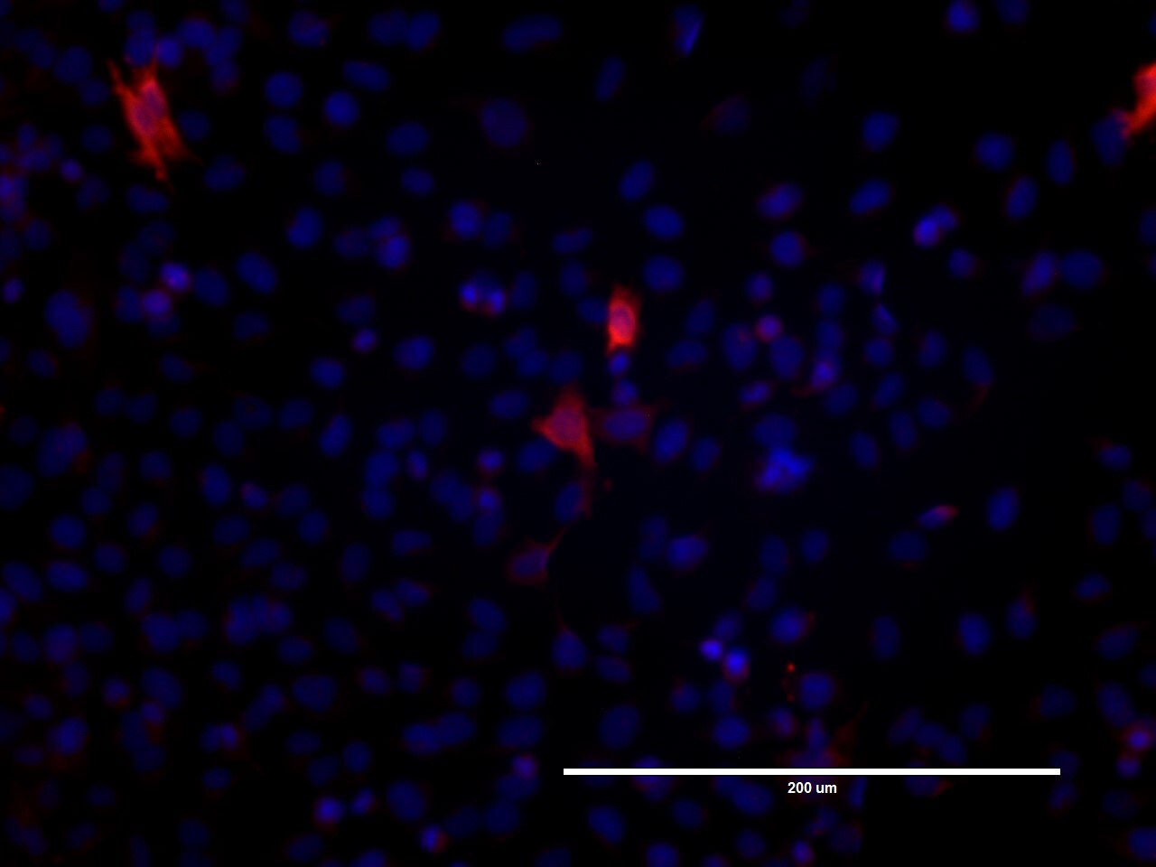
Immunocytochemistry/Immunofluorescence: Gigaxonin Antibody [NBP1-49924] - U251 cells after 48hrs.
Immunocytochemistry/Immunofluorescence: Gigaxonin Antibody [NBP1-49924] - Transfection with wild type Gigaxonin. Image from verified customer review.
Western Blot: Gigaxonin Antibody [NBP1-49924] -
Western Blot: Gigaxonin Antibody [NBP1-49924] - Design of proteomic bait to determine the binding partners of GIG.(A) GIG is a BTB/Kelch protein, & these domains are separated by an intervening BACK domain. GIG is thought to dimerize through its BTB domain [16]. The BTB domain has also been shown to bind to E3 ligases, potentially for ubiquitin tagging of Kelch targets. To identify potential substrates for degradation/modification (Kelch binding partners) we expressed flag-tagged full-length GIG (B) or its BTB domain (C). (D) Sequence of proteomic baits with key regions identified by color. The constructs expressed included a single flag tag & linker (grey), Biotag (black), BTB domain (blue), BACK domain (red), & Kelch domains (green). The location of the truncation for the BTB bait shown in (C) is indicated by the black line at amino acid 268, towards the N-terminus of the BACK domain. (E) Flag-tagged constructs were expressed in HEK293 cells & affinity purified with an anti-Flag antibody (abbreviated as F). The membrane was then probed with anti-Flag tag antibodies. Untransfected cells (NC) were included to control for non-specific binding to the antibody or beads. Flag-tagged GIG was ~62 kDa, while the BTB domain was ~34 kDa & forms a dimer in the immunoprecipitation sample that was ~70 kDa. Image collected & cropped by CiteAb from the following publication (https://pubmed.ncbi.nlm.nih.gov/26460568), licensed under a CC-BY license. Not internally tested by Novus Biologicals.Applications for Gigaxonin Antibody - BSA Free
Application
Recommended Usage
Immunocytochemistry/ Immunofluorescence
Valdiated from a verified customer review
Immunoprecipitation
2-5 ug/mg lysate
Reviewed Applications
Read 1 review rated 4 using NBP1-49924 in the following applications:
Formulation, Preparation, and Storage
Purification
Immunogen affinity purified
Formulation
Tris-Citrate/Phosphate (pH 7.0 - 8.0)
Format
BSA Free
Preservative
0.09% Sodium Azide
Concentration
1.0 mg/ml
Shipping
The product is shipped with polar packs. Upon receipt, store it immediately at the temperature recommended below.
Stability & Storage
Store at 4C. Do not freeze.
Background: Gigaxonin
Alternate Names
FLJ38059, GAN1KLHL16Kelch-like protein 16, giant axonal neuropathy (gigaxonin), gigaxonin
Entrez Gene IDs
8139 (Human)
Gene Symbol
GAN
UniProt
Additional Gigaxonin Products
Product Documents for Gigaxonin Antibody - BSA Free
Certificate of Analysis
To download a Certificate of Analysis, please enter a lot or batch number in the search box below.
Product Specific Notices for Gigaxonin Antibody - BSA Free
This product is for research use only and is not approved for use in humans or in clinical diagnosis. Primary Antibodies are guaranteed for 1 year from date of receipt.
Citations for Gigaxonin Antibody - BSA Free
 Powered by Bioz
Customer Reviews for Gigaxonin Antibody - BSA Free (1)
4 out of 5
1 Customer Rating
Have you used Gigaxonin Antibody - BSA Free?
Submit a review and receive an Amazon gift card!
$25/€18/£15/$25CAN/¥2500 Yen for a review with an image
$10/€7/£6/$10CAN/¥1110 Yen for a review without an image
Submit a review
Customer Images
Showing
1
-
1 of
1 review
Showing All
Filter By:
-
Application: ImmunocytochemistrySample Tested: U251 glioma cell lineSpecies: HumanVerified Customer | Posted 06/14/2023U251 cells after 48hrs. transfection with wild type gigaxonin
 Bio-Techne ResponseThis review was submitted through the legacy Novus Innovators Program, reflecting a new species or application tested on a primary antibody.
There are no reviews that match your criteria.
Protocols
Find general support by application which include: protocols, troubleshooting, illustrated assays, videos and webinars.
- Appropriate Fixation of IHC/ICC Samples
- Cellular Response to Hypoxia Protocols
- ClariTSA™ Fluorophore Kits
- Detection & Visualization of Antibody Binding
- ICC Cell Smear Protocol for Suspension Cells
- ICC Immunocytochemistry Protocol Videos
- ICC for Adherent Cells
- Immunocytochemistry (ICC) Protocol
- Immunocytochemistry Troubleshooting
- Immunofluorescence of Organoids Embedded in Cultrex Basement Membrane Extract
- Immunohistochemistry (IHC) and Immunocytochemistry (ICC) Protocols
- Immunoprecipitation Protocol
- Preparing Samples for IHC/ICC Experiments
- Preventing Non-Specific Staining (Non-Specific Binding)
- Primary Antibody Selection & Optimization
- Protocol for VisUCyte™ HRP Polymer Detection Reagent
- Protocol for the Fluorescent ICC Staining of Cell Smears - Graphic
- Protocol for the Fluorescent ICC Staining of Cultured Cells on Coverslips - Graphic
- Protocol for the Preparation and Fluorescent ICC Staining of Cells on Coverslips
- Protocol for the Preparation and Fluorescent ICC Staining of Non-adherent Cells
- Protocol for the Preparation and Fluorescent ICC Staining of Stem Cells on Coverslips
- Protocol for the Preparation of a Cell Smear for Non-adherent Cell ICC - Graphic
- R&D Systems Quality Control Western Blot Protocol
- TUNEL and Active Caspase-3 Detection by IHC/ICC Protocol
- The Importance of IHC/ICC Controls
- Troubleshooting Guide: Western Blot Figures
- Western Blot Conditions
- Western Blot Protocol
- Western Blot Protocol for Cell Lysates
- Western Blot Troubleshooting
- Western Blot Troubleshooting Guide
- View all Protocols, Troubleshooting, Illustrated assays and Webinars
Loading...