GLI-1 Antibody - BSA Free
Novus Biologicals | Catalog # NB600-600
![Knockdown Validated: GLI-1 Antibody - BSA Free [NB600-600]](https://resources.rndsystems.com/images/products/GLI-1-Antibody-Immunohistochemistry-Paraffin-NB600-600-img0005.jpg "Immunohistochemistry: GLI-1 Antibody - BSA Free [NB600-600]")
Key Product Details
Validated by
Knockout/Knockdown, Biological Validation
Species Reactivity
Validated:
Human, Mouse, Canine
Cited:
Human, Mouse, Porcine, Canine
Applications
Validated:
Immunohistochemistry, Immunohistochemistry-Paraffin, Western Blot, Immunoblotting, ELISA, Flow (Intracellular), Immunocytochemistry/ Immunofluorescence, Chromatin Immunoprecipitation (ChIP), Knockdown Validated
Cited:
Immunohistochemistry-Paraffin, Western Blot, Flow Cytometry, Immunocytochemistry/ Immunofluorescence, Chromatin Immunoprecipitation (ChIP), Chemotaxis, IF/IHC
Label
Unconjugated
Antibody Source
Polyclonal Rabbit IgG
Format
BSA Free
Loading...
Product Specifications
Immunogen
A synthetic peptide made toward the C-terminus portion of the human GLI-1 protein (between residues 800-850) [Uniprot: P08151]
Reactivity Notes
Canine reactivity reported from a verified customer review.
Localization
Cytoplasmic and Nuclear
Specificity
No reaction occurs with human or mouse Gli2 or Gli3.
Clonality
Polyclonal
Host
Rabbit
Isotype
IgG
Scientific Data Images for GLI-1 Antibody - BSA Free
![Immunohistochemistry-Paraffin: GLI-1 Antibody - BSA Free [NB600-600]](https://resources.rndsystems.com/images/products/GLI-1-Antibody-Immunohistochemistry-Paraffin-NB600-600-img0003.jpg "Immunohistochemistry-Paraffin: GLI-1 Antibody - BSA Free [NB600-600]")
Immunohistochemistry-Paraffin: GLI-1 Antibody - BSA Free [NB600-600]
Immunohistochemistry-Paraffin: GLI-1 Antibody [NB600-600] - Analysis of a FFPE mouse brain section using GLI-1 antibody at 1:600 dilution.![Immunohistochemistry-Paraffin: GLI-1 Antibody - BSA Free [NB600-600]](https://resources.rndsystems.com/images/products/GLI-1-Antibody-Immunohistochemistry-Paraffin-NB600-600-img0004.jpg "Immunohistochemistry-Paraffin: GLI-1 Antibody - BSA Free [NB600-600]")
Immunohistochemistry-Paraffin: GLI-1 Antibody - BSA Free [NB600-600]
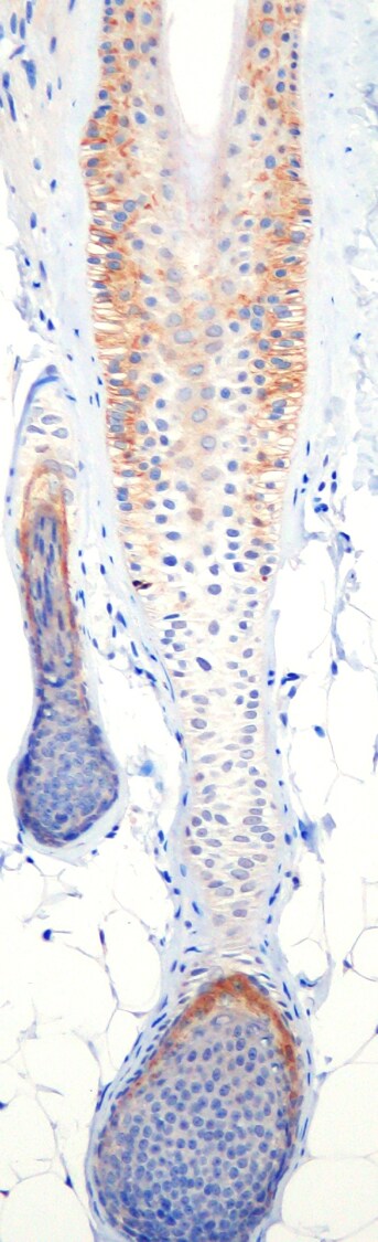
Immunohistochemistry-Paraffin: GLI-1 Antibody [NB600-600] - Staining in canine hair follicle. Image from a verified customer review.![Flow (Intracellular): GLI-1 Antibody - BSA Free [NB600-600]](https://resources.rndsystems.com/images/products/GLI-1-Antibody-Flow-Intracellular-NB600-600-img0002.jpg "Flow (Intracellular): GLI-1 Antibody - BSA Free [NB600-600]")
Flow (Intracellular): GLI-1 Antibody - BSA Free [NB600-600]
Flow (Intracellular): GLI-1 Antibody [NB600-600] - Staining for GLI-1 in untreated human B cell lines (BCWM.1, MWCL-1 and RPCI-WM1) using anti-GLI-1 antibody. Rabbit IgG Isotype Control (Cat# NBP2-36463) was used as a negative control. Image courtesy of Sherine Elsawa, Northern Illinois University.
Immunohistochemistry-Paraffin: GLI-1 Antibody - BSA Free [NB600-600] -
Immunohistochemistry-Paraffin: GLI-1 Antibody - BSA Free [NB600-600] - Paracrine signaling of tumour-derived SHH in CAFs promotes lymphangiogenesis in vivo(A) Growth properties of subcutaneous tumours in mice derived from Caov3 SHH-OE or Caov3 NC cells alone or co-injected with CAFs. Kaplan-Meier plot of time to tumour progression, as evidenced by survival rates over time (n=5). (B) Bright field images of subcutaneous tumours from each group (n=5) 20 days after tumour implantation. (C) Graphical representation of the weights of subcutaneous tumours from each group 20 days after tumour implantation. (D) Immunohistochemical staining for Ki-67, alpha -SMA, Gli-1 & LYVE1 in tumour sections from mice in each group. (E) Graphical representation of the number of LYVE1+ vessels counted from IHC images in panel D. (*P<0.05, **P<0.01, ***P<0.001). Image collected & cropped by CiteAb from the following publication (https://www.oncotarget.com/lookup/doi/10.18632/oncotarget.18621), licensed under a CC-BY license. Not internally tested by Novus Biologicals.
Immunohistochemistry-Paraffin: GLI-1 Antibody - BSA Free [NB600-600] -
Immunohistochemistry-Paraffin: GLI-1 Antibody - BSA Free [NB600-600] - Inhibition of Hh signaling in CAFs blocks its effects on lymphangiogenesis in vivo & in vitro(A) Representative images & statistical analyses of migration of LECs in a co-culture invasion system with CAFs stably transfected with shNC or Gli-shRNA lentivirus in the presence of rSHH (1 μg/ml). (B) Representative images & statistical analyses of capillary tube formation of LECs treated with supernatants from cultures of CAFs stably transfected with shNC or Gli-shRNA lentivirus in the presence of rSHH (1 μg/ml). (C) Bright field images & graphical representation of the weights of tumours of subcutaneous tumours from mice (n=5) bearing Caov3 SHH-OE cells co-injected with CAFs stably transfected with shNC or Gli-shRNA lentivirus 20 days after tumour implantation. (D) Growth properties of subcutaneous tumours in mice derived from Caov3 SHH-OE cells co-injected with CAFs stably transfected with shNC or Gli-shRNA lentivirus. Kaplan-Meier plot of time to tumour progression, as evidenced by survival rates over time. (E) IHC staining for Ki-67, alpha -SMA, Gli-1 & LYVE1 in tumour sections from mice in the two groups. (F) Graphical representation of the number of LYVE1+ vessels counted from IHC images in panel E. (**P<0.01, ***P<0.001). Image collected & cropped by CiteAb from the following publication (https://www.oncotarget.com/lookup/doi/10.18632/oncotarget.18621), licensed under a CC-BY license. Not internally tested by Novus Biologicals.
Western Blot: GLI-1 Antibody - BSA Free [NB600-600] -
Comparison of SHH and GLI-1 expression in the normal mammary gland and MGT tissues. A For western blotting, the blots were cut prior to hybridisation with antibodies. SHH (35 kDa) and GLI-1 (100–140 kDa) protein levels in the normal mammary gland and benign and malignant mammary gland tissues. Beta-actin was used as the loading control. B Relative quantitation of SHH and (C) GLI-1 proteins were normalized to that of the control group. The mean and standard error of the mean are used to express quantitative data. (*, **, #: p <.05). SHH, sonic hedgehog; GLI-1, glioma-associated oncogene 1; MGT, mammary gland tumour Image collected and cropped by CiteAb from the following open publication (https://pubmed.ncbi.nlm.nih.gov/37932728), licensed under a CC-BY license. Not internally tested by Novus Biologicals.Applications for GLI-1 Antibody - BSA Free
Application
Recommended Usage
Chromatin Immunoprecipitation (ChIP)
1:10-1:500
ELISA
1:500 - 1:2000
Flow (Intracellular)
1-2 ug/million cells
Immunoblotting
1 - 2 ug/ml
Immunocytochemistry/ Immunofluorescence
1 ug/ml
Immunohistochemistry
1:200
Immunohistochemistry-Paraffin
1:200
Western Blot
1 - 2 ug/ml
Application Notes
Use in ICC/IF reported in scientific literature (PMID:35226587) Prior to immunostaining paraffin tissues, antigen retrieval with sodium citrate buffer (pH 6.0) is recommended. Flow was reported in a customer review. IHC-P reported from a verified customer review. Use in immunoblotting reported in scientific literature (PMID: 28167297). Use in FLOW reported in scientific literature (PMID: 26238488)..
Reviewed Applications
Read 1 review rated 5 using NB600-600 in the following applications:
Flow Cytometry Panel Builder
Bio-Techne Knows Flow Cytometry
Save time and reduce costly mistakes by quickly finding compatible reagents using the Panel Builder Tool.
Advanced Features
- Spectra Viewer - Custom analysis of spectra from multiple fluorochromes
- Spillover Popups - Visualize the spectra of individual fluorochromes
- Antigen Density Selector - Match fluorochrome brightness with antigen density

Formulation, Preparation, and Storage
Purification
Immunogen affinity purified
Formulation
PBS
Format
BSA Free
Preservative
0.02% Sodium Azide
Concentration
1 mg/ml
Shipping
The product is shipped with polar packs. Upon receipt, store it immediately at the temperature recommended below.
Stability & Storage
Store at 4C short term. Aliquot and store at -20C long term. Avoid freeze-thaw cycles.
Background: GLI-1
Long Name
Glioma-Associated Oncogene Homolog 1 [Zinc Finger Protein]
Alternate Names
GLI1
Gene Symbol
GLI1
UniProt
Additional GLI-1 Products
Product Documents for GLI-1 Antibody - BSA Free
Certificate of Analysis
To download a Certificate of Analysis, please enter a lot or batch number in the search box below.
Product Specific Notices for GLI-1 Antibody - BSA Free
This product is for research use only and is not approved for use in humans or in clinical diagnosis. Primary Antibodies are guaranteed for 1 year from date of receipt.
Related Research Areas
Citations for GLI-1 Antibody - BSA Free
 Powered by Bioz
Customer Reviews for GLI-1 Antibody - BSA Free (1)
5 out of 5
1 Customer Rating
Have you used GLI-1 Antibody - BSA Free?
Submit a review and receive an Amazon gift card!
$25/€18/£15/$25CAN/¥2500 Yen for a review with an image
$10/€7/£6/$10CAN/¥1110 Yen for a review without an image
Submit a review
Customer Images
Showing
1
-
1 of
1 review
Showing All
Filter By:
-
Application: Immunohistochemistry-ParaffinSample Tested: Skin (hair follicle)Species: CanineVerified Customer | Posted 06/12/2019Immunoreactivity for Gli-1 is observed mainly in the isthmus region of the hair follicle.Antibody dilution = 1:600. 3, 3'-diaminobenzidine (DAB) chromogen and counterstaining with Mayer’s hematoxylin.

There are no reviews that match your criteria.
Protocols
View specific protocols for GLI-1 Antibody - BSA Free (NB600-600):
Immunohistochemistry-Paraffin Embedded Sections
Antigen Unmasking:
Bring slides to a boil in 10 mM sodium citrate buffer (pH 6.0) then maintain at a sub-boiling temperature for 10 minutes. Cool slides on bench-top for 30 minutes.
Staining:
1. Wash sections in deionized water three times for 5 minutes each.
2. Wash sections in wash buffer for 5 minutes.
3. Block each section with 100-400 ul blocking solution for 1 hour at room temperature.
4. Remove blocking solution and add 100-400 ul diluted primary antibody. Incubate overnight at 4C.
5. Remove antibody solution and wash sections in wash buffer three times for 5 minutes each.
6. Add 100-400 ul biotinylated diluted secondary antibody. Incubate 30 minutes at room temperature.
7. Remove secondary antibody solution and wash sections three times with wash buffer for 5 minutes each.
8. Add 100-400 ul Streptavidin-HRP reagent to each section and incubate for 30 minutes at room temperature.
9. Wash sections three times in wash buffer for 5 minutes each.
10. Add 100-400 ul DAB substrate to each section and monitor staining closely.
11. As soon as the sections develop, immerse slides in deionized water.
12. Counterstain sections in hematoxylin.
13. Wash sections in deionized water two times for 5 minutes each.
14. Dehydrate sections.
15. Mount coverslips.
Western Blot Protocol
1. Perform SDS-PAGE on samples to be analyzed, loading 40 ug of total protein per lane.
2. Transfer proteins to membrane according to the instructions provided by the manufacturer of the membrane and transfer apparatus.
3. Stain according to standard Ponceau S procedure (or similar product) to assess transfer success, and mark molecular weight standards where appropriate.
4. Rinse the blot.
5. Block the membrane using standard blocking buffer for at least 1 hour.
6. Wash the membrane in wash buffer three times for 10 minutes each.
7. Dilute primary antibody in blocking buffer and incubate 1 hour at room temperature.
8. Wash the membrane in wash buffer three times for 10 minutes each.
9. Apply the diluted HRP conjugated secondary antibody in blocking buffer (as per manufacturers instructions) and incubate 1 hour at room temperature.
10. Wash the blot in wash buffer three times for 10 minutes each (this step can be repeated as required to reduce background).
11. Apply the detection reagent of choice in accordance with the manufacturers instructions.
**Note: Tween-20 can be added to the blocking or antibody dilution buffer at a final concentration of 0.05-0.2%.
Find general support by application which include: protocols, troubleshooting, illustrated assays, videos and webinars.
- 7-Amino Actinomycin D (7-AAD) Cell Viability Flow Cytometry Protocol
- Antigen Retrieval Protocol (PIER)
- Antigen Retrieval for Frozen Sections Protocol
- Appropriate Fixation of IHC/ICC Samples
- Cellular Response to Hypoxia Protocols
- ChIP Protocol Video
- Chromatin Immunoprecipitation (ChIP) Protocol
- Chromatin Immunoprecipitation Protocol
- Chromogenic IHC Staining of Formalin-Fixed Paraffin-Embedded (FFPE) Tissue Protocol
- Chromogenic Immunohistochemistry Staining of Frozen Tissue
- ClariTSA™ Fluorophore Kits
- Detection & Visualization of Antibody Binding
- ELISA Sample Preparation & Collection Guide
- ELISA Troubleshooting Guide
- Extracellular Membrane Flow Cytometry Protocol
- Flow Cytometry Protocol for Cell Surface Markers
- Flow Cytometry Protocol for Staining Membrane Associated Proteins
- Flow Cytometry Staining Protocols
- Flow Cytometry Troubleshooting Guide
- Fluorescent IHC Staining of Frozen Tissue Protocol
- Graphic Protocol for Heat-induced Epitope Retrieval
- Graphic Protocol for the Preparation and Fluorescent IHC Staining of Frozen Tissue Sections
- Graphic Protocol for the Preparation and Fluorescent IHC Staining of Paraffin-embedded Tissue Sections
- Graphic Protocol for the Preparation of Gelatin-coated Slides for Histological Tissue Sections
- How to Run an R&D Systems DuoSet ELISA
- How to Run an R&D Systems Quantikine ELISA
- How to Run an R&D Systems Quantikine™ QuicKit™ ELISA
- ICC Cell Smear Protocol for Suspension Cells
- ICC Immunocytochemistry Protocol Videos
- ICC for Adherent Cells
- IHC Sample Preparation (Frozen sections vs Paraffin)
- Immunocytochemistry (ICC) Protocol
- Immunocytochemistry Troubleshooting
- Immunofluorescence of Organoids Embedded in Cultrex Basement Membrane Extract
- Immunofluorescent IHC Staining of Formalin-Fixed Paraffin-Embedded (FFPE) Tissue Protocol
- Immunohistochemistry (IHC) and Immunocytochemistry (ICC) Protocols
- Immunohistochemistry Frozen Troubleshooting
- Immunohistochemistry Paraffin Troubleshooting
- Intracellular Flow Cytometry Protocol Using Alcohol (Methanol)
- Intracellular Flow Cytometry Protocol Using Detergents
- Intracellular Nuclear Staining Flow Cytometry Protocol Using Detergents
- Intracellular Staining Flow Cytometry Protocol Using Alcohol Permeabilization
- Intracellular Staining Flow Cytometry Protocol Using Detergents to Permeabilize Cells
- Preparing Samples for IHC/ICC Experiments
- Preventing Non-Specific Staining (Non-Specific Binding)
- Primary Antibody Selection & Optimization
- Propidium Iodide Cell Viability Flow Cytometry Protocol
- Protocol for Heat-Induced Epitope Retrieval (HIER)
- Protocol for Liperfluo
- Protocol for Making a 4% Formaldehyde Solution in PBS
- Protocol for VisUCyte™ HRP Polymer Detection Reagent
- Protocol for the Characterization of Human Th22 Cells
- Protocol for the Characterization of Human Th9 Cells
- Protocol for the Fluorescent ICC Staining of Cell Smears - Graphic
- Protocol for the Fluorescent ICC Staining of Cultured Cells on Coverslips - Graphic
- Protocol for the Preparation & Fixation of Cells on Coverslips
- Protocol for the Preparation and Chromogenic IHC Staining of Frozen Tissue Sections
- Protocol for the Preparation and Chromogenic IHC Staining of Frozen Tissue Sections - Graphic
- Protocol for the Preparation and Chromogenic IHC Staining of Paraffin-embedded Tissue Sections
- Protocol for the Preparation and Chromogenic IHC Staining of Paraffin-embedded Tissue Sections - Graphic
- Protocol for the Preparation and Fluorescent ICC Staining of Cells on Coverslips
- Protocol for the Preparation and Fluorescent ICC Staining of Non-adherent Cells
- Protocol for the Preparation and Fluorescent ICC Staining of Stem Cells on Coverslips
- Protocol for the Preparation and Fluorescent IHC Staining of Frozen Tissue Sections
- Protocol for the Preparation and Fluorescent IHC Staining of Paraffin-embedded Tissue Sections
- Protocol for the Preparation of Gelatin-coated Slides for Histological Tissue Sections
- Protocol for the Preparation of a Cell Smear for Non-adherent Cell ICC - Graphic
- Protocol: Annexin V and PI Staining by Flow Cytometry
- Protocol: Annexin V and PI Staining for Apoptosis by Flow Cytometry
- Quantikine HS ELISA Kit Assay Principle, Alkaline Phosphatase
- Quantikine HS ELISA Kit Principle, Streptavidin-HRP Polymer
- R&D Systems Quality Control Western Blot Protocol
- Sandwich ELISA (Colorimetric) – Biotin/Streptavidin Detection Protocol
- Sandwich ELISA (Colorimetric) – Direct Detection Protocol
- TUNEL and Active Caspase-3 Detection by IHC/ICC Protocol
- The Importance of IHC/ICC Controls
- Troubleshooting Guide: ELISA
- Troubleshooting Guide: Fluorokine Flow Cytometry Kits
- Troubleshooting Guide: Immunohistochemistry
- Troubleshooting Guide: Western Blot Figures
- Western Blot Conditions
- Western Blot Protocol
- Western Blot Protocol for Cell Lysates
- Western Blot Troubleshooting
- Western Blot Troubleshooting Guide
- View all Protocols, Troubleshooting, Illustrated assays and Webinars
Loading...