HDAC1 Antibody (4W3X7) - Azide and BSA Free
Novus Biologicals | Catalog # NBP3-15566
![Western Blot: HDAC1 Antibody (4W3X7)Azide and BSA Free [NBP3-15566]](https://resources.rndsystems.com/images/products/HDAC1-Antibody-2W4I5-Western-Blot-NBP3-15566-img0002.jpg "Western Blot: HDAC1 Antibody (4W3X7)Azide and BSA Free [NBP3-15566]")
Loading...
Key Product Details
Species Reactivity
Human, Mouse
Applications
Immunohistochemistry, Immunohistochemistry-Paraffin, Western Blot, Chromatin Immunoprecipitation (ChIP)
Label
Unconjugated
Antibody Source
Monoclonal Mouse IgG1 kappa Clone # 4W3X7
Format
Azide and BSA Free
Loading...
Product Specifications
Immunogen
Recombinant fusion protein containing a sequence corresponding to amino acids 393-482 of human HDAC1 (Q13547). SGDEDEDDPDKRISICSSDKRIACEEEFSDSEEEGEGGRKNSSNFKKAKRVKTEDEKEKDPEEKKEVTEEEKTKEEKPEAKGVKEEVKLA
Clonality
Monoclonal
Host
Mouse
Isotype
IgG1 kappa
Scientific Data Images for HDAC1 Antibody (4W3X7) - Azide and BSA Free
Western Blot: HDAC1 Antibody (4W3X7)Azide and BSA Free [NBP3-15566]
Western Blot: HDAC1 Antibody (2W4I5) [NBP3-15566] - Western blot analysis of extracts of various cell lines, using HDAC1 Mouse mAb (NBP3-15566) at 1:1000 dilution. Secondary antibody: HRP Goat Anti-Mouse IgG (H+L) at 1:10000 dilution. Lysates/proteins: 25ug per lane. Blocking buffer: 3% nonfat dry milk in TBST. Detection: ECL Basic Kit. Exposure time: 30s.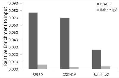
Chromatin Immunoprecipitation: HDAC1 Antibody (2W4I5) [NBP3-15566] - Chromatin immunoprecipitation analysis of extracts of K-562 cells, using HDAC1 antibody (NBP3-15566) and rabbit IgG.The amount of immunoprecipitated DNA was checked by quantitative PCR. Histogram was constructed by the ratios of the immunoprecipitated DNA to the input.
Applications for HDAC1 Antibody (4W3X7) - Azide and BSA Free
Application
Recommended Usage
Chromatin Immunoprecipitation (ChIP)
1:50 - 1:200
Immunohistochemistry
1:50 - 1:200
Western Blot
1:500 - 1:2000
Formulation, Preparation, and Storage
Purification
Affinity purified
Formulation
PBS (pH 7.3), 50% glycerol
Format
Azide and BSA Free
Preservative
0.02% Sodium Azide
Concentration
Please see the vial label for concentration. If unlisted please contact technical services.
Shipping
The product is shipped with polar packs. Upon receipt, store it immediately at the temperature recommended below.
Stability & Storage
Store at -20C. Avoid freeze-thaw cycles.
Background: Histone Deacetylase 1/HDAC1
Long Name
Histone Deacetylase 1
Alternate Names
GON-10, HD1, KDAC1, RPD3L1
Gene Symbol
HDAC1
Additional Histone Deacetylase 1/HDAC1 Products
Product Documents for HDAC1 Antibody (4W3X7) - Azide and BSA Free
Certificate of Analysis
To download a Certificate of Analysis, please enter a lot or batch number in the search box below.
Product Specific Notices for HDAC1 Antibody (4W3X7) - Azide and BSA Free
This product is for research use only and is not approved for use in humans or in clinical diagnosis. Primary Antibodies are guaranteed for 1 year from date of receipt.
⚠ WARNING: This product can expose you to chemicals including Methotrexate, which is known to the State of California to cause reproductive toxicity with developmental effects. For more information, go to www.P65Warnings.ca.govRelated Research Areas
Customer Reviews for HDAC1 Antibody (4W3X7) - Azide and BSA Free
There are currently no reviews for this product. Be the first to review HDAC1 Antibody (4W3X7) - Azide and BSA Free and earn rewards!
Have you used HDAC1 Antibody (4W3X7) - Azide and BSA Free?
Submit a review and receive an Amazon gift card!
$25/€18/£15/$25CAN/¥2500 Yen for a review with an image
$10/€7/£6/$10CAN/¥1110 Yen for a review without an image
Submit a review
Protocols
Find general support by application which include: protocols, troubleshooting, illustrated assays, videos and webinars.
- Antigen Retrieval Protocol (PIER)
- Antigen Retrieval for Frozen Sections Protocol
- Appropriate Fixation of IHC/ICC Samples
- Cellular Response to Hypoxia Protocols
- ChIP Protocol Video
- Chromatin Immunoprecipitation (ChIP) Protocol
- Chromatin Immunoprecipitation Protocol
- Chromogenic IHC Staining of Formalin-Fixed Paraffin-Embedded (FFPE) Tissue Protocol
- Chromogenic Immunohistochemistry Staining of Frozen Tissue
- ClariTSA™ Fluorophore Kits
- Detection & Visualization of Antibody Binding
- Fluorescent IHC Staining of Frozen Tissue Protocol
- Graphic Protocol for Heat-induced Epitope Retrieval
- Graphic Protocol for the Preparation and Fluorescent IHC Staining of Frozen Tissue Sections
- Graphic Protocol for the Preparation and Fluorescent IHC Staining of Paraffin-embedded Tissue Sections
- Graphic Protocol for the Preparation of Gelatin-coated Slides for Histological Tissue Sections
- IHC Sample Preparation (Frozen sections vs Paraffin)
- Immunofluorescent IHC Staining of Formalin-Fixed Paraffin-Embedded (FFPE) Tissue Protocol
- Immunohistochemistry (IHC) and Immunocytochemistry (ICC) Protocols
- Immunohistochemistry Frozen Troubleshooting
- Immunohistochemistry Paraffin Troubleshooting
- Preparing Samples for IHC/ICC Experiments
- Preventing Non-Specific Staining (Non-Specific Binding)
- Primary Antibody Selection & Optimization
- Protocol for Heat-Induced Epitope Retrieval (HIER)
- Protocol for Making a 4% Formaldehyde Solution in PBS
- Protocol for VisUCyte™ HRP Polymer Detection Reagent
- Protocol for the Preparation & Fixation of Cells on Coverslips
- Protocol for the Preparation and Chromogenic IHC Staining of Frozen Tissue Sections
- Protocol for the Preparation and Chromogenic IHC Staining of Frozen Tissue Sections - Graphic
- Protocol for the Preparation and Chromogenic IHC Staining of Paraffin-embedded Tissue Sections
- Protocol for the Preparation and Chromogenic IHC Staining of Paraffin-embedded Tissue Sections - Graphic
- Protocol for the Preparation and Fluorescent IHC Staining of Frozen Tissue Sections
- Protocol for the Preparation and Fluorescent IHC Staining of Paraffin-embedded Tissue Sections
- Protocol for the Preparation of Gelatin-coated Slides for Histological Tissue Sections
- R&D Systems Quality Control Western Blot Protocol
- TUNEL and Active Caspase-3 Detection by IHC/ICC Protocol
- The Importance of IHC/ICC Controls
- Troubleshooting Guide: Immunohistochemistry
- Troubleshooting Guide: Western Blot Figures
- Western Blot Conditions
- Western Blot Protocol
- Western Blot Protocol for Cell Lysates
- Western Blot Troubleshooting
- Western Blot Troubleshooting Guide
- View all Protocols, Troubleshooting, Illustrated assays and Webinars
Loading...