Hepcidin Antimicrobial Peptide Antibody - BSA Free
Novus Biologicals | Catalog # NBP1-59337
![Western Blot: Hepcidin Antimicrobial Peptide Antibody [NBP1-59337]](https://resources.rndsystems.com/images/products/Hepcidin-Antimicrobial-Peptide-Antibody-Western-Blot-NBP1-59337-img0002.jpg "Western Blot: Hepcidin Antimicrobial Peptide Antibody [NBP1-59337]")
Loading...
Key Product Details
Species Reactivity
Validated:
Human, Bovine
Cited:
Human, Mouse, Bovine
Applications
Validated:
Immunohistochemistry, Immunohistochemistry-Paraffin, Western Blot
Cited:
Western Blot
Label
Unconjugated
Antibody Source
Polyclonal Rabbit IgG
Format
BSA Free
Loading...
Product Specifications
Immunogen
Synthetic peptides corresponding to HAMP(hepcidin antimicrobial peptide) The peptide sequence was selected from the N terminal of HAMP. Peptide sequence MALSSQIWAACLLLLLLLASLTSGSVFPQQTGQLAELQPQDRAGARASWM. The peptide sequence for this immunogen was taken from within the described region.
Reactivity Notes
Use in Bovine reported in scientific literature (PMID:31811823).
Clonality
Polyclonal
Host
Rabbit
Isotype
IgG
Theoretical MW
9 kDa.
Disclaimer note: The observed molecular weight of the protein may vary from the listed predicted molecular weight due to post translational modifications, post translation cleavages, relative charges, and other experimental factors.
Disclaimer note: The observed molecular weight of the protein may vary from the listed predicted molecular weight due to post translational modifications, post translation cleavages, relative charges, and other experimental factors.
Scientific Data Images for Hepcidin Antimicrobial Peptide Antibody - BSA Free
Western Blot: Hepcidin Antimicrobial Peptide Antibody [NBP1-59337]
Western Blot: Hepcidin Antimicrobial Peptide Antibody [NBP1-59337] - Human Spleen lysate, concentration 0.2-1 ug/ml.![Immunohistochemistry-Paraffin: Hepcidin Antimicrobial Peptide Antibody [NBP1-59337]](https://resources.rndsystems.com/images/products/Hepcidin-Antimicrobial-Peptide-Antibody-Immunohistochemistry-Paraffin-NBP1-59337-img0003.jpg "Immunohistochemistry-Paraffin: Hepcidin Antimicrobial Peptide Antibody [NBP1-59337]")
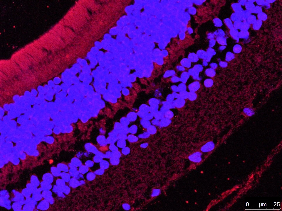
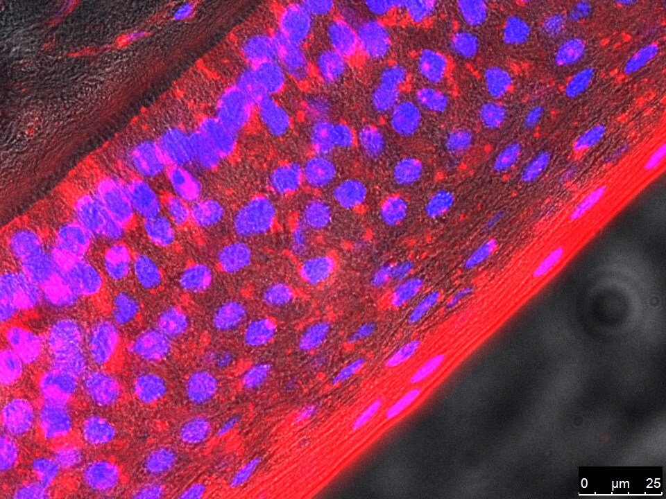
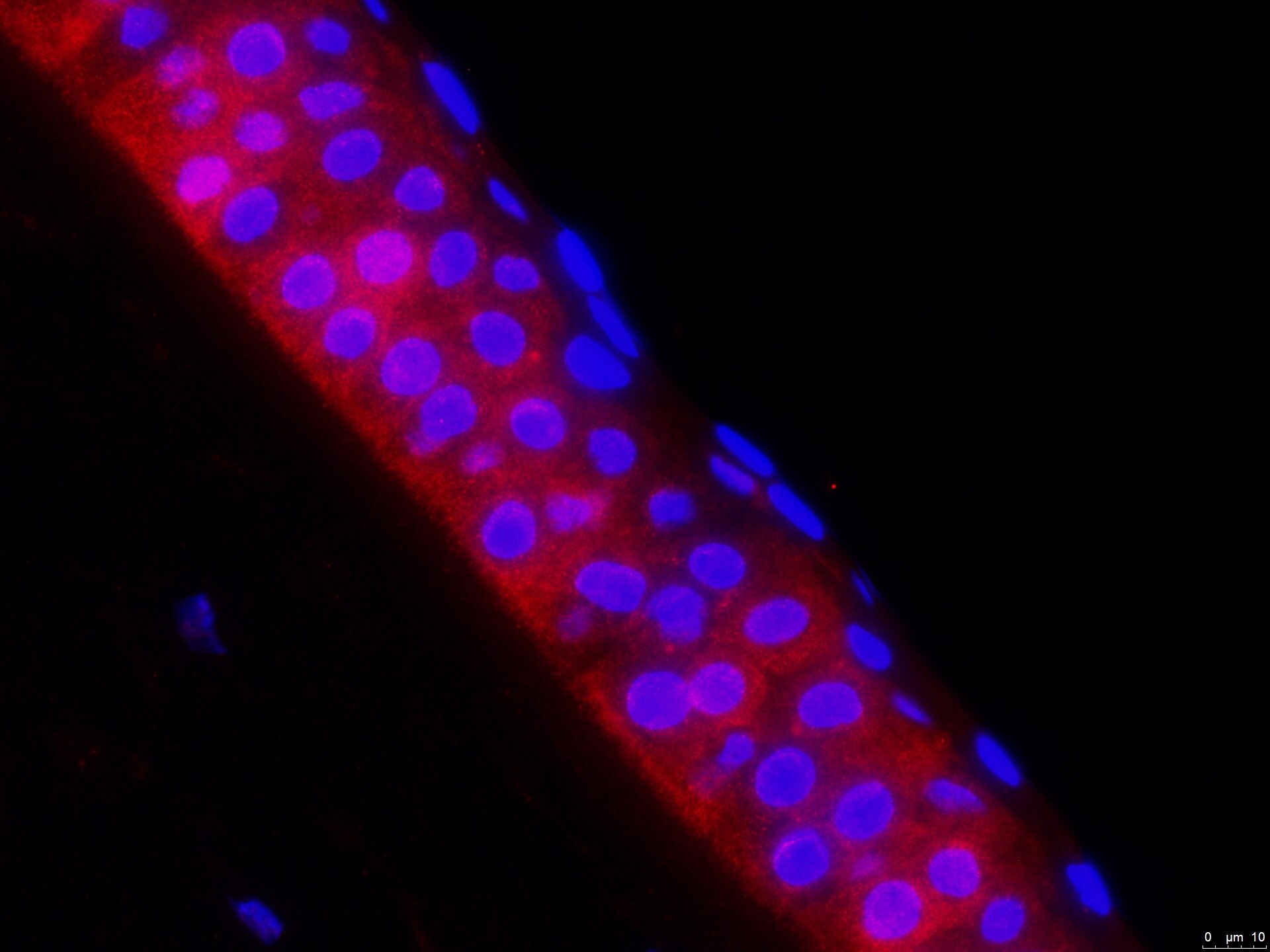
Immunohistochemistry-Paraffin: Hepcidin Antimicrobial Peptide Antibody [NBP1-59337]
Immunohistochemistry-Paraffin: Hepcidin Antimicrobial Peptide Antibody [NBP1-59337] - Paraffin embedded sections of posterior segment of mouse eye, red is Hepcidin and blue is Hoeschst. 4um sections were rehydrated with xylene, followed by a decreasing ethanol concentration gradient (100%, 90%, 70%) and a wash with diH2O. Heat-mediated antigen retrieval performed using EDTA buffer (10mM Trizma Base, 1mM EDTA solution, 0.05% Tween 20, pH 9.0) in an autoclave for 30min. Primary antibody against Hepcidin was diluted 1:10, in blocking solution containing 0.1% BSA, 0.05% Triton X-100, and 5% normal donkey serum in TBS. Sections were incubated for 48hrs at room temperature and then 24hrs at 4C. Tissues were washed with TBS-T (6x5min), and immunoreactivity for Hep was developed. Image from verified customer review.
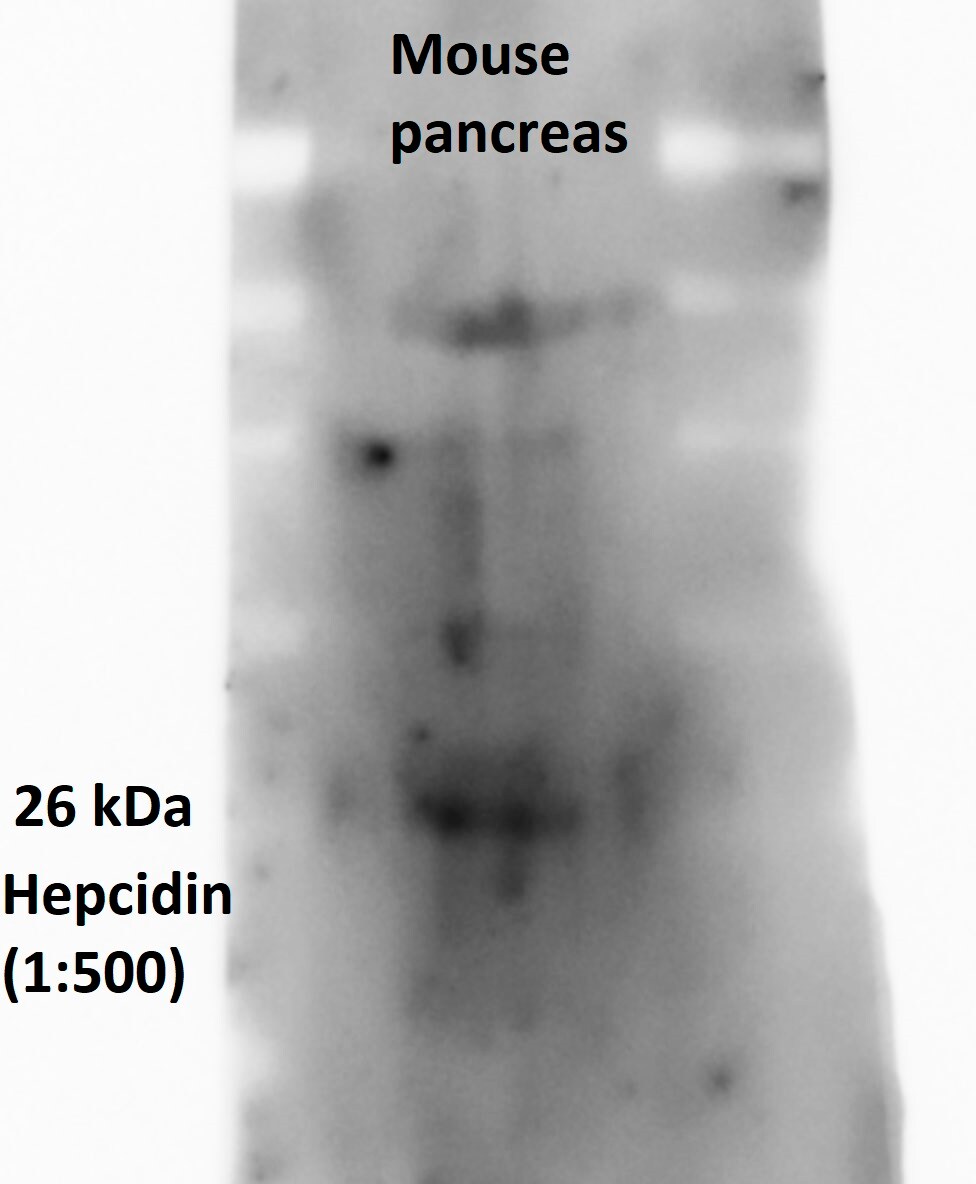
Western Blot: Hepcidin Antimicrobial Peptide Antibody - BSA Free [NBP1-59337] -
Omniscan-induced cells express iron metabolism proteins.(A) Expression of ferroportin and other iron regulatory protein hepcidin by human PBMC treated with 0.5 mM Omniscan and deferiprone for 8 days, as shown by immunocytochemistry staining. Representative images are shown. Deferiprone treatment significantly decreased Omniscan-induced ferroportin expression as shown by western blot analysis (B) left panel. After Omniscan treatment, hepcidin expression was decreased in comparison to untreated cells (A) lower panels, (B) right panels, western blot. Omniscan treatment with deferiprone increased the expression slightly. Representative blots from 3 separate experiments are shown. Values are means +/-SD, obtained from 3 separate experiments, Significance of the data was determined by ANOVA, followed by paired-group comparisons. **p <0.01, compared with control, *p <0.05 (for hepcidin), **p <0.01 (for ferroportin) deferiprone- and Omniscan-treated compared with Omniscan alone. (Scale bars 100 μm for all). Image collected and cropped by CiteAb from the following open publication (https://pubmed.ncbi.nlm.nih.gov/26305890), licensed under a CC0-1.0 license. Not internally tested by Novus Biologicals.Applications for Hepcidin Antimicrobial Peptide Antibody - BSA Free
Application
Recommended Usage
Western Blot
1.0 ug/ml
Application Notes
Immunohistochemistry Paraffin (IHC-P) reported in verified customer review.
Reviewed Applications
Read 6 reviews rated 4.5 using NBP1-59337 in the following applications:
Formulation, Preparation, and Storage
Purification
Affinity purified
Formulation
PBS, 2% Sucrose
Format
BSA Free
Preservative
0.09% Sodium Azide
Concentration
0.5 mg/ml
Shipping
The product is shipped with polar packs. Upon receipt, store it immediately at the temperature recommended below.
Stability & Storage
Store at 4C short term. Aliquot and store at -20C long term. Avoid freeze-thaw cycles.
Background: Hepcidin
Long Name
Hepcidin Antimicrobial Peptide
Alternate Names
HAMP, HEPC, HFE2B, LEAP1, PLTR
Gene Symbol
HAMP
UniProt
Additional Hepcidin Products
Product Documents for Hepcidin Antimicrobial Peptide Antibody - BSA Free
Certificate of Analysis
To download a Certificate of Analysis, please enter a lot or batch number in the search box below.
Product Specific Notices for Hepcidin Antimicrobial Peptide Antibody - BSA Free
This product is for research use only and is not approved for use in humans or in clinical diagnosis. Primary Antibodies are guaranteed for 1 year from date of receipt.
Related Research Areas
Citations for Hepcidin Antimicrobial Peptide Antibody - BSA Free
 Powered by Bioz
Customer Reviews for Hepcidin Antimicrobial Peptide Antibody - BSA Free (6)
4.5 out of 5
6 Customer Ratings
Have you used Hepcidin Antimicrobial Peptide Antibody - BSA Free?
Submit a review and receive an Amazon gift card!
$25/€18/£15/$25CAN/¥2500 Yen for a review with an image
$10/€7/£6/$10CAN/¥1110 Yen for a review without an image
Submit a review
Customer Images
Showing
1
-
5 of
6 reviews
Showing All
Filter By:
-
Application: Immunohistochemistry-ParaffinSample Tested: mouse eyeSpecies: MouseVerified Customer | Posted 04/10/2019Paraffin embedded sections of posterior segment of mouse eye, red is Hepcidin and blue is Hoeschst. 4um sections were rehydrated with xylene, followed by a decreasing ethanol concentration gradient (100%, 90%, 70%) and a wash with diH2O. Heat-mediated antigen retrieval performed using EDTA buffer (10mM Trizma Base, 1mM EDTA solution, 0.05% Tween 20, pH 9.0) in an autoclave for 30min. Primary antibody against Hepcidin was diluted 1:10, in blocking solution containing 0.1% BSA, 0.05% Triton X-100, and 5% normal donkey serum in TBS. Sections were incubated for 48hrs at room temperature and then 24hrs at 4C. Tissues were washed with TBS-T (6x5min), and immunoreactivity for Hep was developed. Image from verified customer review.

-
Application: Western BlotSample Tested: Adult pancreasSpecies: MouseVerified Customer | Posted 08/08/2018
-
Application: Immunohistochemistry-ParaffinSample Tested: eyeSpecies: BovineVerified Customer | Posted 08/03/2018
-
Application: Immunohistochemistry-ParaffinSample Tested: human eyeSpecies: HumanVerified Customer | Posted 07/13/2018
-
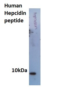
Application: Western BlotSample Tested: Hepcidin peptideSpecies: HumanVerified Customer | Posted 07/11/2018
-
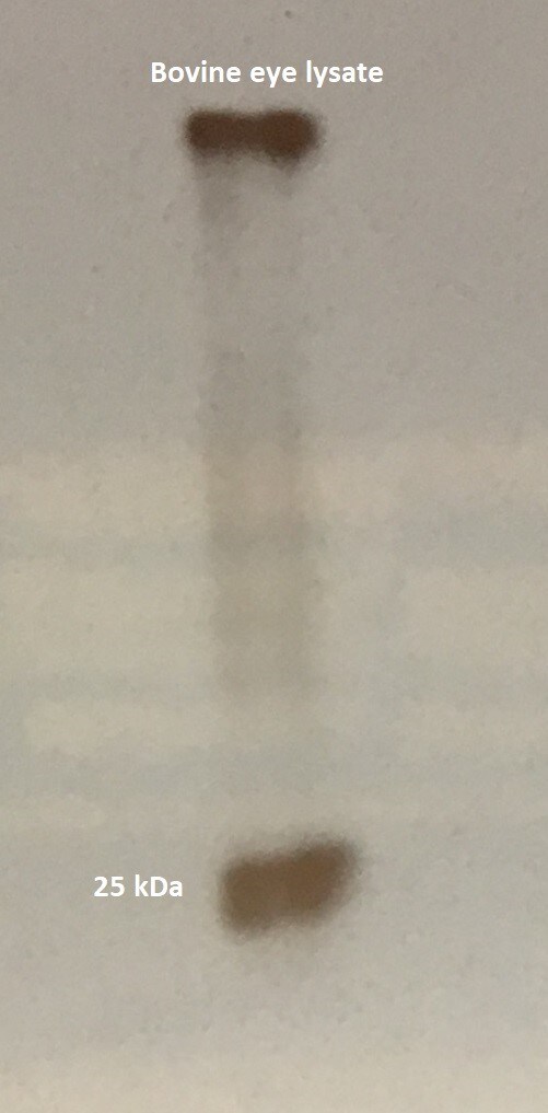
Application: Western BlotSample Tested: Adult eye and muscle tissue (labeled E and MSpecies: BovineVerified Customer | Posted 06/29/20181:500
There are no reviews that match your criteria.
Protocols
Find general support by application which include: protocols, troubleshooting, illustrated assays, videos and webinars.
- Antigen Retrieval Protocol (PIER)
- Antigen Retrieval for Frozen Sections Protocol
- Appropriate Fixation of IHC/ICC Samples
- Cellular Response to Hypoxia Protocols
- Chromogenic IHC Staining of Formalin-Fixed Paraffin-Embedded (FFPE) Tissue Protocol
- Chromogenic Immunohistochemistry Staining of Frozen Tissue
- ClariTSA™ Fluorophore Kits
- Detection & Visualization of Antibody Binding
- Fluorescent IHC Staining of Frozen Tissue Protocol
- Graphic Protocol for Heat-induced Epitope Retrieval
- Graphic Protocol for the Preparation and Fluorescent IHC Staining of Frozen Tissue Sections
- Graphic Protocol for the Preparation and Fluorescent IHC Staining of Paraffin-embedded Tissue Sections
- Graphic Protocol for the Preparation of Gelatin-coated Slides for Histological Tissue Sections
- IHC Sample Preparation (Frozen sections vs Paraffin)
- Immunofluorescent IHC Staining of Formalin-Fixed Paraffin-Embedded (FFPE) Tissue Protocol
- Immunohistochemistry (IHC) and Immunocytochemistry (ICC) Protocols
- Immunohistochemistry Frozen Troubleshooting
- Immunohistochemistry Paraffin Troubleshooting
- Preparing Samples for IHC/ICC Experiments
- Preventing Non-Specific Staining (Non-Specific Binding)
- Primary Antibody Selection & Optimization
- Protocol for Heat-Induced Epitope Retrieval (HIER)
- Protocol for Making a 4% Formaldehyde Solution in PBS
- Protocol for VisUCyte™ HRP Polymer Detection Reagent
- Protocol for the Preparation & Fixation of Cells on Coverslips
- Protocol for the Preparation and Chromogenic IHC Staining of Frozen Tissue Sections
- Protocol for the Preparation and Chromogenic IHC Staining of Frozen Tissue Sections - Graphic
- Protocol for the Preparation and Chromogenic IHC Staining of Paraffin-embedded Tissue Sections
- Protocol for the Preparation and Chromogenic IHC Staining of Paraffin-embedded Tissue Sections - Graphic
- Protocol for the Preparation and Fluorescent IHC Staining of Frozen Tissue Sections
- Protocol for the Preparation and Fluorescent IHC Staining of Paraffin-embedded Tissue Sections
- Protocol for the Preparation of Gelatin-coated Slides for Histological Tissue Sections
- R&D Systems Quality Control Western Blot Protocol
- TUNEL and Active Caspase-3 Detection by IHC/ICC Protocol
- The Importance of IHC/ICC Controls
- Troubleshooting Guide: Immunohistochemistry
- Troubleshooting Guide: Western Blot Figures
- Western Blot Conditions
- Western Blot Protocol
- Western Blot Protocol for Cell Lysates
- Western Blot Troubleshooting
- Western Blot Troubleshooting Guide
- View all Protocols, Troubleshooting, Illustrated assays and Webinars
Loading...