HIP1 Antibody (1B11) - BSA Free
Novus Biologicals | Catalog # NB300-204
![Western Blot: HIP1 Antibody (1B11) [NB300-204]](https://resources.rndsystems.com/images/products/HIP1-Antibody-1B11-Western-Blot-NB300-204-img0002.jpg "Western Blot: HIP1 Antibody (1B11) [NB300-204]")
Loading...
Key Product Details
Species Reactivity
Validated:
Human, Mouse, Bacteria, Rat (Negative)
Cited:
Human, Mouse, Bacteria
Applications
Validated:
Western Blot, Immunocytochemistry/ Immunofluorescence, Immunoprecipitation
Cited:
Western Blot, Immunocytochemistry/ Immunofluorescence, Immunoprecipitation
Label
Unconjugated
Antibody Source
Monoclonal Mouse IgG1 kappa Clone # 1B11
Format
BSA Free
Loading...
Product Specifications
Immunogen
Human 3'-HIP1 (~65 kDa)
Reactivity Notes
Does not appear to work in rat.
Localization
Localization: Cytoplasmic
Specificity
NB300-204 is specific for human HIP1. Does not cross-react with HIP1r.
Clonality
Monoclonal
Host
Mouse
Isotype
IgG1 kappa
Theoretical MW
115 kDa.
Disclaimer note: The observed molecular weight of the protein may vary from the listed predicted molecular weight due to post translational modifications, post translation cleavages, relative charges, and other experimental factors.
Disclaimer note: The observed molecular weight of the protein may vary from the listed predicted molecular weight due to post translational modifications, post translation cleavages, relative charges, and other experimental factors.
Scientific Data Images for HIP1 Antibody (1B11) - BSA Free
Western Blot: HIP1 Antibody (1B11) [NB300-204]
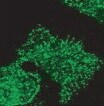
Western Blot: HIP1 Antibody (1B11) [NB300-204] - HeLa whole cell extracts.![Immunocytochemistry/ Immunofluorescence: HIP1 Antibody (1B11) [NB300-204]](https://resources.rndsystems.com/images/products/HIP1-Antibody-1B11-Immunocytochemistry-Immunofluorescence-NB300-204-img0003.jpg "Immunocytochemistry/ Immunofluorescence: HIP1 Antibody (1B11) [NB300-204]")
Immunocytochemistry/ Immunofluorescence: HIP1 Antibody (1B11) [NB300-204]
Immunocytochemistry/Immunofluorescence: HIP1 Antibody (1B11) [NB300-204] - Human pluripotent embryonal carcinoma cells stained with HIP1 antibody. ICC/IF image submitted by a verified customer review.Applications for HIP1 Antibody (1B11) - BSA Free
Application
Recommended Usage
Immunocytochemistry/ Immunofluorescence
1:100
Immunoprecipitation
reported in scientific literature (PMID 23272104)
Western Blot
1:1000
Application Notes
In Western blot, a band is observed at ~115 kDa. The observed molecular weight of the protein may vary from the listed predicted molecular weight due to post translational modifications, post translation cleavages, relative charges, and other experimental factors.
Reviewed Applications
Read 1 review rated 5 using NB300-204 in the following applications:
Formulation, Preparation, and Storage
Purification
Unpurified
Formulation
Ascites
Format
BSA Free
Preservative
0.1% Sodium Azide
Concentration
This product is unpurified. The exact concentration of antibody is not quantifiable.
Shipping
The product is shipped with polar packs. Upon receipt, store it immediately at the temperature recommended below.
Stability & Storage
Store at -20C. Avoid freeze-thaw cycles.
Background: HIP1
Alternate Names
HIP-1, HIP-I, huntingtin interacting protein 1, huntingtin-interacting protein 1, Huntingtin-interacting protein I, ILWEQ, MGC126506
Gene Symbol
HIP1
UniProt
Additional HIP1 Products
Product Documents for HIP1 Antibody (1B11) - BSA Free
Certificate of Analysis
To download a Certificate of Analysis, please enter a lot or batch number in the search box below.
Product Specific Notices for HIP1 Antibody (1B11) - BSA Free
This product is for research use only and is not approved for use in humans or in clinical diagnosis. Primary Antibodies are guaranteed for 1 year from date of receipt.
Citations for HIP1 Antibody (1B11) - BSA Free
 Powered by Bioz
Customer Reviews for HIP1 Antibody (1B11) - BSA Free (1)
5 out of 5
1 Customer Rating
Have you used HIP1 Antibody (1B11) - BSA Free?
Submit a review and receive an Amazon gift card!
$25/€18/£15/$25CAN/¥2500 Yen for a review with an image
$10/€7/£6/$10CAN/¥1110 Yen for a review without an image
Submit a review
Customer Images
Showing
1
-
1 of
1 review
Showing All
Filter By:
-
Application: ImmunocytochemistrySample Tested: Human pluripotent embryonal carcinoma cellsSpecies: HumanVerified Customer | Posted 11/11/2021HIP1 Antibody. Human pluripotent embryonal carcinoma cells.

There are no reviews that match your criteria.
Protocols
View specific protocols for HIP1 Antibody (1B11) - BSA Free (NB300-204):
HIP1 Antibody (1B11):
Western Blot Procedure
1. Run 25 ug/lane of protein (ie: HeLa whole cell extract) on a 10% SDS-PAGE gel.
2. Transfer the proteins to nitrocellulose.
3. Block the membrane with a standard blocking buffer (5% NFDM in TBST), overnight at 4 deg C.
4. Dilute the anti-HIP1 in 1% NFDM in TBST and incubate for 1hour at RT.
5. Wash the membrane, 3x 5-10 minutes.
6. Dilute the secondary antibody [anti-mouse IgG conjugated to HRP] in 1% NFDM in TBST and incubate for 1hour at RT.
7. Wash the membrane, 3x 5-10 minutes.
8. Detect the protein-antibody complex via ECL.
Find general support by application which include: protocols, troubleshooting, illustrated assays, videos and webinars.
- Appropriate Fixation of IHC/ICC Samples
- Cellular Response to Hypoxia Protocols
- ClariTSA™ Fluorophore Kits
- Detection & Visualization of Antibody Binding
- ICC Cell Smear Protocol for Suspension Cells
- ICC Immunocytochemistry Protocol Videos
- ICC for Adherent Cells
- Immunocytochemistry (ICC) Protocol
- Immunocytochemistry Troubleshooting
- Immunofluorescence of Organoids Embedded in Cultrex Basement Membrane Extract
- Immunohistochemistry (IHC) and Immunocytochemistry (ICC) Protocols
- Immunoprecipitation Protocol
- Preparing Samples for IHC/ICC Experiments
- Preventing Non-Specific Staining (Non-Specific Binding)
- Primary Antibody Selection & Optimization
- Protocol for VisUCyte™ HRP Polymer Detection Reagent
- Protocol for the Fluorescent ICC Staining of Cell Smears - Graphic
- Protocol for the Fluorescent ICC Staining of Cultured Cells on Coverslips - Graphic
- Protocol for the Preparation and Fluorescent ICC Staining of Cells on Coverslips
- Protocol for the Preparation and Fluorescent ICC Staining of Non-adherent Cells
- Protocol for the Preparation and Fluorescent ICC Staining of Stem Cells on Coverslips
- Protocol for the Preparation of a Cell Smear for Non-adherent Cell ICC - Graphic
- R&D Systems Quality Control Western Blot Protocol
- TUNEL and Active Caspase-3 Detection by IHC/ICC Protocol
- The Importance of IHC/ICC Controls
- Troubleshooting Guide: Western Blot Figures
- Western Blot Conditions
- Western Blot Protocol
- Western Blot Protocol for Cell Lysates
- Western Blot Troubleshooting
- Western Blot Troubleshooting Guide
- View all Protocols, Troubleshooting, Illustrated assays and Webinars
Loading...