Histone H3 [Trimethyl Lys27] Antibody (BT164) - Chimeric - Azide and BSA Free
Novus Biologicals | Catalog # NBP3-12020
Recombinant Monoclonal Antibody
Key Product Details
Species Reactivity
Human
Applications
Immunohistochemistry, Western Blot, ELISA, Immunocytochemistry/ Immunofluorescence, Immunoprecipitation
Label
Unconjugated
Antibody Source
Recombinant Monoclonal Rabbit IgG Kappa Clone # BT164
Format
Azide and BSA Free
Loading...
Product Specifications
Immunogen
BT164 is a mice-derrived lupus antibody that was established by selection on apoptotic chromatin.
Modification
Trimethyl Lys 27
Specificity
The epitope of BT164 was mapped in the N-terminal tail of histone H3 (27-KSAPAT-32) and included the apoptosis-induced trimethylation of K27.
Clonality
Monoclonal
Host
Rabbit
Isotype
IgG Kappa
Scientific Data Images
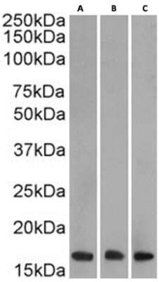
Western Blot: Histone H3 [Trimethyl Lys27] Antibody (BT164) - Chimeric [NBP3-12020] - Nuclear lysate of HeLa(A) (0.0003ug/ml), Jurkat(B) (0.0003ug/ml) and K562(C) (0.01ug/ml) (35ug protein in RIPA buffer) were resolved on a SDS PAGE gel and blots were probed with the chimeric rabbit version of BT164 (NBP3-12020) before detection using an anti-rabbit secondary antibody. A primary incubation of 1h was used and protein was detected by chemiluminescence.
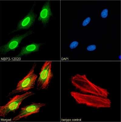
Immunocytochemistry/Immunofluorescence: Histone H3 [Trimethyl Lys27] Antibody (BT164) - Chimeric [NBP3-12020] - Immunofluorescence analysis of paraformaldehyde fixed HeLa cells on Shi-fix(TM) coverslips stained with the chimeric rabbit IgG version of BT164 (NBP3-12020) at 10 ug/ml for 1h followed by Alexa Fluor(R) 488 secondary antibody (2 ug/ml), showing membrane staining. The nuclear stain is DAPI (blue) and the actin stain is phalloidin (red). Panels show from left-right, top-bottom NBP3-12020, DAPI, merged channels and an isotype control. The isotype control was an unknown specificity antibody followed by staining with Alexa Fluor(R) 488 secondary antibody.
![Histone H3 [Trimethyl Lys27] Antibody (BT164) - Chimeric - Azide and BSA Free](https://resources.rndsystems.com/images/products/nbp3-12020_rabbit-monoclonal-histone-h3-trimethyl-lys27-antibody-bt164-chimeric-7420252211303.png "Flow Cytometry: Histone H3 [Trimethyl Lys27] Antibody (BT164) - Chimeric - Azide and BSA Free [NBP3-12020] -")
Flow Cytometry: Histone H3 [Trimethyl Lys27] Antibody (BT164) - Chimeric - Azide and BSA Free [NBP3-12020] -
Flow Cytometry: Histone H3 [Trimethyl Lys27] Antibody (BT164) - Chimeric - Azide and BSA Free [NBP3-12020] - HeLa cells were fixed using 2% PFA and stained with anti-unknown specificity antibody (isotype control, black line) or NBP3-12020 (blue line) at a dilution of 1:100 for 1h at RT. After washing, the bound antibody was detected using a goat anti-rabbit IgG AlexaFluor 488 antibody at a dilution of 1:1000 and cells analyzed using a FACSCanto flow-cytometer.Applications
Application
Recommended Usage
ELISA
Optimal dilutions of this antibody should be experimentally determined.
Immunocytochemistry/ Immunofluorescence
Optimal dilutions of this antibody should be experimentally determined.
Immunohistochemistry
Optimal dilutions of this antibody should be experimentally determined.
Immunoprecipitation
Optimal dilutions of this antibody should be experimentally determined.
Western Blot
Optimal dilutions of this antibody should be experimentally determined.
Application Notes
This chimeric rabbit antibody was made using the variable domain sequences of the original Mouse IgG2b format, for improved compatibility with existing reagents, assays and techniques.
Formulation, Preparation, and Storage
Purification
Protein A purified
Formulation
PBS
Format
Azide and BSA Free
Preservative
0.02% Proclin 300
Concentration
1 mg/ml
Shipping
The product is shipped with polar packs. Upon receipt, store it immediately at the temperature recommended below.
Stability & Storage
Store at 4C for up to 3 months. For longer storage, aliquot and store at -20C.
Background: Histone H3
Histones are nuclear proteins responsible for the nucleosome structure of the chromosomal fiber in eukaryotes. Changes in chromatin structure play a large role in the regulation of transcription. The chromatin fibers are compacted through the interaction of a linker histone, H1, with the DNA between the nucleosomes to form higher order chromatin structures.
Common histone modifications include methylation of lysine and arginine, acetylation of lysine, phosphorylation of threonine and serine, and sumoylation, biotinylation, and ubiquitylation of lysine. Posttranslational modifications such as acetylation of core histones regulates gene expression, thus altering protein function and regulation (1). Histone H3 is primarily acetylated at lysines 9, 14, 18, and 23 and have a theoretical molecular weight of 15 kDa. Acetylation at lysine 9 and 14 appears to control histone deposition, chromatin assembly and active transcription. Methylation of arginine residues within histone H3 has also been linked to transcription regulation. Histone H3 has been linked to various types of cancer as a biomarker through the aberrant expression of histone deacetylase (HDAC) enzymes and changes to chromatins (2-4).
References
1. Zhang, Y. X., Akumuo, R. C., Espana, R. A., Yan, C. X., Gao, W. J., & Li, Y. C. (2018). The histone demethylase KDM6B in the medial prefrontal cortex epigenetically regulates cocaine reward memory. Neuropharmacology, 141, 113-125. doi:10.1016/j.neuropharm.2018.08.030
2. Nandakumar, V., Hansen, N., Glenn, H. L., Han, J. H., Helland, S., Hernandez, K,...Meldrum, D. R. (2016). Vorinostat differentially alters 3D nuclear structure of cancer and non-cancerous esophageal cells. Sci Rep, 6, 30593. doi:10.1038/srep30593
3. Zhou, M., Li, Y., Lin, S., Chen, Y., Qian, Y., Zhao, Z., & Fan, H. (2019). H3K9me3, H3K36me3, and H4K20me3 Expression Correlates with Patient Outcome in Esophageal Squamous Cell Carcinoma as Epigenetic Markers. Dig Dis Sci, 64(8), 2147-2157. doi:10.1007/s10620-019-05529-2
4. Li, Y., Guo, D., Sun, R., Chen, P., Qian, Q., & Fan, H. (2019). Methylation Patterns of Lys9 and Lys27 on Histone H3 Correlate with Patient Outcome in Gastric Cancer. Dig Dis Sci, 64(2), 439-446. doi:10.1007/s10620-018-5341-8
Alternate Names
H3F3A
Gene Symbol
H3C14
Additional Histone H3 Products
Product Documents
Certificate of Analysis
To download a Certificate of Analysis, please enter a lot or batch number in the search box below.
Product Specific Notices
This product is for research use only and is not approved for use in humans or in clinical diagnosis. Primary Antibodies are guaranteed for 1 year from date of receipt.
Related Research Areas
Customer Reviews
There are currently no reviews for this product. Be the first to review Histone H3 [Trimethyl Lys27] Antibody (BT164) - Chimeric - Azide and BSA Free and earn rewards!
Have you used Histone H3 [Trimethyl Lys27] Antibody (BT164) - Chimeric - Azide and BSA Free?
Submit a review and receive an Amazon gift card!
$25/€18/£15/$25CAN/¥2500 Yen for a review with an image
$10/€7/£6/$10CAN/¥1110 Yen for a review without an image
Submit a review
Protocols
Find general support by application which include: protocols, troubleshooting, illustrated assays, videos and webinars.
- Antigen Retrieval Protocol (PIER)
- Antigen Retrieval for Frozen Sections Protocol
- Appropriate Fixation of IHC/ICC Samples
- Cellular Response to Hypoxia Protocols
- Chromogenic IHC Staining of Formalin-Fixed Paraffin-Embedded (FFPE) Tissue Protocol
- Chromogenic Immunohistochemistry Staining of Frozen Tissue
- ClariTSA™ Fluorophore Kits
- Detection & Visualization of Antibody Binding
- ELISA Sample Preparation & Collection Guide
- ELISA Troubleshooting Guide
- Fluorescent IHC Staining of Frozen Tissue Protocol
- Graphic Protocol for Heat-induced Epitope Retrieval
- Graphic Protocol for the Preparation and Fluorescent IHC Staining of Frozen Tissue Sections
- Graphic Protocol for the Preparation and Fluorescent IHC Staining of Paraffin-embedded Tissue Sections
- Graphic Protocol for the Preparation of Gelatin-coated Slides for Histological Tissue Sections
- How to Run an R&D Systems DuoSet ELISA
- How to Run an R&D Systems Quantikine ELISA
- How to Run an R&D Systems Quantikine™ QuicKit™ ELISA
- ICC Cell Smear Protocol for Suspension Cells
- ICC Immunocytochemistry Protocol Videos
- ICC for Adherent Cells
- IHC Sample Preparation (Frozen sections vs Paraffin)
- Immunocytochemistry (ICC) Protocol
- Immunocytochemistry Troubleshooting
- Immunofluorescence of Organoids Embedded in Cultrex Basement Membrane Extract
- Immunofluorescent IHC Staining of Formalin-Fixed Paraffin-Embedded (FFPE) Tissue Protocol
- Immunohistochemistry (IHC) and Immunocytochemistry (ICC) Protocols
- Immunohistochemistry Frozen Troubleshooting
- Immunohistochemistry Paraffin Troubleshooting
- Immunoprecipitation Protocol
- Preparing Samples for IHC/ICC Experiments
- Preventing Non-Specific Staining (Non-Specific Binding)
- Primary Antibody Selection & Optimization
- Protocol for Heat-Induced Epitope Retrieval (HIER)
- Protocol for Making a 4% Formaldehyde Solution in PBS
- Protocol for VisUCyte™ HRP Polymer Detection Reagent
- Protocol for the Fluorescent ICC Staining of Cell Smears - Graphic
- Protocol for the Fluorescent ICC Staining of Cultured Cells on Coverslips - Graphic
- Protocol for the Preparation & Fixation of Cells on Coverslips
- Protocol for the Preparation and Chromogenic IHC Staining of Frozen Tissue Sections
- Protocol for the Preparation and Chromogenic IHC Staining of Frozen Tissue Sections - Graphic
- Protocol for the Preparation and Chromogenic IHC Staining of Paraffin-embedded Tissue Sections
- Protocol for the Preparation and Chromogenic IHC Staining of Paraffin-embedded Tissue Sections - Graphic
- Protocol for the Preparation and Fluorescent ICC Staining of Cells on Coverslips
- Protocol for the Preparation and Fluorescent ICC Staining of Non-adherent Cells
- Protocol for the Preparation and Fluorescent ICC Staining of Stem Cells on Coverslips
- Protocol for the Preparation and Fluorescent IHC Staining of Frozen Tissue Sections
- Protocol for the Preparation and Fluorescent IHC Staining of Paraffin-embedded Tissue Sections
- Protocol for the Preparation of Gelatin-coated Slides for Histological Tissue Sections
- Protocol for the Preparation of a Cell Smear for Non-adherent Cell ICC - Graphic
- Quantikine HS ELISA Kit Assay Principle, Alkaline Phosphatase
- Quantikine HS ELISA Kit Principle, Streptavidin-HRP Polymer
- R&D Systems Quality Control Western Blot Protocol
- Sandwich ELISA (Colorimetric) – Biotin/Streptavidin Detection Protocol
- Sandwich ELISA (Colorimetric) – Direct Detection Protocol
- TUNEL and Active Caspase-3 Detection by IHC/ICC Protocol
- The Importance of IHC/ICC Controls
- Troubleshooting Guide: ELISA
- Troubleshooting Guide: Immunohistochemistry
- Troubleshooting Guide: Western Blot Figures
- Western Blot Conditions
- Western Blot Protocol
- Western Blot Protocol for Cell Lysates
- Western Blot Troubleshooting
- Western Blot Troubleshooting Guide
- View all Protocols, Troubleshooting, Illustrated assays and Webinars
Loading...