Histone H3 [Asym-dimethyl Arg2] Antibody - BSA Free
Novus Biologicals | Catalog # NB21-1002
Key Product Details
Species Reactivity
Validated:
Human, Mouse, C. elegans
Cited:
Human, Mouse
Predicted:
Chicken (100%), Drosophila (100%), Plant (100%), Rat (100%), Xenopus (100%). Backed by our 100% Guarantee.
Applications
Validated:
Western Blot, Immunocytochemistry/ Immunofluorescence, Chromatin Immunoprecipitation (ChIP), Dot Blot
Cited:
Western Blot, Chemotaxis
Label
Unconjugated
Antibody Source
Polyclonal Rabbit IgG
Format
BSA Free
Loading...
Product Specifications
Immunogen
This Histone H3 [Asym-dimethyl Arg2] antibody was raised against synthetic dimethylated peptide surrounding Arginine 2 of human Histone H3 [Asym-dimethyl Arg2] [Swiss Prot# Q71DI3].
Reactivity Notes
Predicted to react with many species including rat, chicken, Xenopus, Drosophila, and plant based on 100% sequence homology.
Modification
Asym-dimethyl Arg2
Localization
Nucleus. Chromosome.
Clonality
Polyclonal
Host
Rabbit
Isotype
IgG
Theoretical MW
15 kDa.
Disclaimer note: The observed molecular weight of the protein may vary from the listed predicted molecular weight due to post translational modifications, post translation cleavages, relative charges, and other experimental factors.
Disclaimer note: The observed molecular weight of the protein may vary from the listed predicted molecular weight due to post translational modifications, post translation cleavages, relative charges, and other experimental factors.
Scientific Data Images for Histone H3 [Asym-dimethyl Arg2] Antibody - BSA Free
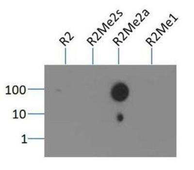
Dot Blot: Histone H3 [Asym-dimethyl Arg2] Antibody [NB21-1002] - Analysis of Histone H3 [Asym-dimethyl Arg2] in picomoles of peptide.
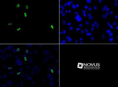
Immunocytochemistry/Immunofluorescence: Histone H3 [Asym-dimethyl Arg2] Antibody [NB21-1002] - Histone H3 [Asym-dimethyl Arg2] antibody was tested in HeLa cells with DyLight 488 (green). Nuclei were counterstained with DAPI (blue).
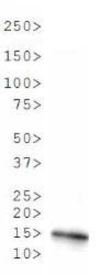
Western Blot: Histone H3 [Asym-dimethyl Arg2] Antibody [NB21-1002] - Analysis of Histone H3 [Asym-dimethyl Arg2] in C. elegans embryo lysate. Observed molecular weight is ~15 kDa.
![Histone H3 [Asym-dimethyl Arg2] Antibody - BSA Free](https://resources.rndsystems.com/images/products/nb21-1002_rabbit-polyclonal-histone-h3-asym-dimethyl-arg2-antibody-western-blot-132202615271923.jpg "Western Blot: Histone H3 [Asym-dimethyl Arg2] Antibody - BSA Free [NB21-1002] -")
Western Blot: Histone H3 [Asym-dimethyl Arg2] Antibody - BSA Free [NB21-1002] -
FBXW17/protein arginine N-methyltransferase 6 (PRMT6) signaling involves in cigarette smoke extract (CSE)-induced lung epithelial inflammation and apoptosis. (A) Vector, FBXW17-FLAG plasmids, scramble small hairpin RNA (shRNA), and FBXW17 shRNA were transfected into MLE12 cells separately. Annexin V and PI kits were used to detect the rate of apoptotic cells. Before detection, cells were treated with CSE for 6 h. The “percentage of cells +/- SD" values were shown in all the quadrants of flow cytometry. (B) The plotted data of apoptosis in each group are presented. (C) MLE12 cells were transfected with either overexpressed or knockdown plasmids of FBXW17. Before collection, the cells were treated with 5% CSE for 6 h. Cell lysate was conducted with immunoblotting with indicated antibodies. (D) The plotted PRMT6, H3R2me2a, and Bcl-2 protein expression were shown. (E) The relative protein expression of H3K4me3, Bax, TNF-alpha, IL-1 beta, and COX-2 were presented. Data were represented by n = 3 separate experiments. The graph shows mean +/- SD. “∗" denotes p < 0.05 between indicated groups. Image collected and cropped by CiteAb from the following open publication (https://pubmed.ncbi.nlm.nih.gov/33959602), licensed under a CC-BY license. Not internally tested by Novus Biologicals.Applications for Histone H3 [Asym-dimethyl Arg2] Antibody - BSA Free
Application
Recommended Usage
Dot Blot
1:1000
Immunocytochemistry/ Immunofluorescence
1:50-1:100
Western Blot
1:500
Application Notes
This Histone H3 [Asym-dimethyl Arg2] antibodiy is useful for Immunocytochemistry/Immunofluorescence, Dot Blot, and Western Blot where a band is seen ~15 kDa in C. elegans embryo lysate. In ICC/IF, nuclear staining was observed in HeLa cells.
Formulation, Preparation, and Storage
Purification
Immunogen affinity purified
Formulation
PBS and 30% Glycerol
Format
BSA Free
Preservative
0.05% Sodium Azide
Concentration
0.71 mg/ml
Shipping
The product is shipped with polar packs. Upon receipt, store it immediately at the temperature recommended below.
Stability & Storage
Store at 4C short term. Aliquot and store at -20C long term. Avoid freeze-thaw cycles.
Background: Histone H3
Histones are nuclear proteins responsible for the nucleosome structure of the chromosomal fiber in eukaryotes. Changes in chromatin structure play a large role in the regulation of transcription. The chromatin fibers are compacted through the interaction of a linker histone, H1, with the DNA between the nucleosomes to form higher order chromatin structures.
Common histone modifications include methylation of lysine and arginine, acetylation of lysine, phosphorylation of threonine and serine, and sumoylation, biotinylation, and ubiquitylation of lysine. Posttranslational modifications such as acetylation of core histones regulates gene expression, thus altering protein function and regulation (1). Histone H3 is primarily acetylated at lysines 9, 14, 18, and 23 and have a theoretical molecular weight of 15 kDa. Acetylation at lysine 9 and 14 appears to control histone deposition, chromatin assembly and active transcription. Methylation of arginine residues within histone H3 has also been linked to transcription regulation. Histone H3 has been linked to various types of cancer as a biomarker through the aberrant expression of histone deacetylase (HDAC) enzymes and changes to chromatins (2-4).
References
1. Zhang, Y. X., Akumuo, R. C., Espana, R. A., Yan, C. X., Gao, W. J., & Li, Y. C. (2018). The histone demethylase KDM6B in the medial prefrontal cortex epigenetically regulates cocaine reward memory. Neuropharmacology, 141, 113-125. doi:10.1016/j.neuropharm.2018.08.030
2. Nandakumar, V., Hansen, N., Glenn, H. L., Han, J. H., Helland, S., Hernandez, K,...Meldrum, D. R. (2016). Vorinostat differentially alters 3D nuclear structure of cancer and non-cancerous esophageal cells. Sci Rep, 6, 30593. doi:10.1038/srep30593
3. Zhou, M., Li, Y., Lin, S., Chen, Y., Qian, Y., Zhao, Z., & Fan, H. (2019). H3K9me3, H3K36me3, and H4K20me3 Expression Correlates with Patient Outcome in Esophageal Squamous Cell Carcinoma as Epigenetic Markers. Dig Dis Sci, 64(8), 2147-2157. doi:10.1007/s10620-019-05529-2
4. Li, Y., Guo, D., Sun, R., Chen, P., Qian, Q., & Fan, H. (2019). Methylation Patterns of Lys9 and Lys27 on Histone H3 Correlate with Patient Outcome in Gastric Cancer. Dig Dis Sci, 64(2), 439-446. doi:10.1007/s10620-018-5341-8
Alternate Names
H3F3A, H3R2Me2a
Gene Symbol
H3C14
UniProt
Additional Histone H3 Products
Product Documents for Histone H3 [Asym-dimethyl Arg2] Antibody - BSA Free
Certificate of Analysis
To download a Certificate of Analysis, please enter a lot or batch number in the search box below.
Product Specific Notices for Histone H3 [Asym-dimethyl Arg2] Antibody - BSA Free
Epi-Plus antibody production in collaboration with Rockland Immunochemicals Inc.
This product is for research use only and is not approved for use in humans or in clinical diagnosis. Primary Antibodies are guaranteed for 1 year from date of receipt.
Related Research Areas
Citations for Histone H3 [Asym-dimethyl Arg2] Antibody - BSA Free
 Powered by Bioz
Customer Reviews for Histone H3 [Asym-dimethyl Arg2] Antibody - BSA Free
There are currently no reviews for this product. Be the first to review Histone H3 [Asym-dimethyl Arg2] Antibody - BSA Free and earn rewards!
Have you used Histone H3 [Asym-dimethyl Arg2] Antibody - BSA Free?
Submit a review and receive an Amazon gift card!
$25/€18/£15/$25CAN/¥2500 Yen for a review with an image
$10/€7/£6/$10CAN/¥1110 Yen for a review without an image
Submit a review
Protocols
View specific protocols for Histone H3 [Asym-dimethyl Arg2] Antibody - BSA Free (NB21-1002):
Histone H3 [Asym-dimethyl Arg2] Antibody:
Immunocytochemistry Protocol
Culture cells to appropriate density in 35 mm culture dishes or 6-well plates.
1. Remove culture medium and add 10% formalin to the dish. Fix at room temperature for 30 minutes.
2. Remove the formalin and add ice cold methanol. Incubate for 5-10 minutes.
3. Remove methanol and add washing solution (i.e. PBS). Be sure to not let the specimen dry out. Wash three times for 10 minutes.
4. To block nonspecific antibody binding incubate in 10% normal goat serum from 1 hour to overnight at room temperature.
5. Add primary antibody at appropriate dilution and incubate at room temperature from 2 hours to overnight at room temperature.
6. Remove primary antibody and replace with washing solution. Wash three times for 10 minutes.
7. Add secondary antibody at appropriate dilution. Incubate for 1 hour at room temperature.
8. Remove antibody and replace with wash solution, then wash for 10 minutes. Add Hoechst 33258 to wash solution at 1:25,0000 and incubate for 10 minutes. Wash a third time for 10 minutes.
9. Cells can be viewed directly after washing. The plates can also be stored in PBS containing Azide covered in Parafilm (TM). Cells can also be cover-slipped using Fluoromount, with appropriate sealing.
*The above information is only intended as a guide. The researcher should determine what protocol best meets their needs. Please follow safe laboratory procedures.
Find general support by application which include: protocols, troubleshooting, illustrated assays, videos and webinars.
- Appropriate Fixation of IHC/ICC Samples
- Cellular Response to Hypoxia Protocols
- ChIP Protocol Video
- Chromatin Immunoprecipitation (ChIP) Protocol
- Chromatin Immunoprecipitation Protocol
- ClariTSA™ Fluorophore Kits
- Detection & Visualization of Antibody Binding
- ICC Cell Smear Protocol for Suspension Cells
- ICC Immunocytochemistry Protocol Videos
- ICC for Adherent Cells
- Immunocytochemistry (ICC) Protocol
- Immunocytochemistry Troubleshooting
- Immunofluorescence of Organoids Embedded in Cultrex Basement Membrane Extract
- Immunohistochemistry (IHC) and Immunocytochemistry (ICC) Protocols
- Preparing Samples for IHC/ICC Experiments
- Preventing Non-Specific Staining (Non-Specific Binding)
- Primary Antibody Selection & Optimization
- Protocol for VisUCyte™ HRP Polymer Detection Reagent
- Protocol for the Fluorescent ICC Staining of Cell Smears - Graphic
- Protocol for the Fluorescent ICC Staining of Cultured Cells on Coverslips - Graphic
- Protocol for the Preparation and Fluorescent ICC Staining of Cells on Coverslips
- Protocol for the Preparation and Fluorescent ICC Staining of Non-adherent Cells
- Protocol for the Preparation and Fluorescent ICC Staining of Stem Cells on Coverslips
- Protocol for the Preparation of a Cell Smear for Non-adherent Cell ICC - Graphic
- R&D Systems Quality Control Western Blot Protocol
- TUNEL and Active Caspase-3 Detection by IHC/ICC Protocol
- The Importance of IHC/ICC Controls
- Troubleshooting Guide: Western Blot Figures
- Western Blot Conditions
- Western Blot Protocol
- Western Blot Protocol for Cell Lysates
- Western Blot Troubleshooting
- Western Blot Troubleshooting Guide
- View all Protocols, Troubleshooting, Illustrated assays and Webinars
FAQs for Histone H3 [Asym-dimethyl Arg2] Antibody - BSA Free
Showing
1
-
1 of
1 FAQ
Showing All
-
Q: I received H3R2me2a antibody from your company. I would like to use your antibody for western blotting. May I know the best blocking reagents (5% milk or 5%BSA) for this antibody?
A: When we generated our Western blot data with NB21-1002 we used a 4-15% gradient gel, and ran this in Tris-glycine buffer for one hour at 100V. The proteins were transferred to a 0.22uM nitrocellulose membrane, and this was blocked for one hour at room temperature with 3% BSA in TBS-T (1x TBS, 0.1% Tween-20). NB21-1002 was diluted to 1:1000 in blocking buffer and incubated at 4C overnight. The secondary antibody was incubated for one hour at room temperature.
Loading...