Histone H4 [p Ser1] Antibody - BSA Free
Novus Biologicals | Catalog # NB21-2000
![Western Blot: Histone H4 [p Ser1] AntibodyBSA Free [NB21-2000]](https://resources.rndsystems.com/images/products/Histone-H4-[p-Ser1]-Antibody-Western-Blot-NB21-2000-img0004.jpg "Western Blot: Histone H4 [p Ser1] AntibodyBSA Free [NB21-2000]")
Key Product Details
Species Reactivity
Validated:
Human, Mouse, Rat, Mammal
Cited:
Human
Applications
Validated:
Western Blot, Immunocytochemistry/ Immunofluorescence, Chromatin Immunoprecipitation (ChIP), Dot Blot
Cited:
Chemotaxis
Label
Unconjugated
Antibody Source
Polyclonal Rabbit IgG
Format
BSA Free
Loading...
Product Specifications
Immunogen
Synthetic phosphorylated peptide surrounding Serine 1 of human Histone H4 [Swiss Prot# P62805].
Reactivity Notes
Predicted to react with most mammal species.
Modification
p Ser1
Localization
Nucleus.
Marker
H4pS1
Clonality
Polyclonal
Host
Rabbit
Isotype
IgG
Scientific Data Images for Histone H4 [p Ser1] Antibody - BSA Free
Western Blot: Histone H4 [p Ser1] AntibodyBSA Free [NB21-2000]
Western Blot: Histone H4 [p Ser1] Antibody [NB21-2000] - Analysis of Histone H4 pS1 in HeLa histone prep.![Immunocytochemistry/ Immunofluorescence: Histone H4 [p Ser1] Antibody - BSA Free [NB21-2000]](https://resources.rndsystems.com/images/products/Histone-H4-[p-Ser1]-Antibody-Immunocytochemistry-Immunofluorescence-NB21-2000-img0009.jpg "Immunocytochemistry/ Immunofluorescence: Histone H4 [p Ser1] Antibody - BSA Free [NB21-2000]")
Immunocytochemistry/ Immunofluorescence: Histone H4 [p Ser1] Antibody - BSA Free [NB21-2000]
Immunocytochemistry/Immunofluorescence: Histone H4 [p Ser1] Antibody [NB21-2000] - Histone H4 pS1 antibody was tested in nonmitotic and telophase HeLa cells with FITC (green). Nuclei and alpha-tubulin were counterstained with DAPI (blue) and DyLight 550 (red)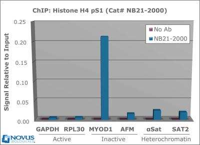
Chromatin Immunoprecipitation: Histone H4 [p Ser1] Antibody [NB21-2000] - 2 ug of NB21-2000 was used to IP DNA from fixed HeLa cells alongside a no antibody (No Ab) control. DNA was measured by qRT-PCR and normalized to total input (input=1).
![Immunocytochemistry/ Immunofluorescence: Histone H4 [p Ser1] Antibody - BSA Free [NB21-2000]](https://resources.rndsystems.com/images/products/Histone-H4-[p-Ser1]-Antibody-Immunocytochemistry-Immunofluorescence-NB21-2000-img0008.jpg "Immunocytochemistry/ Immunofluorescence: Histone H4 [p Ser1] Antibody - BSA Free [NB21-2000]")
Immunocytochemistry/ Immunofluorescence: Histone H4 [p Ser1] Antibody - BSA Free [NB21-2000]
Immunocytochemistry/Immunofluorescence: Histone H4 [p Ser1] Antibody [NB21-2000] - Histone H4 pS1 antibody was tested in in nonmitotic, prophase, and telophase HeLa cells with FITC (green). Nuclei and alpha-tubulin were counterstained with DAPI (blue) and DyLight 550 (red).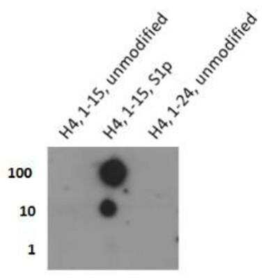
Dot Blot: Histone H4 [p Ser1] Antibody [NB21-2000] - Analysis of Histone H4 pS1 in picomoles of peptide.

Histone H4 [p Ser1] Antibody [NB21-2000]
Applications for Histone H4 [p Ser1] Antibody - BSA Free
Application
Recommended Usage
Chromatin Immunoprecipitation (ChIP)
2-5 ug per million cells
Dot Blot
0.1 ug/ml
Immunocytochemistry/ Immunofluorescence
1:50
Western Blot
1:1000
Application Notes
This H4pS1 antibody is useful for ICC/IF, ChIP, Dot blot, and Western blot where a band is detected ~13 kDa.
Formulation, Preparation, and Storage
Purification
Immunogen affinity purified
Formulation
PBS and 30% Glycerol
Format
BSA Free
Preservative
0.05% Sodium Azide
Concentration
0.89 mg/ml
Shipping
The product is shipped with polar packs. Upon receipt, store it immediately at the temperature recommended below.
Stability & Storage
Store at 4C short term. Aliquot and store at -20C long term. Avoid freeze-thaw cycles.
Background: Histone H4
Alternate Names
H4, H4/M, H4FM, H4M, HIST1H4I, HIST4H4, Histone Cluster 1 H4, Histone Cluster 1 H4i, Histone Cluster 4 H4, H4S1p
Gene Symbol
H4-16
UniProt
Additional Histone H4 Products
Product Documents for Histone H4 [p Ser1] Antibody - BSA Free
Certificate of Analysis
To download a Certificate of Analysis, please enter a lot or batch number in the search box below.
Product Specific Notices for Histone H4 [p Ser1] Antibody - BSA Free
Epi-Plus antibody production in collaboration with Rockland Immunochemicals Inc.
This product is for research use only and is not approved for use in humans or in clinical diagnosis. Primary Antibodies are guaranteed for 1 year from date of receipt.
Related Research Areas
Citations for Histone H4 [p Ser1] Antibody - BSA Free
 Powered by Bioz
Customer Reviews for Histone H4 [p Ser1] Antibody - BSA Free
There are currently no reviews for this product. Be the first to review Histone H4 [p Ser1] Antibody - BSA Free and earn rewards!
Have you used Histone H4 [p Ser1] Antibody - BSA Free?
Submit a review and receive an Amazon gift card!
$25/€18/£15/$25CAN/¥2500 Yen for a review with an image
$10/€7/£6/$10CAN/¥1110 Yen for a review without an image
Submit a review
Protocols
View specific protocols for Histone H4 [p Ser1] Antibody - BSA Free (NB21-2000):
Histone H4 [p Ser1] Antibody:
Chromatin Immunoprecipitation Protocol
Cell Fixation and Preparation
1. Begin with a cell culture that has reached 80% confluency.
2. Add formaldehyde to a final concentration of 1% in growth media and incubate for 10 minutes at room temperature.
3. Add glycine to reach a final concentration of 125 mM in the media. Incubate for 5 minutes at room temperature.
4. Remove all media and wash twice with 20 mL of ice cold PBS.
5. Add 2 mL of ice cold PBS with protease inhibitors*. Scrape cells into microcentrifuge tube.
6. Spin cells at 4 C for 5 minutes at 800 x g.
7. Remove supernatant and resuspend cells in 750 ul of RIPA lysis buffer containing protease inhibitors* per 10,000,000 cells (enough for 10 IPs). Incubate at 4 C for 15 minutes.
8. Spin cells at 4 C for 5 minutes at 800 x g.
DNA Shearing by Sonication
1. Sonicate crosslinked DNA to fragments sizes of 200-1000 base pairs. Keep samples ice cold to prevent denaturing of chromatin. Conditions for fragmenting must be empirically derived, and vary depending on equipment, cell type, cell density, and cross-linking efficiency.
2. Centrifuge samples to remove debris at 4 C for 10 minutes at 12,500 x g. Remove supernatant and transfer to a new tube. Discard pellet. Set aside 75 ul of sample for input fraction, which will not go through the subsequent IP steps. The remaining sample can be moved into 75 ul aliquots, each of which is sufficient for a single IP. Although it is preferable to proceed directly to the following steps, sheared chromatin can now be frozen at -80 C for up to 1 month.
a. Optional: Test the efficiency of the shearing by running 5-10 ul of sample on a 2% agarose gel after reversal of crosslinking, RNase treatment (0.5 mg/ml) and proteinase K treatment (0.1 ug/ul) as described below.
Chromatin Immunoprecipitation
Recommended controls include: No antibody negative control OR normal IgG negative control, positive control antibody.
1. Dilute each IP sample 1:10 by adding 75 ul sheared chromatin to 675 ul dilution buffer, along with appropriate protease inhibitors*. Save undiluted input fraction for step 10.
2. Add 25 ul of fully suspended protein A/G magnetic bead slurry. Do not allow the beads to dry.
3. Add antibody of interest to the diluted sample. For best results, incubate tubes with rotation at 4 C overnight. Alternatively, incubate at room temperature for 1-2 hours.
4. Pellet magnetic beads with a magnetic separator and remove the supernatant.
5. Add 750 ul cold low salt buffer and wash for 5 minutes with rotation. Pellet beads with separator and discard supernatant.
6. Add 750 ul cold high salt buffer and wash for 5 minutes with rotation. Pellet beads with separator and discard supernatant.
7. Add 750 ul cold LiCl buffer and wash for 5 minutes with rotation. Pellet beads with separator and discard supernatant.
8. Add 750 ul cold TE buffer and wash for 5 minutes with rotation. Pellet beads with separator and discard supernatant.
9. Elute complex by adding 200 ul elution buffer and agitate at RT for 15 minutes. Pellet beads with separator and discard beads. Keep the supernatant.
10. Add 8 ul of 5M NaCl and 2 ul of proteinase K (10 ug/ul) to each sample to reverse cross-linking. For the input fraction, add 125 ul of elution buffer along with NaCl and proteinase K. Incubate at 62 C for 4 hours or overnight. Incubate at 95 C for 10 minutes to deactivate proteinase K.
a. Optional: To decrease time, this step may also be performed at 95 C for 20 minutes without proteinase K treatment.
DNA Purification and Amplification
1. DNA can now be purified with either a commercially available column kit or by phenol/chloroform extraction.
2. Perform real-time PCR with 2 ul of purified DNA and primers (catalog numbers NBP1-71650, NBP1-71651, NBP1-71652, NBP1-71653, NBP1-71654 and NBP1-71655) per reaction. Dilute input fraction to 1% before PCR. Normalize all IPs and no antibody control IP to adjusted input fraction.
a. Ex. Raw input CT=30, adjusted input: 30 - 6.44 = 23.4
b. Antibody CT=28
c. Normalized signal relative to input: 2 ^ (23.4-28) = 0.04
Buffers
Dilution Buffer: 0.01% SDS, 1.1% Triton X-100, 1.2 mM EDTA, 16.7 mM Tris-HCL (pH8.1), 167 mM NaCl
Low Salt Wash Buffer: 0.1% SDS, 1% Triton X-100, 2mM EDTA, 20mM Tris-HCl, pH 8.1, 150mM NaCl.
High Salt Wash Buffer: 0.1% SDS, 1% Triton X-100, 2mM EDTA, 20mM Tris-HCl, pH 8.1, 500mM NaCl.
LiCl Wash Buffer: 250mM LiCl, 1% NP-40, 1% Na-deoxycholate, 1mM EDTA, 10mM Tris, pH 8.1.
TE Buffer: 10mM Tris-HCL pH 8.1, 1 mM EDTA
Elution buffer: 1% SDS, 100mM NaHCO3
* Please note that protease inhibitors have variable half-lives and should be freshly prepared as applicable.
Western Blot Protocol
1. Perform SDS-PAGE on samples to be analyzed, loading 40 ug of total protein per lane.
2. Transfer proteins to membrane according to the instructions provided by the manufacturer of the membrane and transfer apparatus.
3. Stain according to standard Ponceau S procedure (or similar product) to assess transfer success, and mark molecular weight standards where appropriate.
4. Rinse the blot.
5. Block the membrane using standard blocking buffer for at least 1 hour.
6. Wash the membrane in wash buffer three times for 10 minutes each.
7. Dilute primary antibody in blocking buffer and incubate 1 hour at room temperature.
8. Wash the membrane in wash buffer three times for 10 minutes each.
9. Apply the diluted HRP conjugated secondary antibody in blocking buffer (as per manufacturers instructions) and incubate 1 hour at room temperature.
10. Wash the blot in wash buffer three times for 10 minutes each (this step can be repeated as required to reduce background).
11. Apply the detection reagent of choice in accordance with the manufacturers instructions.
Note: Tween-20 can be added to the blocking or antibody dilution buffer at a final concentration of 0.05-0.2%.
Immunocytochemistry Protocol
Culture cells to appropriate density in 35 mm culture dishes or 6-well plates.
1. Remove culture medium and add 10% formalin to the dish. Fix at room temperature for 30 minutes.
2. Remove the formalin and add ice cold methanol. Incubate for 5-10 minutes.
3. Remove methanol and add washing solution (i.e. PBS). Be sure to not let the specimen dry out. Wash three times for 10 minutes.
4. To block nonspecific antibody binding incubate in 10% normal goat serum from 1 hour to overnight at room temperature.
5. Add primary antibody at appropriate dilution and incubate at room temperature from 2 hours to overnight at room temperature.
6. Remove primary antibody and replace with washing solution. Wash three times for 10 minutes.
7. Add secondary antibody at appropriate dilution. Incubate for 1 hour at room temperature.
8. Remove antibody and replace with wash solution, then wash for 10 minutes. Add Hoechst 33258 to wash solution at 1:25,0000 and incubate for 10 minutes. Wash a third time for 10 minutes.
9. Cells can be viewed directly after washing. The plates can also be stored in PBS containing Azide covered in Parafilm (TM). Cells can also be cover-slipped using Fluoromount, with appropriate sealing.
*The above information is only intended as a guide. The researcher should determine what protocol best meets their needs. Please follow safe laboratory procedures.
Find general support by application which include: protocols, troubleshooting, illustrated assays, videos and webinars.
- Appropriate Fixation of IHC/ICC Samples
- Cellular Response to Hypoxia Protocols
- ChIP Protocol Video
- Chromatin Immunoprecipitation (ChIP) Protocol
- Chromatin Immunoprecipitation Protocol
- ClariTSA™ Fluorophore Kits
- Detection & Visualization of Antibody Binding
- ICC Cell Smear Protocol for Suspension Cells
- ICC Immunocytochemistry Protocol Videos
- ICC for Adherent Cells
- Immunocytochemistry (ICC) Protocol
- Immunocytochemistry Troubleshooting
- Immunofluorescence of Organoids Embedded in Cultrex Basement Membrane Extract
- Immunohistochemistry (IHC) and Immunocytochemistry (ICC) Protocols
- Preparing Samples for IHC/ICC Experiments
- Preventing Non-Specific Staining (Non-Specific Binding)
- Primary Antibody Selection & Optimization
- Protocol for VisUCyte™ HRP Polymer Detection Reagent
- Protocol for the Fluorescent ICC Staining of Cell Smears - Graphic
- Protocol for the Fluorescent ICC Staining of Cultured Cells on Coverslips - Graphic
- Protocol for the Preparation and Fluorescent ICC Staining of Cells on Coverslips
- Protocol for the Preparation and Fluorescent ICC Staining of Non-adherent Cells
- Protocol for the Preparation and Fluorescent ICC Staining of Stem Cells on Coverslips
- Protocol for the Preparation of a Cell Smear for Non-adherent Cell ICC - Graphic
- R&D Systems Quality Control Western Blot Protocol
- TUNEL and Active Caspase-3 Detection by IHC/ICC Protocol
- The Importance of IHC/ICC Controls
- Troubleshooting Guide: Western Blot Figures
- Western Blot Conditions
- Western Blot Protocol
- Western Blot Protocol for Cell Lysates
- Western Blot Troubleshooting
- Western Blot Troubleshooting Guide
- View all Protocols, Troubleshooting, Illustrated assays and Webinars
Loading...
Associated Pathways
