Histone H4 [ac Lys16] Antibody - BSA Free
Novus Biologicals | Catalog # NB21-2077
![Western Blot: Histone H4 [ac Lys16] AntibodyBSA Free [NB21-2077]](https://resources.rndsystems.com/images/products/Histone-H4-[ac-Lys16]-Antibody-Western-Blot-NB21-2077-img0008.jpg "Western Blot: Histone H4 [ac Lys16] AntibodyBSA Free [NB21-2077]")
Key Product Details
Species Reactivity
Validated:
Human, Mouse, Rat, Porcine, Mammal
Cited:
Mouse
Applications
Validated:
Western Blot, Immunocytochemistry/ Immunofluorescence, Chromatin Immunoprecipitation (Negative), Dot Blot
Cited:
Western Blot
Label
Unconjugated
Antibody Source
Polyclonal Rabbit IgG
Format
BSA Free
Loading...
Product Specifications
Immunogen
Synthetic acetylated peptide surrounding Lysine 16 of human Histone H4 [Swiss Prot P62805].
Reactivity Notes
Predicted to react with most mammal species.
Modification
ac Lys16
Localization
Nucleus. Chromosome.
Marker
H4k16ac
Clonality
Polyclonal
Host
Rabbit
Isotype
IgG
Scientific Data Images for Histone H4 [ac Lys16] Antibody - BSA Free
Western Blot: Histone H4 [ac Lys16] AntibodyBSA Free [NB21-2077]
Western Blot: Histone H4 [ac Lys16] Antibody [NB21-2077] - Western Blot using NB21-2077 of porcine whole ovarian homogenate. Image submitted via verified customer review.![Immunocytochemistry/ Immunofluorescence: Histone H4 [ac Lys16] Antibody - BSA Free [NB21-2077]](https://resources.rndsystems.com/images/products/Histone-H4-[ac--Lys16]-Antibody-Immunocytochemistry-Immunofluorescence-NB21-2077-img0005.jpg "Immunocytochemistry/ Immunofluorescence: Histone H4 [ac Lys16] Antibody - BSA Free [NB21-2077]")
Immunocytochemistry/ Immunofluorescence: Histone H4 [ac Lys16] Antibody - BSA Free [NB21-2077]
Immunocytochemistry/Immunofluorescence: Histone H4 [ac Lys16] Antibody [NB21-2077] - Histone H4 K16ac antibody was tested in HeLa cells with FITC (green). Nuclei and alpha-tubulin were counterstained with DAPI (blue) and Dylight 550 (red).![Western Blot: Histone H4 [ac Lys16] AntibodyBSA Free [NB21-2077]](https://resources.rndsystems.com/images/products/Histone-H4-[ac-Lys16]-Antibody-Western-Blot-NB21-2077-img0002.jpg "Western Blot: Histone H4 [ac Lys16] AntibodyBSA Free [NB21-2077]")
Western Blot: Histone H4 [ac Lys16] AntibodyBSA Free [NB21-2077]
Western Blot: Histone H4 [ac Lys16] Antibody [NB21-2077] - WB analysis of H4K16ac in 1) HeLa histone prep lysate and 2) NIH 3T3 histone prep lysate.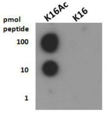
Dot Blot: Histone H4 [ac Lys16] Antibody [NB21-2077] - Dot blot analysis of H4K16ac in picomoles of protein.

Histone H4 [ac Lys16] Antibody [NB21-2077]
Applications for Histone H4 [ac Lys16] Antibody - BSA Free
Application
Recommended Usage
Dot Blot
1:1000
Immunocytochemistry/ Immunofluorescence
1:100
Western Blot
1:1000
Application Notes
This H4K16ac antibody is useful for Western Blot, Immunocytochemistry/Immunofluorescence, and Dot Blot. In Western Blot, a band is seen ~13kDa in HeLa histone prep and NIH 3T3 histone prep. In ICC/IF, nuclear staining was observed in HeLa cells. This antibody is not applicable for Chromatin Immunoprecipitation.
Reviewed Applications
Read 1 review rated 5 using NB21-2077 in the following applications:
Formulation, Preparation, and Storage
Purification
Immunogen affinity purified
Formulation
PBS and 30% Glycerol
Format
BSA Free
Preservative
0.05% Sodium Azide
Concentration
0.62 mg/ml
Shipping
The product is shipped with polar packs. Upon receipt, store it immediately at the temperature recommended below.
Stability & Storage
Store at 4C short term. Aliquot and store at -20C long term. Avoid freeze-thaw cycles.
Background: Histone H4
Alternate Names
H4, H4/M, H4FM, H4M, HIST1H4I, HIST4H4, Histone Cluster 1 H4, Histone Cluster 1 H4i, Histone Cluster 4 H4, H4K16ac
Gene Symbol
H4-16
UniProt
Additional Histone H4 Products
Product Documents for Histone H4 [ac Lys16] Antibody - BSA Free
Certificate of Analysis
To download a Certificate of Analysis, please enter a lot or batch number in the search box below.
Product Specific Notices for Histone H4 [ac Lys16] Antibody - BSA Free
EpiPlus antibodies are produced in collaboration with Rockland Immunochemicals Inc.
This product is for research use only and is not approved for use in humans or in clinical diagnosis. Primary Antibodies are guaranteed for 1 year from date of receipt.
Related Research Areas
Citations for Histone H4 [ac Lys16] Antibody - BSA Free
 Powered by Bioz
Customer Reviews for Histone H4 [ac Lys16] Antibody - BSA Free (1)
5 out of 5
1 Customer Rating
Have you used Histone H4 [ac Lys16] Antibody - BSA Free?
Submit a review and receive an Amazon gift card!
$25/€18/£15/$25CAN/¥2500 Yen for a review with an image
$10/€7/£6/$10CAN/¥1110 Yen for a review without an image
Submit a review
Customer Images
![Histone H4 [ac Lys16] Antibody - BSA Free NB21-2077](https://resources.rndsystems.com/images/reviews/review_nb21-2077_26021.png)
Showing
1
-
1 of
1 review
Showing All
Filter By:
-
Application: Western BlotSample Tested: Ovary tissueSpecies: PigVerified Customer | Posted 11/17/2016H4K16AC on porcine whole ovarian homogenateSample Information: Cell Line or Tissue: Ovary Species: Pig Treatment: None. Run on control tissue Controls: Positive Control: None Negative Control: Primary only, secondary only, and secondary with IgG all negative Loading Control (please attach additional images if applicable): Ponceau S stain Total Protein Loaded: 40 ug/well Electrophoresis: Gel Percentage: Gradient, 4-20% Voltage: 50V then 90V Time: 5 min then 1 hr Membrane Transfer: Method (Submersion/Semi-dry): Semi-dry using iBlot2 system Membrane Type (PVDF/Nitrocellulose): Nitrocellulose Time: 7 min (iBlot2 Protocol 0) Voltage: Click here to enter text. Blocking: Blocking Solution: 5% BSA in.2% PBST Time: 1.5 hours Primary Antibody: Dilution: 1:1000 Diluent Buffer: 5% BSA in.2% PBST Incubation Time: overnight Incubation Temperature: 4°C Washing Conditions: Wash Solution:.2% PBST Time and Repetitions: 3X, 10 minutes each Secondary Antibody Manufacturer and Catalog #: Cell Signaling, #7074 Secondary description: goat anti-rabbit Dilution: 1:1000 Diluent Buffer: 5% BSA in.2% PBST Incubation Time: 1 hour Incubation Temperature: room temperature Detection Method: Detection: SignalFire ECL reagent (Cell Signaling) Procedure: ECL on blot 3 min Development Time: 5 min Molecular weight of band(s): 13 kDa
![Histone H4 [ac Lys16] Antibody - BSA Free NB21-2077](data:image/png;base64,R0lGODlhAQABAAD/ACwAAAAAAQABAAACADs=)
There are no reviews that match your criteria.
Protocols
View specific protocols for Histone H4 [ac Lys16] Antibody - BSA Free (NB21-2077):
Histone H4 [ac Lys16] Antibody:
Western Blot Protocol
1. Perform SDS-PAGE on samples to be analyzed, loading 10 ug of histone preps per lane.
2. Transfer proteins to membrane according to the instructions provided by the manufacturer of the membrane and transfer apparatus.
3. Stain according to standard Ponceau S procedure (or similar product) to assess transfer success, and mark molecular weight standards where appropriate.
4. Rinse the blot.
5. Block the membrane using standard blocking buffer for at least 1 hour.
6. Wash the membrane in wash buffer three times for 10 minutes each.
7. Dilute primary antibody in blocking buffer and incubate 1 hour at room temperature.
8. Wash the membrane in wash buffer three times for 10 minutes each.
9. Apply the diluted HRP conjugated secondary antibody in blocking buffer (as per manufacturers instructions) and incubate 1 hour at room temperature.
10. Wash the blot in wash buffer three times for 10 minutes each (this step can be repeated as required to reduce background).
11. Apply the detection reagent of choice in accordance with the manufacturers instructions.
Note: Tween-20 can be added to the blocking or antibody dilution buffer at a final concentration of 0.05-0.2%.
Immunocytochemistry Protocol
Culture cells to appropriate density in 35 mm culture dishes or 6-well plates.
1. Remove culture medium and add 10% formalin to the dish. Fix at room temperature for 30 minutes.
2. Remove the formalin and add ice cold methanol. Incubate for 5-10 minutes.
3. Remove methanol and add washing solution (i.e. PBS). Be sure to not let the specimen dry out. Wash three times for 10 minutes.
4. To block nonspecific antibody binding incubate in 10% normal goat serum from 1 hour to overnight at room temperature.
5. Add primary antibody at appropriate dilution and incubate at room temperature from 2 hours to overnight at room temperature.
6. Remove primary antibody and replace with washing solution. Wash three times for 10 minutes.
7. Add secondary antibody at appropriate dilution. Incubate for 1 hour at room temperature.
8. Remove antibody and replace with wash solution, then wash for 10 minutes. Add Hoechst 33258 to wash solution at 1:25,0000 and incubate for 10 minutes. Wash a third time for 10 minutes.
9. Cells can be viewed directly after washing. The plates can also be stored in PBS containing Azide covered in Parafilm (TM). Cells can also be cover-slipped using Fluoromount, with appropriate sealing.
*The above information is only intended as a guide. The researcher should determine what protocol best meets their needs. Please follow safe laboratory procedures.
Find general support by application which include: protocols, troubleshooting, illustrated assays, videos and webinars.
- Appropriate Fixation of IHC/ICC Samples
- Cellular Response to Hypoxia Protocols
- ClariTSA™ Fluorophore Kits
- Detection & Visualization of Antibody Binding
- ICC Cell Smear Protocol for Suspension Cells
- ICC Immunocytochemistry Protocol Videos
- ICC for Adherent Cells
- Immunocytochemistry (ICC) Protocol
- Immunocytochemistry Troubleshooting
- Immunofluorescence of Organoids Embedded in Cultrex Basement Membrane Extract
- Immunohistochemistry (IHC) and Immunocytochemistry (ICC) Protocols
- Preparing Samples for IHC/ICC Experiments
- Preventing Non-Specific Staining (Non-Specific Binding)
- Primary Antibody Selection & Optimization
- Protocol for VisUCyte™ HRP Polymer Detection Reagent
- Protocol for the Fluorescent ICC Staining of Cell Smears - Graphic
- Protocol for the Fluorescent ICC Staining of Cultured Cells on Coverslips - Graphic
- Protocol for the Preparation and Fluorescent ICC Staining of Cells on Coverslips
- Protocol for the Preparation and Fluorescent ICC Staining of Non-adherent Cells
- Protocol for the Preparation and Fluorescent ICC Staining of Stem Cells on Coverslips
- Protocol for the Preparation of a Cell Smear for Non-adherent Cell ICC - Graphic
- R&D Systems Quality Control Western Blot Protocol
- TUNEL and Active Caspase-3 Detection by IHC/ICC Protocol
- The Importance of IHC/ICC Controls
- Troubleshooting Guide: Western Blot Figures
- Western Blot Conditions
- Western Blot Protocol
- Western Blot Protocol for Cell Lysates
- Western Blot Troubleshooting
- Western Blot Troubleshooting Guide
- View all Protocols, Troubleshooting, Illustrated assays and Webinars
Loading...
Associated Pathways
