HNF-3 alpha/FoxA1 Antibody (3B11NB) - BSA Free
Novus Biologicals | Catalog # NBP2-45269
Key Product Details
Species Reactivity
Validated:
Human
Cited:
Human, Mouse
Predicted:
Mouse (98%), Rat (96%). Backed by our 100% Guarantee.
Applications
Validated:
Immunohistochemistry, Immunohistochemistry-Paraffin, Western Blot, Flow Cytometry, Immunocytochemistry/ Immunofluorescence
Cited:
Immunohistochemistry-Paraffin, IF/IHC
Label
Unconjugated
Antibody Source
Monoclonal Mouse IgG2b Kappa Clone # 3B11NB
Format
BSA Free
Loading...
Product Specifications
Immunogen
Partial recombinant human FOXA1 protein (between amino acids 350-500) [Uniprot: P55317].
Clonality
Monoclonal
Host
Mouse
Isotype
IgG2b Kappa
Scientific Data Images for HNF-3 alpha/FoxA1 Antibody (3B11NB) - BSA Free
![Western Blot: HNF-3 alpha/FoxA1 Antibody (3B11NB)BSA Free [NBP2-45269]](https://resources.rndsystems.com/images/products/HNF-3-alpha-FoxA1-Antibody-3B11NB-Western-Blot-NBP2-45269-img0005.jpg "Western Blot: HNF-3 alpha/FoxA1 Antibody (3B11NB)BSA Free [NBP2-45269]")
Western Blot: HNF-3 alpha/FoxA1 Antibody (3B11NB)BSA Free [NBP2-45269]
Western Blot: HNF-3 alpha/FoxA1 Antibody (3B11NB) [NBP2-45269] - Whole cell protein from PC3 (1) and MCF7 (2) was separated on a 7.5% gel by SDS-PAGE, transferred to PVDF membrane and blocked in 5% non-fat milk in TBST. The membrane was probed with 4 ug/ml anti-hFOXA1 (3B11NB) in 1% milk and detected with an anti-mouse HRP secondary antibody using chemiluminescence.![Immunocytochemistry/ Immunofluorescence: HNF-3 alpha/FoxA1 Antibody (3B11NB) - BSA Free [NBP2-45269]](https://resources.rndsystems.com/images/products/HNF-3-alpha-FoxA1-Antibody-3B11NB-Immunocytochemistry-Immunofluorescence-NBP2-45269-img0004.jpg "Immunocytochemistry/ Immunofluorescence: HNF-3 alpha/FoxA1 Antibody (3B11NB) - BSA Free [NBP2-45269]")
Immunocytochemistry/ Immunofluorescence: HNF-3 alpha/FoxA1 Antibody (3B11NB) - BSA Free [NBP2-45269]
Immunocytochemistry/Immunofluorescence: HNF-3 alpha/FoxA1 Antibody (3B11NB) [NBP2-45269] - FOXA1 (3B11NB) antibody was tested in HepG2 cells at a 1:25 dilution against DyLight 488 (Green) using a 40X objective. Actin and nuclei were counterstained against Phalloidin 568 (Red) and DAPI (Blue), respectively![Immunohistochemistry-Paraffin: HNF-3 alpha/FoxA1 Antibody (3B11NB) - BSA Free [NBP2-45269]](https://resources.rndsystems.com/images/products/HNF-3-alpha-FoxA1-Antibody-3B11NB-Immunohistochemistry-Paraffin-NBP2-45269-img0003.jpg "Immunohistochemistry-Paraffin: HNF-3 alpha/FoxA1 Antibody (3B11NB) - BSA Free [NBP2-45269]")
Immunohistochemistry-Paraffin: HNF-3 alpha/FoxA1 Antibody (3B11NB) - BSA Free [NBP2-45269]
Immunohistochemistry-Paraffin: HNF-3 alpha/FoxA1 Antibody (3B11NB) [NBP2-45269] - Analysis of FOXA1 (3B11NB) in human prostate.![Flow Cytometry: HNF-3 alpha/FoxA1 Antibody (3B11NB) - BSA Free [NBP2-45269]](https://resources.rndsystems.com/images/products/HNF-3-alpha-FoxA1-Antibody-3B11NB-Flow-Cytometry-NBP2-45269-img0012.jpg "Flow Cytometry: HNF-3 alpha/FoxA1 Antibody (3B11NB) - BSA Free [NBP2-45269]")
Flow Cytometry: HNF-3 alpha/FoxA1 Antibody (3B11NB) - BSA Free [NBP2-45269]
Flow Cytometry: HNF-3 alpha/FoxA1 Antibody (3B11NB) [NBP2-45269] - Analysis using the Azide Free version of NBP2-45269. Staining of LnCAP cells (1 x 10^6 cells/ml) with FOXA1 antibody, clone 3B11NB (orange), shown with the negative control (blue).![Western Blot: HNF-3 alpha/FoxA1 Antibody (3B11NB)BSA Free [NBP2-45269]](https://resources.rndsystems.com/images/products/HNF-3-alpha-FoxA1-Antibody-3B11NB-Western-Blot-NBP2-45269-img0001.jpg "Western Blot: HNF-3 alpha/FoxA1 Antibody (3B11NB)BSA Free [NBP2-45269]")
Western Blot: HNF-3 alpha/FoxA1 Antibody (3B11NB)BSA Free [NBP2-45269]
Western Blot: HNF-3 alpha/FoxA1 Antibody (3B11NB) [NBP2-45269] - Analysis of FOXA1 (3B11NB) in partial recombinant FOXA1 protein and PC3 lysate, respectivetly.![Immunohistochemistry-Paraffin: HNF-3 alpha/FoxA1 Antibody (3B11NB) - BSA Free [NBP2-45269]](https://resources.rndsystems.com/images/products/HNF-3-alpha-FoxA1-Antibody-3B11NB-Immunohistochemistry-Paraffin-NBP2-45269-img0002.jpg "Immunohistochemistry-Paraffin: HNF-3 alpha/FoxA1 Antibody (3B11NB) - BSA Free [NBP2-45269]")
Immunohistochemistry-Paraffin: HNF-3 alpha/FoxA1 Antibody (3B11NB) - BSA Free [NBP2-45269]
Immunohistochemistry-Paraffin: HNF-3 alpha/FoxA1 Antibody (3B11NB) [NBP2-45269] - Analysis of FOXA1 (3B11NB) in human breast adenocarcinoma.![Flow Cytometry: HNF-3 alpha/FoxA1 Antibody (3B11NB) - BSA Free [NBP2-45269]](https://resources.rndsystems.com/images/products/HNF-3-alpha-FoxA1-Antibody-3B11NB-Flow-Cytometry-NBP2-45269-img0011.jpg "Flow Cytometry: HNF-3 alpha/FoxA1 Antibody (3B11NB) - BSA Free [NBP2-45269]")
Flow Cytometry: HNF-3 alpha/FoxA1 Antibody (3B11NB) - BSA Free [NBP2-45269]
Flow Cytometry: HNF-3 alpha/FoxA1 Antibody (3B11NB) [NBP2-45269] - Analysis using the Azide Free version of NBP2-45269. Staining of HepG2 cells (1 x 10^6 cells/ml) with FOXA1 antibody, clone 3B11NB (orange), shown with the negative control (blue). [NBP2-45269]")
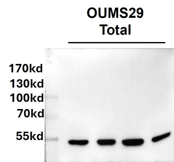
Western Blot: Mouse Monoclonal HNF-3 alpha/FoxA1 Antibody (3B11NB) [NBP2-45269]
Western Blot: Mouse Monoclonal HNF-3 alpha/FoxA1 Antibody (3B11NB) [NBP2-45269] - 40 ug protein from OUMS29 cell lysates were loaded into gels for western blot experiment. The PVDF membranes were probed with this antibody at 1ug/ml concentration. Bands for HNF-3 alpha/FoxA1 appeared around 53kDa. Image from a verified customer review.Applications for HNF-3 alpha/FoxA1 Antibody (3B11NB) - BSA Free
Application
Recommended Usage
Flow Cytometry
25 ug/ml
Immunocytochemistry/ Immunofluorescence
1:25 - 1:50
Immunohistochemistry
5 ug/ml
Immunohistochemistry-Paraffin
5 ug/ml
Western Blot
2-4 ug/ml
Reviewed Applications
Read 1 review rated 4 using NBP2-45269 in the following applications:
Flow Cytometry Panel Builder
Bio-Techne Knows Flow Cytometry
Save time and reduce costly mistakes by quickly finding compatible reagents using the Panel Builder Tool.
Advanced Features
- Spectra Viewer - Custom analysis of spectra from multiple fluorochromes
- Spillover Popups - Visualize the spectra of individual fluorochromes
- Antigen Density Selector - Match fluorochrome brightness with antigen density

Formulation, Preparation, and Storage
Purification
Protein G purified
Formulation
PBS
Format
BSA Free
Preservative
0.02% Sodium Azide
Concentration
1.0 mg/ml
Shipping
The product is shipped with polar packs. Upon receipt, store it immediately at the temperature recommended below.
Stability & Storage
Store at 4C short term. Aliquot and store at -20C long term. Avoid freeze-thaw cycles.
Background: HNF-3 alpha/FoxA1
Long Name
Hepatocyte Nuclear Factor-3 alpha/Forkhead Box Protein A1
Alternate Names
FoxA1, HNF3 alpha, TCF3A
Gene Symbol
FOXA1
UniProt
Additional HNF-3 alpha/FoxA1 Products
Product Documents for HNF-3 alpha/FoxA1 Antibody (3B11NB) - BSA Free
Certificate of Analysis
To download a Certificate of Analysis, please enter a lot or batch number in the search box below.
Product Specific Notices for HNF-3 alpha/FoxA1 Antibody (3B11NB) - BSA Free
This product is for research use only and is not approved for use in humans or in clinical diagnosis. Primary Antibodies are guaranteed for 1 year from date of receipt.
Related Research Areas
Citations for HNF-3 alpha/FoxA1 Antibody (3B11NB) - BSA Free
 Powered by Bioz
Customer Reviews for HNF-3 alpha/FoxA1 Antibody (3B11NB) - BSA Free (1)
4 out of 5
1 Customer Rating
Have you used HNF-3 alpha/FoxA1 Antibody (3B11NB) - BSA Free?
Submit a review and receive an Amazon gift card!
$25/€18/£15/$25CAN/¥2500 Yen for a review with an image
$10/€7/£6/$10CAN/¥1110 Yen for a review without an image
Submit a review
Customer Images
Showing
1
-
1 of
1 review
Showing All
Filter By:
-
Application: Western BlotSample Tested: OUMS29 Human hepatocyte lineSpecies: HumanVerified Customer | Posted 11/27/2024Hnf3alpha in OUMS29 cells40 ug protein from OUMS29 cell lysates were loaded into gels for western blot experiment. The PVDF membranes were probed with this antibody at 1ug/ml concentration. Bands for Hnf3alpha appeared around 53kDa.

There are no reviews that match your criteria.
Protocols
View specific protocols for HNF-3 alpha/FoxA1 Antibody (3B11NB) - BSA Free (NBP2-45269):
HNF-3 alpha/FoxA1 Antibody (3B11NB):
Immunocytochemistry Protocol
Culture cells to appropriate density in 35 mm culture dishes or 6-well plates.
1. Remove culture medium and add 10% formalin to the dish. Fix at room temperature for 30 minutes.
2. Remove the formalin and add ice cold methanol. Incubate for 5-10 minutes.
3. Remove methanol and add washing solution (i.e. PBS). Be sure to not let the specimen dry out. Wash three times for 10 minutes.
4. To block nonspecific antibody binding incubate in 10% normal goat serum from 1 hour to overnight at room temperature.
5. Add primary antibody at appropriate dilution and incubate at room temperature from 2 hours to overnight at room temperature.
6. Remove primary antibody and replace with washing solution. Wash three times for 10 minutes.
7. Add secondary antibody at appropriate dilution. Incubate for 1 hour at room temperature.
8. Remove antibody and replace with wash solution, then wash for 10 minutes. Add Hoechst 33258 to wash solution at 1:25,0000 and incubate for 10 minutes. Wash a third time for 10 minutes.
9. Cells can be viewed directly after washing. The plates can also be stored in PBS containing Azide covered in Parafilm (TM). Cells can also be cover-slipped using Fluoromount, with appropriate sealing.
*The above information is only intended as a guide. The researcher should determine what protocol best meets their needs. Please follow safe laboratory
HNF-3 alpha/FoxA1 Antibody (3B11NB):
1. Deparaffinize the tissue sections by immersing the slides in Xylene with two changes for 10 min each. Sections should not get dried at any stage from this point.
2. Rehydrate the tissue sections by immersing the slides in decreasing grades of ethanol as follows:
a. Immerse in 100% ethanol with 2 changes for 5 minutes each
b. Immerse in 95% ethanol with 2 changes for 5 minutes each
c. Immerse in 90% ethanol for 5 minutes
d. Immerse in 70% ethanol for 5 minutes
e. Immerse in 50% ethanol for 5 minutes
f. Immerse in distilled water for 5 minutes
3. Antigen Retrieval (Microwave Method):
a. Immerse the slides in a microwave compatible tray containing 10 mM Sodium Citrate buffer (pH 6.0) with 0.05% Tween 20.
b. Boil the slides and maintain the sub-boiling temperature for 5 minutes in the microwave. Thereafter, take out the tray very carefully and cool it at room temperature (RT) for about 30 minutes.
c. Wash the slides 3 times, 3 minutes each by immersing them in TBST (Tris Buffered Saline having 0.05% Tween 20).
4. Quenching of Endogenous Peroxidase:
a. Incubate the slides in 3% hydrogen peroxide prepared in methanol for 15 minutes (at RT, in dark conditions).
b. Wash the slides in TBST 3 times, 3 minutes each.
5. Protein Blocking:
a. Incubate the sections with background sniper solution at RT for 15 minutes (Biocare Medicals, USA).
b. Wash the sections 3 times, 3 min each by immersing the slides in TBST.
6. Primary Antibody:
a. Dilute the primary antibody at 5ug/ml concentration using PBS as a diluent.
b. Incubate the sections with diluted primary antibody for 90 minutes at RT in a humidified chamber.
c. Thereafter, wash the slides 4 times, 5 minutes each with TBST.
7. Probe (Secondary Reagent):
a. Incubate with MACH 1 Mouse probe for 15 minutes at RT.
b. Incubate for 30 min at room temperature with HRP-Polymer (Biocare Medical, USA).
c. Wash the slides with TBST 4 times, 5 minutes each
8. Chromogen:
a. Mix 32ul of DAB Chromogen with 1 ml of DAB substrate buffer (Biocare Medical, USA).
b. Apply 200ul DAB mixture/section and incubate at RT in dark conditions (few seconds - 5 minutes).
c. As soon as an appropriate color develops, rinse the slides with deionized water (2-3 brief rinses).
9. Counter stain:
a. Counter stain with Hematoxylin for 30 seconds (Vector Labs, USA).
b. Wash in deionized water for 1-2 minutes to clear the extra stain.
c. Incubate the slides in bluing solution or Scott's water twice for 2 minutes each time.
10. Dehydrate the sections in increasing grades of alcohols:
a. 50% alcohol for 1 minute
b. 70% for 1 minute
c. 90% for 1 minute
d. 95% for 1 minute
e. 100% for 1 minute
f. Xylene with 2 changes for 2 minutes each
11. Mount with DPX mount and cover-slip glass (Fisher Scientific, USA), carefully not allowing any air bubbles to enter.
NOTE:- This protocol is provided as a reference tool only. Depending upon the type of tissues /tissue processing and reagents employed, the end user will need to optimize the final conditions for achieving an expected staining.
HNF-3 alpha/FoxA1 Antibody (3B11NB):
1. Perform SDS-PAGE on samples to be analyzed, loading 25 ug of total protein per lane.
2. Transfer proteins to membrane according to the instructions provided by the manufacturer of the membrane and transfer apparatus.
3. Stain according to standard Ponceau S procedure (or similar product) to assess transfer success, and mark molecular weight standards where appropriate.
4. Rinse the blot.
5. Block the membrane using standard blocking buffer for at least 1 hour.
6. Wash the membrane in wash buffer three times for 10 minutes each.
7. Dilute anti-FOXA1 primary antibody in blocking buffer and incubate 1 hour at room temperature.
8. Wash the membrane in wash buffer three times for 10 minutes each.
9. Apply the diluted HRP conjugated secondary antibody in blocking buffer (as per manufacturers instructions) and incubate 1 hour at room temperature.
10. Wash the blot in wash buffer three times for 10 minutes each (this step can be repeated as required to reduce background).
11. Apply the detection reagent of choice in accordance with the manufacturers instructions.
Note: Tween-20 can be added to the blocking or antibody dilution buffer at a final concentration of 0.05-0.2%.
Find general support by application which include: protocols, troubleshooting, illustrated assays, videos and webinars.
- 7-Amino Actinomycin D (7-AAD) Cell Viability Flow Cytometry Protocol
- Antigen Retrieval Protocol (PIER)
- Antigen Retrieval for Frozen Sections Protocol
- Appropriate Fixation of IHC/ICC Samples
- Cellular Response to Hypoxia Protocols
- Chromogenic IHC Staining of Formalin-Fixed Paraffin-Embedded (FFPE) Tissue Protocol
- Chromogenic Immunohistochemistry Staining of Frozen Tissue
- ClariTSA™ Fluorophore Kits
- Detection & Visualization of Antibody Binding
- Extracellular Membrane Flow Cytometry Protocol
- Flow Cytometry Protocol for Cell Surface Markers
- Flow Cytometry Protocol for Staining Membrane Associated Proteins
- Flow Cytometry Staining Protocols
- Flow Cytometry Troubleshooting Guide
- Fluorescent IHC Staining of Frozen Tissue Protocol
- Graphic Protocol for Heat-induced Epitope Retrieval
- Graphic Protocol for the Preparation and Fluorescent IHC Staining of Frozen Tissue Sections
- Graphic Protocol for the Preparation and Fluorescent IHC Staining of Paraffin-embedded Tissue Sections
- Graphic Protocol for the Preparation of Gelatin-coated Slides for Histological Tissue Sections
- ICC Cell Smear Protocol for Suspension Cells
- ICC Immunocytochemistry Protocol Videos
- ICC for Adherent Cells
- IHC Sample Preparation (Frozen sections vs Paraffin)
- Immunocytochemistry (ICC) Protocol
- Immunocytochemistry Troubleshooting
- Immunofluorescence of Organoids Embedded in Cultrex Basement Membrane Extract
- Immunofluorescent IHC Staining of Formalin-Fixed Paraffin-Embedded (FFPE) Tissue Protocol
- Immunohistochemistry (IHC) and Immunocytochemistry (ICC) Protocols
- Immunohistochemistry Frozen Troubleshooting
- Immunohistochemistry Paraffin Troubleshooting
- Intracellular Flow Cytometry Protocol Using Alcohol (Methanol)
- Intracellular Flow Cytometry Protocol Using Detergents
- Intracellular Nuclear Staining Flow Cytometry Protocol Using Detergents
- Intracellular Staining Flow Cytometry Protocol Using Alcohol Permeabilization
- Intracellular Staining Flow Cytometry Protocol Using Detergents to Permeabilize Cells
- Preparing Samples for IHC/ICC Experiments
- Preventing Non-Specific Staining (Non-Specific Binding)
- Primary Antibody Selection & Optimization
- Propidium Iodide Cell Viability Flow Cytometry Protocol
- Protocol for Heat-Induced Epitope Retrieval (HIER)
- Protocol for Liperfluo
- Protocol for Making a 4% Formaldehyde Solution in PBS
- Protocol for VisUCyte™ HRP Polymer Detection Reagent
- Protocol for the Characterization of Human Th22 Cells
- Protocol for the Characterization of Human Th9 Cells
- Protocol for the Fluorescent ICC Staining of Cell Smears - Graphic
- Protocol for the Fluorescent ICC Staining of Cultured Cells on Coverslips - Graphic
- Protocol for the Preparation & Fixation of Cells on Coverslips
- Protocol for the Preparation and Chromogenic IHC Staining of Frozen Tissue Sections
- Protocol for the Preparation and Chromogenic IHC Staining of Frozen Tissue Sections - Graphic
- Protocol for the Preparation and Chromogenic IHC Staining of Paraffin-embedded Tissue Sections
- Protocol for the Preparation and Chromogenic IHC Staining of Paraffin-embedded Tissue Sections - Graphic
- Protocol for the Preparation and Fluorescent ICC Staining of Cells on Coverslips
- Protocol for the Preparation and Fluorescent ICC Staining of Non-adherent Cells
- Protocol for the Preparation and Fluorescent ICC Staining of Stem Cells on Coverslips
- Protocol for the Preparation and Fluorescent IHC Staining of Frozen Tissue Sections
- Protocol for the Preparation and Fluorescent IHC Staining of Paraffin-embedded Tissue Sections
- Protocol for the Preparation of Gelatin-coated Slides for Histological Tissue Sections
- Protocol for the Preparation of a Cell Smear for Non-adherent Cell ICC - Graphic
- Protocol: Annexin V and PI Staining by Flow Cytometry
- Protocol: Annexin V and PI Staining for Apoptosis by Flow Cytometry
- R&D Systems Quality Control Western Blot Protocol
- TUNEL and Active Caspase-3 Detection by IHC/ICC Protocol
- The Importance of IHC/ICC Controls
- Troubleshooting Guide: Fluorokine Flow Cytometry Kits
- Troubleshooting Guide: Immunohistochemistry
- Troubleshooting Guide: Western Blot Figures
- Western Blot Conditions
- Western Blot Protocol
- Western Blot Protocol for Cell Lysates
- Western Blot Troubleshooting
- Western Blot Troubleshooting Guide
- View all Protocols, Troubleshooting, Illustrated assays and Webinars
Loading...
Associated Pathways
