HSPA8/HSC71/Hsc70 Antibody (13D3)
Novus Biologicals | Catalog # NB120-2788
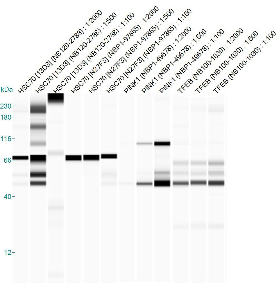
![Western Blot: HSPA8/HSC71/Hsc70 Antibody (13D3) [NB120-2788]](https://resources.rndsystems.com/images/products/Hsp70-Hsc70-Antibody-13D3-Western-Blot-NB120-2788-img0019.jpg "Western Blot: HSPA8/HSC71/Hsc70 Antibody (13D3) [NB120-2788]")
Key Product Details
Species Reactivity
Validated:
Cited:
Applications
Validated:
Cited:
Label
Antibody Source
Product Specifications
Immunogen
Reactivity Notes
Specificity
Clonality
Host
Isotype
Theoretical MW
Disclaimer note: The observed molecular weight of the protein may vary from the listed predicted molecular weight due to post translational modifications, post translation cleavages, relative charges, and other experimental factors.
Scientific Data Images for HSPA8/HSC71/Hsc70 Antibody (13D3)
Western Blot: HSPA8/HSC71/Hsc70 Antibody (13D3) [NB120-2788]
Western Blot: Hsp70 / Hsc70 Antibody (13D3) [NB120-2788] - Proteins were transferred to a PVDF membrane and blocked with 5% BSA/TBST for at least 1 hour. The membrane was probed with a HSP70 monoclonal antibody at a dilution of 1:1000 overnight at 4C on a rocking platform, washed in TBS-0.1%Tween 20, and probed with a goat anti-mouse IgM-HRP secondary antibody at a dilution of 1:20,000 for at least 1 hour.![Immunocytochemistry/ Immunofluorescence: HSPA8/HSC71/Hsc70 Antibody (13D3) [NB120-2788]](https://resources.rndsystems.com/images/products/Hsp70-Hsc70-Antibody-13D3-Immunocytochemistry-Immunofluorescence-NB120-2788-img0013.jpg "Immunocytochemistry/ Immunofluorescence: HSPA8/HSC71/Hsc70 Antibody (13D3) [NB120-2788]")
Immunocytochemistry/ Immunofluorescence: HSPA8/HSC71/Hsc70 Antibody (13D3) [NB120-2788]
Immunocytochemistry/Immunofluorescence: Hsp70 / Hsc70 Antibody (13D3) [NB120-2788] - Analysis of HSC70 using Monoclonal antibody (13D3) shows staining in NIH-3T3 cells. HSC70 staining (green), F-Actin staining with Phalloidin (red) and nuclei with DAPI (blue) is shown. Cells were grown on chamber slides and fixed with formaldehyde prior to staining. Cells were probed without (control) or with or an antibody recognizing HSC70 at a dilution of 1:20-1:200 over night at 4C, washed with PBS and incubated with a DyLight-488 conjugated.![Immunohistochemistry-Paraffin: HSPA8/HSC71/Hsc70 Antibody (13D3) [NB120-2788]](https://resources.rndsystems.com/images/products/Hsp70-Hsc70-Antibody-13D3-Immunohistochemistry-Paraffin-NB120-2788-img0022.jpg "Immunohistochemistry-Paraffin: HSPA8/HSC71/Hsc70 Antibody (13D3) [NB120-2788]")
Immunohistochemistry-Paraffin: HSPA8/HSC71/Hsc70 Antibody (13D3) [NB120-2788]
Immunohistochemistry-Paraffin: Hsp70 / Hsc70 Antibody (13D3) [NB120-2788] - Both normal and cancer biopsies of deparaffinized human Prostate carcinoma tissue.![Immunocytochemistry/ Immunofluorescence: HSPA8/HSC71/Hsc70 Antibody (13D3) [NB120-2788]](https://resources.rndsystems.com/images/products/Hsp70-Hsc70-Antibody-13D3-Immunocytochemistry-Immunofluorescence-NB120-2788-img0010.jpg "Immunocytochemistry/ Immunofluorescence: HSPA8/HSC71/Hsc70 Antibody (13D3) [NB120-2788]")
Immunocytochemistry/ Immunofluorescence: HSPA8/HSC71/Hsc70 Antibody (13D3) [NB120-2788]
Immunocytochemistry/Immunofluorescence: Hsp70 / Hsc70 Antibody (13D3) [NB120-2788] - Formalin fixed cells were permeabilized with 0.1% Triton X-100 in TBS for 10 minutes at room temperature and blocked with 1% Blocker BSA for 15 minutes at room temperature. Cells were probed with a HSP70 monoclonal antibody at a dilution of 1:50 for at least 1 hour at room temperature, washed with PBS, and incubated with DyLight 488 goat anti-mouse IgG secondary antibody at a dilution of 1:400 for 30 minutes at room temperature. Nuclei (blue) were stained with Hoechst 33342 dye.![Immunocytochemistry/ Immunofluorescence: HSPA8/HSC71/Hsc70 Antibody (13D3) [NB120-2788]](https://resources.rndsystems.com/images/products/Hsp70-Hsc70-Antibody-13D3-Immunocytochemistry-Immunofluorescence-NB120-2788-img0011.jpg "Immunocytochemistry/ Immunofluorescence: HSPA8/HSC71/Hsc70 Antibody (13D3) [NB120-2788]")
Immunocytochemistry/ Immunofluorescence: HSPA8/HSC71/Hsc70 Antibody (13D3) [NB120-2788]
Immunocytochemistry/Immunofluorescence: Hsp70 / Hsc70 Antibody (13D3) [NB120-2788] - Analysis of HSC70 using HSC70 Monoclonal antibody (13D3) shows staining in HeLa cells. HSC70 staining (green), F-Actin staining with Phalloidin (red) and nuclei with DAPI (blue) is shown. Cells were grown on chamber slides and fixed with formaldehyde prior to staining. Cells were probed without (control) or with or an antibody recognizing HSC70 at a dilution of 1:20-1:200 over night at 4C, washed with PBS and incubated with a DyLight-488 conjugated.![Immunocytochemistry/ Immunofluorescence: HSPA8/HSC71/Hsc70 Antibody (13D3) [NB120-2788]](https://resources.rndsystems.com/images/products/Hsp70-Hsc70-Antibody-13D3-Immunocytochemistry-Immunofluorescence-NB120-2788-img0012.jpg "Immunocytochemistry/ Immunofluorescence: HSPA8/HSC71/Hsc70 Antibody (13D3) [NB120-2788]")
Immunocytochemistry/ Immunofluorescence: HSPA8/HSC71/Hsc70 Antibody (13D3) [NB120-2788]
Immunocytochemistry/Immunofluorescence: Hsp70 / Hsc70 Antibody (13D3) [NB120-2788] - Analysis of HSC70 using HSC70 Monoclonal antibody (13D3) shows staining in MCF-7 cells. HSC70 staining (green), F-Actin staining with Phalloidin (red) and nuclei with DAPI (blue) is shown. Cells were grown on chamber slides and fixed with formaldehyde prior to staining. Cells were probed without (control) or with or an antibody recognizing HSC70 at a dilution of 1:20-1:200 C, washed with PBS and incubated with a DyLight-488 conjugated.![Immunohistochemistry-Paraffin: HSPA8/HSC71/Hsc70 Antibody (13D3) [NB120-2788]](https://resources.rndsystems.com/images/products/Hsp70-Hsc70-Antibody-13D3-Immunohistochemistry-Paraffin-NB120-2788-img0020.jpg "Immunohistochemistry-Paraffin: HSPA8/HSC71/Hsc70 Antibody (13D3) [NB120-2788]")
Immunohistochemistry-Paraffin: HSPA8/HSC71/Hsc70 Antibody (13D3) [NB120-2788]
Immunohistochemistry-Paraffin: Hsp70 / Hsc70 Antibody (13D3) [NB120-2788] - Both normal and cancer biopsies of deparaffinized human Tonsil tissue.![Immunohistochemistry-Paraffin: HSPA8/HSC71/Hsc70 Antibody (13D3) [NB120-2788]](https://resources.rndsystems.com/images/products/Hsp70-Hsc70-Antibody-13D3-Immunohistochemistry-Paraffin-NB120-2788-img0021.jpg "Immunohistochemistry-Paraffin: HSPA8/HSC71/Hsc70 Antibody (13D3) [NB120-2788]")
Immunohistochemistry-Paraffin: HSPA8/HSC71/Hsc70 Antibody (13D3) [NB120-2788]
Immunohistochemistry-Paraffin: Hsp70 / Hsc70 Antibody (13D3) [NB120-2788] - Both normal and cancer biopsies of deparaffinized human Testis tissue.![Immunoprecipitation: HSPA8/HSC71/Hsc70 Antibody (13D3) [NB120-2788]](https://resources.rndsystems.com/images/products/Hsp70-Hsc70-Antibody-13D3-Immunoprecipitation-NB120-2788-img0023.jpg "Immunoprecipitation: HSPA8/HSC71/Hsc70 Antibody (13D3) [NB120-2788]")
Immunoprecipitation: HSPA8/HSC71/Hsc70 Antibody (13D3) [NB120-2788]
Immunoprecipitation: Hsp70 / Hsc70 Antibody (13D3) [NB120-2788] - Analysis of HSC70 was performed on HeLa cells. Antigen-antibody complexes were formed by incubating 500ug whole cell lysate with 2ul of HSC70 monoclonal antibody overnight on a rocking platform at 4C. The immune complexes were captured on 50ul Protein A/G Plus Agarose, washed extensively, and eluted with Lane Marker Reducing Sample Buffer. Samples were resolved on a 4-20% Tris-HCl polyacrylamide gel, transferred to a PVDF membrane, and blocked with 5% BSA/TBST for at least 1 hour. The membrane was probed with a HSC70 monoclonal antibody at a dilution of 1:1000 overnight rotating at 4C, washed in TBST, and probed with goat anti-mouse IgM-HRP secondary antibody for at least 1 hour. Chemiluminescent detection was performed using SuperSignal West Dura. [NB120-2788] -")
Western Blot: HSPA8/HSC71/Hsc70 Antibody (13D3) [NB120-2788] -
HSPA8 interacts with CAV‐1 through the KIFSN motif. A,B) The interaction between HSPA8 and CAV1 in RKO cells was determined by co‐IP assays. C,D) The interaction between myc‐HSPA8 and HA‐CAV1 in HEK293T cells was determined by co‐IP assays. E) KFERQ‐like motifs within the protein sequence of CAV1 were identified using KFERQ finder software v0.8. F) Molecular docking of 3D structures predicts the binding of the CAV1 KIFSN motif (yellow) with HSPA8. G) A schematic representation of HA‐CAV1 with full length (WT) and KIFSN motif deletion (Del). H,I) The interaction between myc‐HSPA8 and HA‐CAV1 (WT or Del) in HEK293T cells was determined by co‐IP assays. J) The interaction between HSPA8 and HA‐CAV1 with the S168A or S168D mutation in HEK293T cells was determined by co‐IP assays. K) Potential kinases to phosphorylate the CAV1 S168 site predicted by NetPhos 3.1. L) The interaction between HA‐CAV1 and p38 in HEK293T cells transfected with siNC or siMAPK14 was determined by co‐IP assays. Image collected and cropped by CiteAb from the following open publication (https://pubmed.ncbi.nlm.nih.gov/37973552), licensed under a CC-BY license. Not internally tested by Novus Biologicals. [NB120-2788] -")
Western Blot: HSPA8/HSC71/Hsc70 Antibody (13D3) [NB120-2788] -
HSPA8 activates EMT by abrogating CAV1‐mediated inhibition of the beta ‐catenin/Wnt pathway in BRAF V600E CRC cells. A) The interactions between CAV1 and several proteins, including LAMP2A, beta ‐catenin, and LC3, in HEK293T cells were determined by co‐IP assays. B) The interaction between CAV1 and beta ‐catenin in RKO cells stably expressing shScramble or shHSPA8 was determined by co‐IP assays. C) Immunoblotting analysis of beta ‐catenin protein expression in the cytoplasm or nucleus in RKO cells stably expressing shScramble or shHSPA8. GAPDH is used as cytoplasmic marker and Histone 3 (H3) is a nuclear marker. D,E) Immunofluorescence assays display the subcellular localization of beta ‐catenin in RKO cells transfected with shScramble, shHSPA8, shScramble+siCAV1, or shHSPA8+siCAV1. Scale bar: 10 um. F) Immunoblotting analysis of the expression of HSPA8, CAV1, and ZO‐1 in RKO cells transfected with shScramble, shHSPA8, shScramble+siCAV1, or shHSPA8+siCAV1. G,H) Transwell assays showing the cell migration and invasion of RKO cells transfected with shScramble, shHSPA8, or shHSPA8+siCAV1. Scale bar: 100 um. I,J) Wound healing assay showing the migration of RKO cells transfected with shScramble, shHSPA8, or shHSPA8+siCAV1 after 24 h. Scale bar: 200 um. K) Real‐time qPCR analysis was performed to examine the mRNA expression levels (mean +/- SEM) of MMPs in RKO cells stably expressing shScramble or shHSPA8. ***P < 0.001, **P < 0.01, *P < 0.05, and data are the mean +/- SEM from at least three independent experiments. Image collected and cropped by CiteAb from the following open publication (https://pubmed.ncbi.nlm.nih.gov/37973552), licensed under a CC-BY license. Not internally tested by Novus Biologicals. [NB120-2788] -")
Western Blot: HSPA8/HSC71/Hsc70 Antibody (13D3) [NB120-2788] -
HSPA8 promotes epithelial‐mesenchymal transition in BRAF V600E CRC cells. A) Immunoblotting analysis of RKO cells stably expressing shScramble, shHSPA8, or shHSPA8+OE‐HSPA8. B) Immunoblotting analysis of HT29 cells stably expressing vector or HSPA8. C,D) Immunoblotting analysis of the effects of HSPA8 on the expression of EMT marker proteins in RKO and HT29 cells. E–H) Transwell assay showing the migration and invasion ability of RKO cells transfected with shScramble, shHSPA8, or shHSPA8+OE‐HSPA8 and HT29 cells stably expressing vector or HSPA8. Scale bar: 100 um. I–L) Wound healing assay showing the migration of RKO cells transfected with shScramble, shHSPA8, or shHSPA8+OE‐HSPA8 and HT29 cells stably expressing vector or HSPA8 after 24 h. Scale bar: 200 um. M–P) Transwell assays showing the effects of HSPA8 and Dabrafenib/Encorafenib on cell migration and invasion. Scale bar: 100 um. Q–S) Representative images of the colony formation of the indicated cells and quantification of clone numbers. ***P < 0.001, **P < 0.01, *P < 0.05, and data are the mean +/- SEM from at least three independent experiments. Image collected and cropped by CiteAb from the following open publication (https://pubmed.ncbi.nlm.nih.gov/37973552), licensed under a CC-BY license. Not internally tested by Novus Biologicals. [NB120-2788] -")
Western Blot: HSPA8/HSC71/Hsc70 Antibody (13D3) [NB120-2788] -
Effect of Na2S on the amounts of Nrf2 and CMA-related proteins in AD293 cells. (A) Immunoblot analyses of Nrf2 and beta -actin in cell lysates from AD293 cells treated with Na2S (10 μM) for 4 and 24 h. (B) Quantitative analyses of Nrf2 amounts from the immunoblot results shown in A. (C) Immunoblot analyses of CMA-related proteins (LAMP2A, Hsc70) and beta -actin in cell lysates from AD293 cells treated with Na2S (10 μM) for 24 h. (D) Quantitative analyses of LAMP2A and Hsc70 amounts from the immunoblot results shown in C. (E) Immunoblot analyses of a CMA/mA substrate (MEF2D) and beta -actin in cell lysates from AD293 cells treated with Na2S (10 μM) for 24 h. (F) Quantitative analysis of MEF2D amount from the immunoblot results shown in F. Whole blot images are presented in Figure S5. Amounts of beta -actin were used as internal controls for quantification. Numbers in the columns represent the number of samples. * p < 0.05, *** p < 0.001 (unpaired t-test). Image collected and cropped by CiteAb from the following open publication (https://pubmed.ncbi.nlm.nih.gov/35406792), licensed under a CC-BY license. Not internally tested by Novus Biologicals. [NB120-2788] -")
Western Blot: HSPA8/HSC71/Hsc70 Antibody (13D3) [NB120-2788] -
Lysosomal isolation and mass spectrometry analysis of constitutive resident proteins. (a) Western blot analysis for LAMP1, LAMP2A, CTSD, GBA, and HSC70 in homogenate (Total), cytosol (Cyt), and lysosomes (Lys) with high CMA activity (CMA) or high MA activity (MA) isolated from control or palbociclib‐treated SK‐MEL‐103 cells. Arrows indicate precursor and mature form. Ponceau staining is shown as loading control. (b) Scheme of the hypothetical changes in levels of proteins in lysosomes treated or not with ammonium chloride and leupeptin (N/L) 16 h before isolation. Proteins are classified depending on these changes as degraded (lysosomal substrates) if they accumulate upon N/L treatment, or constitutive (resident proteins) if they do not accumulate upon N/L treatment. (c) Principal component analysis (PCA) of the CMA and MA lysosome samples, n = 3 with two technical replicates each. (d) Changes in abundance of the lysosomal constitutive proteins in CMA and MA lysosomes. Constitutive proteins were defined as those that do not change significantly p > 0.05 in N/L versus vehicle or that have a negative log2 fold change N/L versus vehicle. (e) Top lysosomal resident proteins found at higher or lower levels in CMA and MA lysosomes. (f) Fold change (over control) in protein levels of TOR protein components found in both CMA and MA lysosomes. Image collected and cropped by CiteAb from the following open publication (https://pubmed.ncbi.nlm.nih.gov/36087066), licensed under a CC-BY license. Not internally tested by Novus Biologicals. [NB120-2788] -")
Western Blot: HSPA8/HSC71/Hsc70 Antibody (13D3) [NB120-2788] -
HSPA8 interacts with CAV‐1 through the KIFSN motif. A,B) The interaction between HSPA8 and CAV1 in RKO cells was determined by co‐IP assays. C,D) The interaction between myc‐HSPA8 and HA‐CAV1 in HEK293T cells was determined by co‐IP assays. E) KFERQ‐like motifs within the protein sequence of CAV1 were identified using KFERQ finder software v0.8. F) Molecular docking of 3D structures predicts the binding of the CAV1 KIFSN motif (yellow) with HSPA8. G) A schematic representation of HA‐CAV1 with full length (WT) and KIFSN motif deletion (Del). H,I) The interaction between myc‐HSPA8 and HA‐CAV1 (WT or Del) in HEK293T cells was determined by co‐IP assays. J) The interaction between HSPA8 and HA‐CAV1 with the S168A or S168D mutation in HEK293T cells was determined by co‐IP assays. K) Potential kinases to phosphorylate the CAV1 S168 site predicted by NetPhos 3.1. L) The interaction between HA‐CAV1 and p38 in HEK293T cells transfected with siNC or siMAPK14 was determined by co‐IP assays. Image collected and cropped by CiteAb from the following open publication (https://pubmed.ncbi.nlm.nih.gov/37973552), licensed under a CC-BY license. Not internally tested by Novus Biologicals. [NB120-2788] -")
Western Blot: HSPA8/HSC71/Hsc70 Antibody (13D3) [NB120-2788] -
Effects of ar-turmerone analogs on the expression of CMA-related proteins and mRNAs in SH-SY5Y cells stably expressing GAPDH-HT. (A) Representative immunoblot images of LAMP2A, Hsc70, MEF2D, and beta -tubulin in cells treated with vehicle, 5 μM MPA, or ar-turmerone analogs (20 μM) for 24 h. (B) Quantitative analyses of the immunoreactive bands of LAMP2A, Hsc70, and MEF2D. The band intensities were normalized with the bands of beta -tubulin as an internal control. Data are presented as the mean +/- SEM of four different samples. *p < 0.05, **p < 0.01, ***p < 0.001 vs. vehicle (one-way ANOVA, followed by a post hoc Tukey test). (C) The mRNA amounts of LAMP2A, LAMP2B, LAMP1, HSPA8 (Hsc70), TFEB, and MEF2D were quantified with RT-qPCR. Cells were treated with vehicle, 20 μM A2, or 20 μM A4 for 24 h. The data are presented as the mean +/- SEM of four independent samples. *p < 0.05, **p < 0.01, ***p < 0.001 vs. vehicle (one-way ANOVA, followed by a post hoc Tukey test). Image collected and cropped by CiteAb from the following open publication (https://www.frontiersin.org/articles/10.3389/fcell.2024.1418296/full), licensed under a CC-BY license. Not internally tested by Novus Biologicals. [NB120-2788] -")
Western Blot: HSPA8/HSC71/Hsc70 Antibody (13D3) [NB120-2788] -
Effect of long-term treatment with D-cysteine on the amounts of Nrf2- and CMA-related proteins in cerebellar lysates from ICR mice. (A) Immunoblot analyses of Nrf2, NQO1, LAMP2A, Hsc70, MEF2D, and beta -actin in cerebellar lysates from ICR mice daily treated with saline (Sal) and D-cysteine (100 mg/kg/day) for 10 weeks. Whole blot images are presented in Figure S6. (B) Quantitative analyses of the amounts of Nrf2, NQO1, LAMP2A, Hsc70, and MEF2D shown in A. Amounts of beta -actin were used as internal controls for the quantification. * p < 0.05, ** p < 0.01 vs. saline-treated mice (unpaired t-test, n = 5 in both saline- and D-cysteine-treated mice). Image collected and cropped by CiteAb from the following open publication (https://pubmed.ncbi.nlm.nih.gov/35406792), licensed under a CC-BY license. Not internally tested by Novus Biologicals. [NB120-2788] -")
Knockdown Validated: HSPA8/HSC71/Hsc70 Antibody (13D3) [NB120-2788] -
HSPA8 promotes epithelial‐mesenchymal transition in BRAF V600E CRC cells. A) Immunoblotting analysis of RKO cells stably expressing shScramble, shHSPA8, or shHSPA8+OE‐HSPA8. B) Immunoblotting analysis of HT29 cells stably expressing vector or HSPA8. C,D) Immunoblotting analysis of the effects of HSPA8 on the expression of EMT marker proteins in RKO and HT29 cells. E–H) Transwell assay showing the migration and invasion ability of RKO cells transfected with shScramble, shHSPA8, or shHSPA8+OE‐HSPA8 and HT29 cells stably expressing vector or HSPA8. Scale bar: 100 um. I–L) Wound healing assay showing the migration of RKO cells transfected with shScramble, shHSPA8, or shHSPA8+OE‐HSPA8 and HT29 cells stably expressing vector or HSPA8 after 24 h. Scale bar: 200 um. M–P) Transwell assays showing the effects of HSPA8 and Dabrafenib/Encorafenib on cell migration and invasion. Scale bar: 100 um. Q–S) Representative images of the colony formation of the indicated cells and quantification of clone numbers. ***P < 0.001, **P < 0.01, *P < 0.05, and data are the mean +/- SEM from at least three independent experiments. Image collected and cropped by CiteAb from the following open publication (https://pubmed.ncbi.nlm.nih.gov/37973552), licensed under a CC-BY license. Not internally tested by Novus Biologicals.Applications for HSPA8/HSC71/Hsc70 Antibody (13D3)
Flow Cytometry
Immunocytochemistry/ Immunofluorescence
Immunohistochemistry
Immunohistochemistry-Frozen
Immunohistochemistry-Paraffin
Immunoprecipitation
Simple Western
Western Blot
Reviewed Applications
Read 2 reviews rated 3.5 using NB120-2788 in the following applications:
Flow Cytometry Panel Builder
Bio-Techne Knows Flow Cytometry
Save time and reduce costly mistakes by quickly finding compatible reagents using the Panel Builder Tool.
Advanced Features
- Spectra Viewer - Custom analysis of spectra from multiple fluorochromes
- Spillover Popups - Visualize the spectra of individual fluorochromes
- Antigen Density Selector - Match fluorochrome brightness with antigen density

Formulation, Preparation, and Storage
Purification
Formulation
Preservative
Concentration
Shipping
Stability & Storage
Background: HSPA8/HSC71
Long Name
Alternate Names
Gene Symbol
UniProt
Additional HSPA8/HSC71 Products
Product Documents for HSPA8/HSC71/Hsc70 Antibody (13D3)
Certificate of Analysis
To download a Certificate of Analysis, please enter a lot or batch number in the search box below.
Product Specific Notices for HSPA8/HSC71/Hsc70 Antibody (13D3)
This product is for research use only and is not approved for use in humans or in clinical diagnosis. Primary Antibodies are guaranteed for 1 year from date of receipt.
Related Research Areas
Citations for HSPA8/HSC71/Hsc70 Antibody (13D3)
 Powered by Bioz
Customer Reviews for HSPA8/HSC71/Hsc70 Antibody (13D3) (2)
Have you used HSPA8/HSC71/Hsc70 Antibody (13D3)?
Submit a review and receive an Amazon gift card!
$25/€18/£15/$25CAN/¥2500 Yen for a review with an image
$10/€7/£6/$10CAN/¥1110 Yen for a review without an image
Submit a review
Customer Images
-
Application: Simple WesternSample Tested: whole brain homogenates from miceSpecies: MouseVerified Customer | Posted 01/21/2016Simple Western: Hsp70 expression in mouse brain lysate

-
Application: Western BlotSample Tested: HEK293 cell lysate, Sample Amount: 20 ugSpecies: HumanVerified Customer | Posted 02/04/2009
There are no reviews that match your criteria.
Protocols
Find general support by application which include: protocols, troubleshooting, illustrated assays, videos and webinars.
- 7-Amino Actinomycin D (7-AAD) Cell Viability Flow Cytometry Protocol
- Antigen Retrieval Protocol (PIER)
- Antigen Retrieval for Frozen Sections Protocol
- Appropriate Fixation of IHC/ICC Samples
- Cellular Response to Hypoxia Protocols
- Chromogenic IHC Staining of Formalin-Fixed Paraffin-Embedded (FFPE) Tissue Protocol
- Chromogenic Immunohistochemistry Staining of Frozen Tissue
- ClariTSA™ Fluorophore Kits
- Detection & Visualization of Antibody Binding
- Extracellular Membrane Flow Cytometry Protocol
- Flow Cytometry Protocol for Cell Surface Markers
- Flow Cytometry Protocol for Staining Membrane Associated Proteins
- Flow Cytometry Staining Protocols
- Flow Cytometry Troubleshooting Guide
- Fluorescent IHC Staining of Frozen Tissue Protocol
- Graphic Protocol for Heat-induced Epitope Retrieval
- Graphic Protocol for the Preparation and Fluorescent IHC Staining of Frozen Tissue Sections
- Graphic Protocol for the Preparation and Fluorescent IHC Staining of Paraffin-embedded Tissue Sections
- Graphic Protocol for the Preparation of Gelatin-coated Slides for Histological Tissue Sections
- ICC Cell Smear Protocol for Suspension Cells
- ICC Immunocytochemistry Protocol Videos
- ICC for Adherent Cells
- IHC Sample Preparation (Frozen sections vs Paraffin)
- Immunocytochemistry (ICC) Protocol
- Immunocytochemistry Troubleshooting
- Immunofluorescence of Organoids Embedded in Cultrex Basement Membrane Extract
- Immunofluorescent IHC Staining of Formalin-Fixed Paraffin-Embedded (FFPE) Tissue Protocol
- Immunohistochemistry (IHC) and Immunocytochemistry (ICC) Protocols
- Immunohistochemistry Frozen Troubleshooting
- Immunohistochemistry Paraffin Troubleshooting
- Immunoprecipitation Protocol
- Intracellular Flow Cytometry Protocol Using Alcohol (Methanol)
- Intracellular Flow Cytometry Protocol Using Detergents
- Intracellular Nuclear Staining Flow Cytometry Protocol Using Detergents
- Intracellular Staining Flow Cytometry Protocol Using Alcohol Permeabilization
- Intracellular Staining Flow Cytometry Protocol Using Detergents to Permeabilize Cells
- Preparing Samples for IHC/ICC Experiments
- Preventing Non-Specific Staining (Non-Specific Binding)
- Primary Antibody Selection & Optimization
- Propidium Iodide Cell Viability Flow Cytometry Protocol
- Protocol for Heat-Induced Epitope Retrieval (HIER)
- Protocol for Liperfluo
- Protocol for Making a 4% Formaldehyde Solution in PBS
- Protocol for VisUCyte™ HRP Polymer Detection Reagent
- Protocol for the Characterization of Human Th22 Cells
- Protocol for the Characterization of Human Th9 Cells
- Protocol for the Fluorescent ICC Staining of Cell Smears - Graphic
- Protocol for the Fluorescent ICC Staining of Cultured Cells on Coverslips - Graphic
- Protocol for the Preparation & Fixation of Cells on Coverslips
- Protocol for the Preparation and Chromogenic IHC Staining of Frozen Tissue Sections
- Protocol for the Preparation and Chromogenic IHC Staining of Frozen Tissue Sections - Graphic
- Protocol for the Preparation and Chromogenic IHC Staining of Paraffin-embedded Tissue Sections
- Protocol for the Preparation and Chromogenic IHC Staining of Paraffin-embedded Tissue Sections - Graphic
- Protocol for the Preparation and Fluorescent ICC Staining of Cells on Coverslips
- Protocol for the Preparation and Fluorescent ICC Staining of Non-adherent Cells
- Protocol for the Preparation and Fluorescent ICC Staining of Stem Cells on Coverslips
- Protocol for the Preparation and Fluorescent IHC Staining of Frozen Tissue Sections
- Protocol for the Preparation and Fluorescent IHC Staining of Paraffin-embedded Tissue Sections
- Protocol for the Preparation of Gelatin-coated Slides for Histological Tissue Sections
- Protocol for the Preparation of a Cell Smear for Non-adherent Cell ICC - Graphic
- Protocol: Annexin V and PI Staining by Flow Cytometry
- Protocol: Annexin V and PI Staining for Apoptosis by Flow Cytometry
- R&D Systems Quality Control Western Blot Protocol
- TUNEL and Active Caspase-3 Detection by IHC/ICC Protocol
- The Importance of IHC/ICC Controls
- Troubleshooting Guide: Fluorokine Flow Cytometry Kits
- Troubleshooting Guide: Immunohistochemistry
- Troubleshooting Guide: Western Blot Figures
- Western Blot Conditions
- Western Blot Protocol
- Western Blot Protocol for Cell Lysates
- Western Blot Troubleshooting
- Western Blot Troubleshooting Guide
- View all Protocols, Troubleshooting, Illustrated assays and Webinars
FAQs for HSPA8/HSC71/Hsc70 Antibody (13D3)
-
Q: What is the concentration of this product?
A: As this product is sold as unpurified ascites, the concentration of the antibody has not been determined.
-
Q: Would any endotoxin and azide exist in buffer?
A: The antibody solution contains 0.05% sodium azide. We have not tested this product for endotoxin levels.
-
Q: What is the concentration of this product?
A: As this product is sold as unpurified ascites, the concentration of the antibody has not been determined.
-
Q: Would any endotoxin and azide exist in buffer?
A: The antibody solution contains 0.05% sodium azide. We have not tested this product for endotoxin levels.