Human COUP-TF II/NR2F2 Antibody (H7147)
R&D Systems | Catalog # PP-H7147-00

Loading...
Key Product Details
Validated by
Knockout/Knockdown, Biological Validation
Species Reactivity
Validated:
Human
Cited:
Human, Mouse, Rat, Primate, Primate - Chlorocebus aethiops (African Green Monkey), Primate - Macaca fascicularis (Crab-eating Monkey or Cynomolgus Macaque), Transgenic Mouse
Applications
Validated:
Immunohistochemistry, Western Blot, Immunoprecipitation
Cited:
Immunohistochemistry, Immunohistochemistry-Paraffin, Immunohistochemistry-Frozen, Western Blot, Flow Cytometry, Immunocytochemistry, Immunoprecipitation, Chromatin Immunoprecipitation (ChIP), Bioassay, In Situ Hybridization
Label
Unconjugated
Antibody Source
Monoclonal Mouse IgG2A Clone # H7147
Loading...
Product Specifications
Immunogen
Recombinant human COUP-TF II/NR2F2
Amino Acids 43-64
Amino Acids 43-64
Specificity
This antibody specifically recognizes human COUP-TF II and cross-reacts with rat COUP-TF II. This antibody does not cross-react with human COUP-TF I or EAR2. Not yet tested in other species.
Clonality
Monoclonal
Host
Mouse
Isotype
IgG2A
Scientific Data Images for Human COUP-TF II/NR2F2 Antibody (H7147)
Detection of Human COUP-TF II/NR2F2 by Knockdown Validated
Reduction of COUP-TFII or nucleolin decreases RAR beta 2 transcription in MCF-7 cells.MCF-7 (A) and T47D (B) cells were transfected with control siRNA or an siRNA targeting nucleolin for 48 h. T47D cells were treated with EtOH or 1 µM atRA for 24 h. Q-PCR for nucleolin (NCL) and RARB2. Values are the average of triplicates. C, Western blot showing COUP-TFII and RAR beta 2 expression after transfection with siCOUP-TFII. Values are relative to beta -actin. MCF-7 were transfected with siControl or siCOUP-TFII for 48 h and treated with 1 µM atRA for 6 h. Q-PCR was also performed for RRIG1. P<0.001 * versus control or ** versus atRA. Image collected and cropped by CiteAb from the following publication (https://dx.plos.org/10.1371/journal.pone.0038278), licensed under a CC-BY license. Not internally tested by R&D Systems.
Detection of Human COUP-TF II/NR2F2 by Knockdown Validated
Loss of miR-101 promotes cancer metastasis through de-repression of COUP-TFII expression.(a) LNCaP and PC3 cells were individually treated with 50 nM of miR-101 and miR-27a mimic for 48 h and then invasion assays were performed for an additional 16 h (n=3). *P<0.05 (two-sided Student's t-test) compared with the control group. (b) Representative western blot showed the levels of COUP-TFII, E-cadherin, vimentin and beta -actin in PC3 cells stably knocked down by COUP-TFII. (c) LNCaP and PC3 cells stably knocked-down of COUP-TFII were used to perform cell invasion assay. Invaded cells were stained and counted (lower panel) (n=3). Representative western blot showed the knockdown efficiency of COUP-TFII (upper panel). *: P<0.05 (two-sided Student's t-test) compared with control group. (d) PC3 cells carrying an inducible miR-101 in the absence and presence of doxycycline, and re-expression of COUP-TFII were used to perform invasion assays (n=3). Representative western blot showed the levels of COUP-TFII and beta -actin (left). Invaded cells were counted and results are shown in the right panel. *: P<0.05 (two-sided Student's t-test) (e) 22RV-1 cells were treated with miR-101 inhibitor (antisense RNA) in conjunction with knockdown of COUP-TFII and used for invasion assays (n=3). Representative western blot show the levels of COUP-TFII and beta -actin (left). Invaded cells were counted and result is shown in the right panel. *: P<0.05 (two-sided Student's t-test). (f) LNCaP cells containing a construct with inducible expression of COUP-TFII shRNA and constructs expressing with anti-miR-101 and anti-mir-27a were orthotopically injected into NOD-SCID mouse prostate. In addition these cells also contain a luciferase reporter to detect cancer cells. After the tumour size was bigger than 50 mm3, drinking water with or without doxycycline (1 mg ml−1) was given to the mice for 6 weeks to induce the COUP-TFII shRNA to repress COUP-TFII expression. Representative bioluminescence results show th
Detection of Human COUP-TF II/NR2F2 by Knockdown Validated
FOXM1 and CENPF are downstream targets of COUP-TFII in PCa.(a) Gene expression profile of COUP-TFII knockdown PC3 cells (GSE33182) was downloaded from GEO database and analysed using GenePattern. Genes with P<0.05, FOLD CHANGE >1.5-fold are represented as a heatmap. The gene symbol of FOXM1 and CENPF genes after knockdown of COUP-TFII are annotated in red colour. The others were FOXM1 and CENPF co-regulated genes annotated in black colour. siCtrl: knockdown scramble control; siCII: knockdown of COUP-TFII. (b) A representative western blot shows the level of COUP-TFII in PC3 cells transfected with two different siRNAs against COUP-TFII for 72 h (upper panel). Levels of FOXM1 and CENPF are quantified by RT-qPCR (lower panel) (n=3). *P<0.05 (two-sided Student's t-test) compared with the control group (siCON). (c) A cartoon shows the FOXM1 and CENPF loci. COUP-TFII-binding sites are annotated by a black rectangle (upper panel). ChIP results are shown in the PC3 cells treated with control and two different siRNAs against COUP-TFII for 72 h (lower panel) (n=3). *P<0.05 (two-sided Student's t-test) compared with control. (d) FOXM1 and CENPF promoter activities were measured in PC3 cells carrying an inducible COUP-TFII gene in the absence or the presence of doxycycline for 48 h (n=3). *P<0.05 (two-sided Student's t-test). (e) FOXM1 and CENPF promoter activities were measured in control or stably knocked-down COUP-TFII in PC3 cells for 48 h (n=3). *P<0.05 (two-sided Student's t-test). (f) PC3 cells carrying with inducible COUP-TFII-Flag gene were treated with or without doxycycline for 48 h and then transfected with individual siRNA against FOXM1 and CENPF or double knockdown of FOXM1 and CENPF for an additional 48 h. western blot (left panel) shows the level of COUP-TFII, CENPF and FOXM1 expression. Invasion assay (right panel) was performed by using those cells (n=3). *P<0.05 (two-sided Student's t-test) compared with control. Image collected and cropped by CiteAb from the
Detection of Human COUP-TF II/NR2F2 by Western Blot
Loss of miR-101 promotes cancer metastasis through de-repression of COUP-TFII expression. (d) PC3 cells carrying an inducible miR-101 in the absence&presence of doxycycline,&re-expression of COUP-TFII used to perform invasion assays (n=3). Representative WB showed the levels of COUP-TFII& beta -actin (left). Invaded cells counted&results are shown in the right panel. *: P<0.05 (two-sided Student's t-test). Image collected & cropped by CiteAb from the following open publication (https://pubmed.ncbi.nlm.nih.gov/27108958), licensed under a CC-BY license. Not internally tested by R&D Systems.
Detection of Human COUP-TF II/NR2F2 by Western Blot
FOXM1&CENPF are downstream targets of COUP-TFII in PCa. (f) PC3 cells carrying with inducible COUP-TFII-Flag gene treated with or without doxycycline for 48 h&then transfected with individual siRNA against FOXM1&CENPF or double knockdown of FOXM1&CENPF for an additional 48 h. WB (left panel) shows the level of COUP-TFII, CENPF&FOXM1 expression. Invasion assay (right panel) was performed by using those cells (n=3). *P<0.05 (two-sided Student's t-test) compared with control. Image collected & cropped by CiteAb from the following open publication (https://pubmed.ncbi.nlm.nih.gov/27108958), licensed under a CC-BY license. Not internally tested by R&D Systems.
Detection of Human COUP-TF II/NR2F2 by Western Blot
Loss of miR-101 promotes cancer metastasis through de-repression of COUP-TFII expression.(e) 22RV-1 cells treated with miR-101 inhibitor (antisense RNA) in conjunction with knockdown of COUP-TFII&used for invasion assays (n=3). Representative WB show the levels of COUP-TFII& beta -actin (left). Invaded cells counted&result is shown in the right panel. *: P<0.05 (two-sided Student's t-test). Image collected & cropped by CiteAb from the following open publication (https://pubmed.ncbi.nlm.nih.gov/27108958), licensed under a CC-BY license. Not internally tested by R&D Systems.Applications for Human COUP-TF II/NR2F2 Antibody (H7147)
Application
Recommended Usage
Immunohistochemistry
This antibody can be used at 10 ug/mL with the appropriate secondary reagents to detect human COUP-TF II alpha.
Immunoprecipitation
Optimal dilutions should be determined by each laboratory.
Western Blot
This antibody can be used at 1 μg/mL with the appropriate secondary reagents to detect human COUP-TF II.
Reviewed Applications
Read 6 reviews rated 4.5 using PP-H7147-00 in the following applications:
Formulation, Preparation, and Storage
Purification
Protein A or G purified from ascites
Formulation
A liquid formulation in physiologic saline with 0.1% NaN3.
Shipping
The product is shipped with dry ice or equivalent. Upon receipt, store it immediately at the temperature recommended below.
Stability & Storage
This antibody is stable for greater than six months when held at -20° C in a manual defrost freezer or at -70° C. Upon thawing, the antibody can be stored at 2° - 8° C for at least 1 month without detectable loss of activity. Avoid repeated freeze-thaw cycles.
Background: COUP-TF II/NR2F2
Long Name
COUP Transcription Factor 1/Nuclear Receptor Subfamily 2, Group F, Member 2
Alternate Names
COUPTF II, NR2F2, SVP40
Entrez Gene IDs
7026 (Human)
Gene Symbol
NR2F2
Additional COUP-TF II/NR2F2 Products
Product Documents for Human COUP-TF II/NR2F2 Antibody (H7147)
Certificate of Analysis
To download a Certificate of Analysis, please enter a lot or batch number in the search box below.
Note: Certificate of Analysis not available for kit components.
Product Specific Notices for Human COUP-TF II/NR2F2 Antibody (H7147)
For research use only
Related Research Areas
Citations for Human COUP-TF II/NR2F2 Antibody (H7147)
 Powered by Bioz
Customer Reviews for Human COUP-TF II/NR2F2 Antibody (H7147) (6)
4.5 out of 5
6 Customer Ratings
Have you used Human COUP-TF II/NR2F2 Antibody (H7147)?
Submit a review and receive an Amazon gift card!
$25/€18/£15/$25CAN/¥2500 Yen for a review with an image
$10/€7/£6/$10CAN/¥1110 Yen for a review without an image
Submit a review
Customer Images
Showing
1
-
5 of
6 reviews
Showing All
Filter By:
-
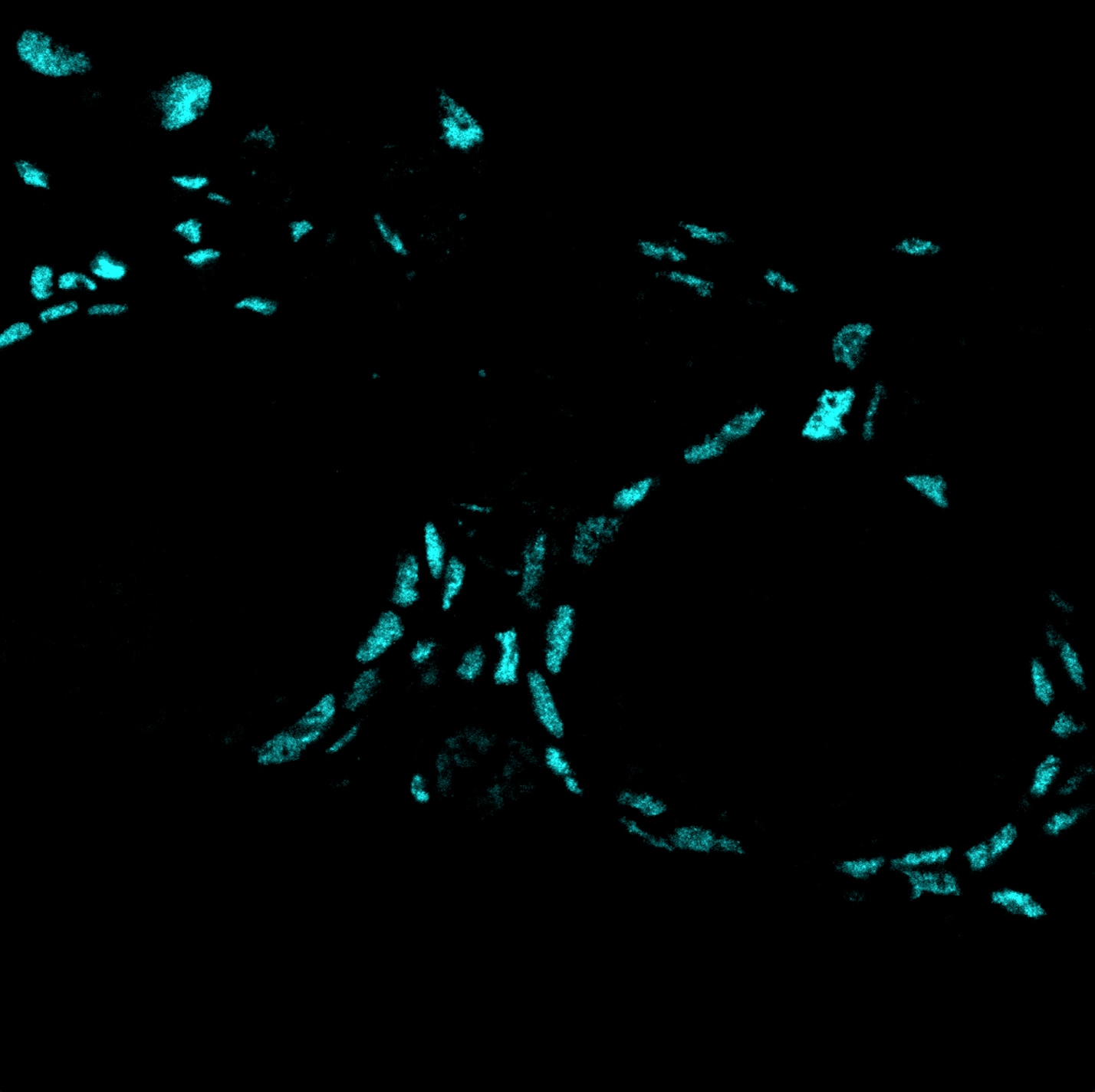
Application: ImmunohistochemistrySample Tested: Ovary (developing oocytes)Species: MouseVerified Customer | Posted 11/15/2024Worked on mouse organoid, concentration 1:200, IHC-P

-
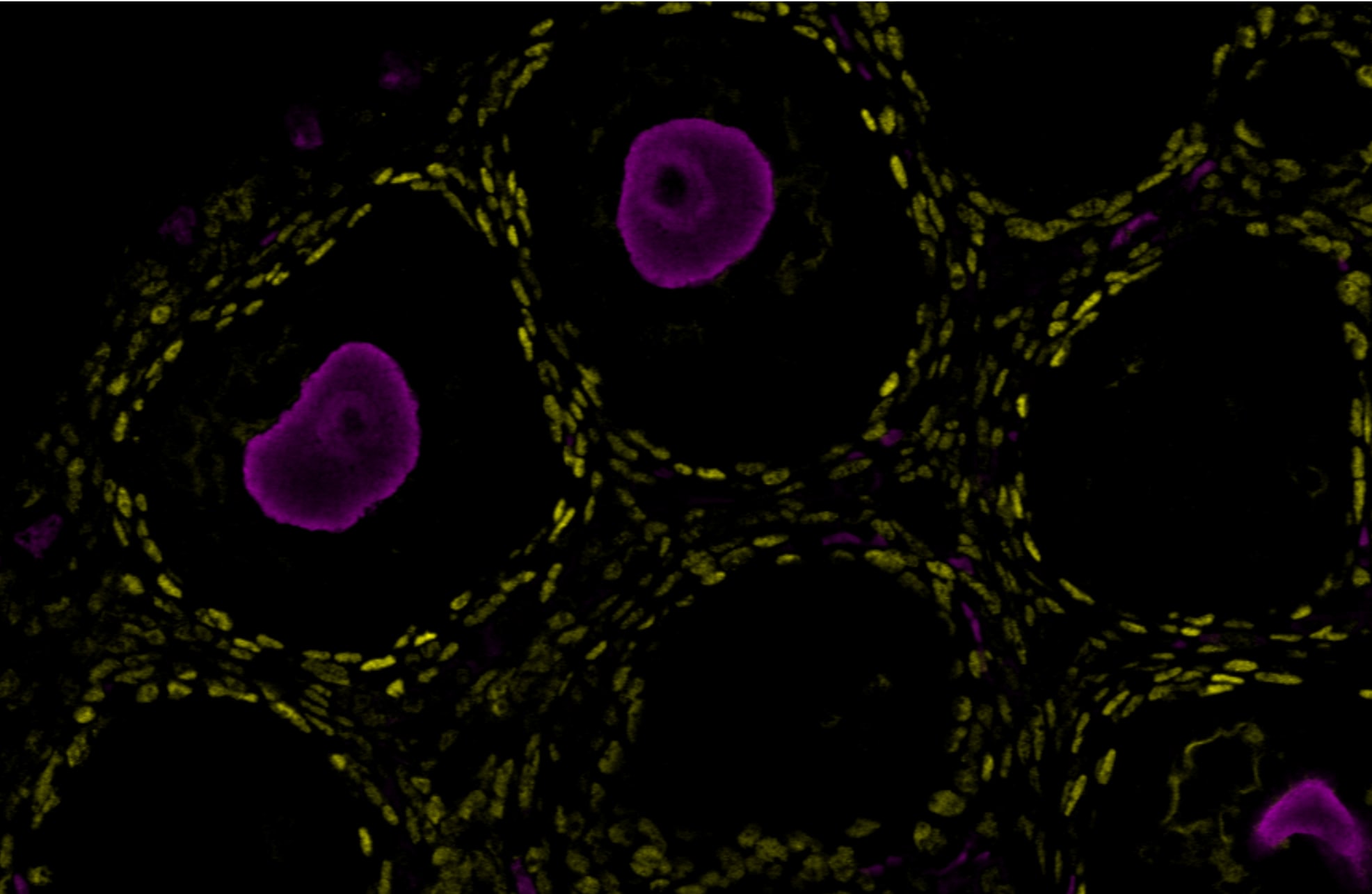
Application: Immunocytochemistry/ImmunofluorescenceSample Tested: Ovary tissueSpecies: MouseVerified Customer | Posted 11/08/2024The antibody was tested on a paraffin-embedded section of a mouse P14 ovary, with successful staining observed at a dilution of 1:200. Yellow (Nr2F2) Pink (Stella)
-
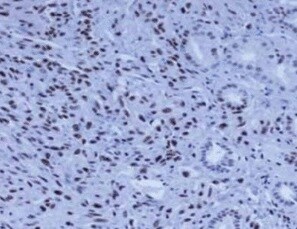
Application: ImmunohistochemistrySample Tested: Kidney angiomyolipomaSpecies: HumanVerified Customer | Posted 08/17/2021
-
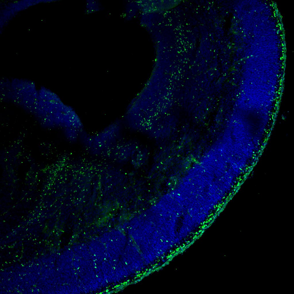
Application: Immunohistochemistry-FrozenSample Tested: E17 coronal brain sectionSpecies: MouseVerified Customer | Posted 05/13/2019E17 frozen mouse embryonic brain section was stained with Coup-TFII antibody (green) and dapi was used for contrast.Brain sections were antigen retrieved with 10mM sodium citrate (pH6) buffer and then blocked with 10% donkey serum in PBST. primary and secondary antibodies were prepared in the same blocking buffer.
-
Application: Immunohistochemistry-ParaffinSample Tested: See PMID 22253897Species: RatVerified Customer | Posted 02/25/2015
-
Application: ImmunofluorescenceSample Tested: See PMID 22615892Species: RatVerified Customer | Posted 02/25/2015
There are no reviews that match your criteria.
Protocols
Find general support by application which include: protocols, troubleshooting, illustrated assays, videos and webinars.
- Antigen Retrieval Protocol (PIER)
- Antigen Retrieval for Frozen Sections Protocol
- Appropriate Fixation of IHC/ICC Samples
- Cellular Response to Hypoxia Protocols
- Chromogenic IHC Staining of Formalin-Fixed Paraffin-Embedded (FFPE) Tissue Protocol
- Chromogenic Immunohistochemistry Staining of Frozen Tissue
- ClariTSA™ Fluorophore Kits
- Detection & Visualization of Antibody Binding
- Fluorescent IHC Staining of Frozen Tissue Protocol
- Graphic Protocol for Heat-induced Epitope Retrieval
- Graphic Protocol for the Preparation and Fluorescent IHC Staining of Frozen Tissue Sections
- Graphic Protocol for the Preparation and Fluorescent IHC Staining of Paraffin-embedded Tissue Sections
- Graphic Protocol for the Preparation of Gelatin-coated Slides for Histological Tissue Sections
- IHC Sample Preparation (Frozen sections vs Paraffin)
- Immunofluorescent IHC Staining of Formalin-Fixed Paraffin-Embedded (FFPE) Tissue Protocol
- Immunohistochemistry (IHC) and Immunocytochemistry (ICC) Protocols
- Immunohistochemistry Frozen Troubleshooting
- Immunohistochemistry Paraffin Troubleshooting
- Immunoprecipitation Protocol
- Preparing Samples for IHC/ICC Experiments
- Preventing Non-Specific Staining (Non-Specific Binding)
- Primary Antibody Selection & Optimization
- Protocol for Heat-Induced Epitope Retrieval (HIER)
- Protocol for Making a 4% Formaldehyde Solution in PBS
- Protocol for VisUCyte™ HRP Polymer Detection Reagent
- Protocol for the Preparation & Fixation of Cells on Coverslips
- Protocol for the Preparation and Chromogenic IHC Staining of Frozen Tissue Sections
- Protocol for the Preparation and Chromogenic IHC Staining of Frozen Tissue Sections - Graphic
- Protocol for the Preparation and Chromogenic IHC Staining of Paraffin-embedded Tissue Sections
- Protocol for the Preparation and Chromogenic IHC Staining of Paraffin-embedded Tissue Sections - Graphic
- Protocol for the Preparation and Fluorescent IHC Staining of Frozen Tissue Sections
- Protocol for the Preparation and Fluorescent IHC Staining of Paraffin-embedded Tissue Sections
- Protocol for the Preparation of Gelatin-coated Slides for Histological Tissue Sections
- R&D Systems Quality Control Western Blot Protocol
- TUNEL and Active Caspase-3 Detection by IHC/ICC Protocol
- The Importance of IHC/ICC Controls
- Troubleshooting Guide: Immunohistochemistry
- Troubleshooting Guide: Western Blot Figures
- Western Blot Conditions
- Western Blot Protocol
- Western Blot Protocol for Cell Lysates
- Western Blot Troubleshooting
- Western Blot Troubleshooting Guide
- View all Protocols, Troubleshooting, Illustrated assays and Webinars
Loading...