Granulocyte Macrophage Colony Stimulating Factor (GM-CSF) is a growth factor produced by a variety of lymphoid cells. It binds to receptor heterodimers consisting of a GM-CSF-specific alpha chain and the common beta chain that is shared by the high-affinity receptors for IL-3 and IL-5.

Key Product Details
Species Reactivity
Validated:
Human
Cited:
Human, Mouse
Applications
Validated:
Western Blot, Neutralization, Immunocytochemistry
Cited:
Immunohistochemistry, Immunohistochemistry-Paraffin, Immunohistochemistry-Frozen, Neutralization, Immunodepletion
Label
Unconjugated
Antibody Source
Monoclonal Mouse IgG1 Clone # 3209
Loading...
Product Specifications
Immunogen
E. coli-derived recombinant human GM-CSF
Ala18-Glu144
Accession # P04141
Ala18-Glu144
Accession # P04141
Specificity
Detects human GM-CSF in Western blots.
Clonality
Monoclonal
Host
Mouse
Isotype
IgG1
Endotoxin Level
<0.10 EU per 1 μg of the antibody by the LAL method.
Scientific Data Images for Human GM-CSF Antibody (3209)
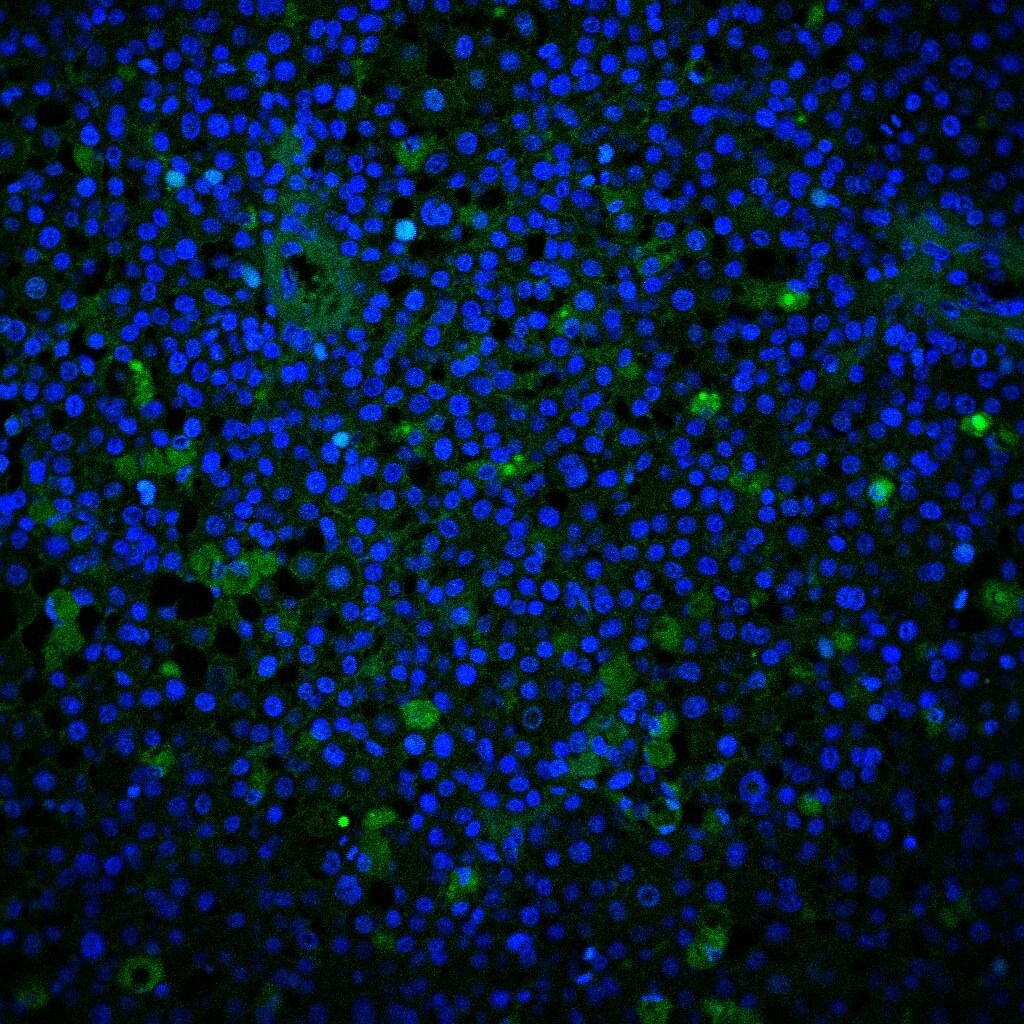
GMCSF in Human PBMCs.
Granulocyte Macrophage Colony Stimulating Factor (GM-CSF) was detected in human peripheral blood mononuclear cells (PBMCs) using Human GM-CSF Monoclonal Antibody (Catalog # MAB215) at 10 µg/mL for 3 hours at room temperature. Cells were stained using the NorthernLights™ 557-conjugated Anti-Mouse IgG Secondary Antibody (red; Catalog # NL007) and counterstained with DAPI (blue). View our protocol for Fluorescent ICC Staining of Non-adherent Cells.
Cell Proliferation Induced by GM‑CSF and Neutralization by Human GM‑CSF Antibody.
Recombinant Human GM-CSF (Catalog # 215-GM) stimulates proliferation in the TF-1 human erythroleukemic cell line in a dose-dependent manner (orange line) as measured by Resazurin (Catalog # AR002). Proliferation elicited by Recombinant Human GM-CSF (0.1 ng/mL) is neutralized (green line) by increasing concentrations of Human GM-CSF Monoclonal Antibody (Catalog # MAB215). The ND50 is typically 0.075-0.45 µg/mL.
Detection of GM-CSF by Flow Cytometry
TC-derived soluble factors promoted neutrophil survival. A. Neutrophils were cultured in a TC-CM or the control medium. At the indicated time points, live cells were evaluated by flow cytometry with FITC-conjugated annexin V and PI. Results were expressed as percentages of live cells (mean ± SEM of five independent experiments); ***p < 0.005; **p < 0.01; *p < 0.05. B. Representative flow cytometric panels of dot plots of PMNs cultured in a TC-CM or control medium and stained with FITC-conjugated annexin V and propidium iodide (PI) at 24 (upper panels) and 48 (lower panels) hours. C. The GM-CSF release by TPC1 and 8505c cells was evaluated by an ELISA in a TC-CM or in the control medium. Results were expressed as mean ± SEM of seven independent experiments; ****p < 0.001; ***p < 0.005. D-F. Neutrophil survival in a TPC1-derived (D-E) or 8505c-derived (F-G) conditioned medium was evaluated in the presence of an anti-GM-CSF blocking antibody or the relative isotype control (10 μg/ml). At 24 hours, live cells were stained with FITC-conjugated annexin V and PI and analyzed by flow cytometry. Figs E and G illustrate representative flow cytometric panels of one out of five independent experiments. The results were expressed as mean ± SEM of five independent experiments; **p < 0.01; *p < 0.05. Image collected and cropped by CiteAb from the following open publication (https://pubmed.ncbi.nlm.nih.gov/29953504), licensed under a CC-BY license. Not internally tested by R&D Systems.
Detection of GM-CSF by Immunohistochemistry
Knockdown of GM-CSF protein levels after siRNA application in cancer cells. HeLa/DLD-1 cells were transfected with control siRNA (1/1*, 2/2*) or GM-CSF siRNA (3/3*, 4/4*) and cultured in the absence or presence of 25 ng/mL HB-EGF. The numbers indicate the culture conditions and the corresponding supernatants (SN) used for ELISA or cell stimulation. (A) Blockade of GM-CSF production in cultures of HeLa/DLD-1 cells transfected with GM-CSF siRNA was confirmed by immunocytochemistry (2/2* vs. 4/4*) and ELISA (left side; 2/2* vs. 4/4*, p < 0.05). (B) SN from GM-CSF-silenced HeLa/DLD-1 did not induce HB-EGF expression in mononuclear phagocytes (Mø), as revealed by flow cytometry (2/2* vs. 4/4*) and ELISA (left side; 2/2* vs. 4/4*, p < 0.05). (C) Mø stimulated with SN from GM-CSF-silenced HeLa/DLD-1 cells released SN less effective at inducing GM-CSF in non-silenced cancer cells, as determined by ELISA (see Methods section; SN2 vs. SN4, p < 0.05). Representative pictures or the means ± SD out of 5 experiments are shown. Image collected and cropped by CiteAb from the following open publication (https://pubmed.ncbi.nlm.nih.gov/20946648), licensed under a CC-BY license. Not internally tested by R&D Systems.
Detection of GM-CSF by Flow Cytometry
TC-derived soluble factors promoted neutrophil survival. A. Neutrophils were cultured in a TC-CM or the control medium. At the indicated time points, live cells were evaluated by flow cytometry with FITC-conjugated annexin V and PI. Results were expressed as percentages of live cells (mean ± SEM of five independent experiments); ***p < 0.005; **p < 0.01; *p < 0.05. B. Representative flow cytometric panels of dot plots of PMNs cultured in a TC-CM or control medium and stained with FITC-conjugated annexin V and propidium iodide (PI) at 24 (upper panels) and 48 (lower panels) hours. C. The GM-CSF release by TPC1 and 8505c cells was evaluated by an ELISA in a TC-CM or in the control medium. Results were expressed as mean ± SEM of seven independent experiments; ****p < 0.001; ***p < 0.005. D-F. Neutrophil survival in a TPC1-derived (D-E) or 8505c-derived (F-G) conditioned medium was evaluated in the presence of an anti-GM-CSF blocking antibody or the relative isotype control (10 μg/ml). At 24 hours, live cells were stained with FITC-conjugated annexin V and PI and analyzed by flow cytometry. Figs E and G illustrate representative flow cytometric panels of one out of five independent experiments. The results were expressed as mean ± SEM of five independent experiments; **p < 0.01; *p < 0.05. Image collected and cropped by CiteAb from the following open publication (https://pubmed.ncbi.nlm.nih.gov/29953504), licensed under a CC-BY license. Not internally tested by R&D Systems.
Human GM-CSF ELISA Standard Curve
Recombinant Human GM‑CSF (Catalog # 215-GM) was serially diluted and captured by Mouse Anti-Human GM‑CSF Monoclonal Antibody (Catalog # MAB615) coated on a Clear Polystyrene Microplate (Catalog # DY990). Mouse Anti-Human GM‑CSF Monoclonal Antibody (Catalog # MAB215) was biotinylated and incubated with the protein captured on the plate. Detection of the standard curve was achieved by incubating Streptavidin-HRP (Catalog # DY998)Applications for Human GM-CSF Antibody (3209)
Application
Recommended Usage
Immunocytochemistry
8-25 µg/mL
Sample: Immersion fixed human peripheral blood mononuclear cells
Sample: Immersion fixed human peripheral blood mononuclear cells
Western Blot
1 µg/mL
Sample: Recombinant Human GM-CSF (Catalog # 215-GM)
under non-reducing conditions only
Sample: Recombinant Human GM-CSF (Catalog # 215-GM)
under non-reducing conditions only
Neutralization
Measured by its ability to neutralize GM‑CSF-induced proliferation in the TF‑1 human erythroleukemic cell line. Kitamura, T. et al. (1989) J. Cell Physiol. 140:323. The Neutralization Dose (ND50) is typically 0.075-0.45 µg/mL in the presence of 0.1 ng/mL Recombinant Human GM‑CSF.
Reviewed Applications
Read 2 reviews rated 4.5 using MAB215 in the following applications:
Formulation, Preparation, and Storage
Purification
Protein A or G purified from hybridoma culture supernatant
Reconstitution
Reconstitute at 0.5 mg/mL in sterile PBS. For liquid material, refer to CoA for concentration.
Loading...
Formulation
Lyophilized from a 0.2 μm filtered solution in PBS with Trehalose. *Small pack size (SP) is supplied either lyophilized or as a 0.2 µm filtered solution in PBS.
Shipping
Lyophilized product is shipped at ambient temperature. Liquid small pack size (-SP) is shipped with polar packs. Upon receipt, store immediately at the temperature recommended below.
Stability & Storage
Use a manual defrost freezer and avoid repeated freeze-thaw cycles.
- 12 months from date of receipt, -20 to -70 °C as supplied.
- 1 month, 2 to 8 °C under sterile conditions after reconstitution.
- 6 months, -20 to -70 °C under sterile conditions after reconstitution.
Calculators
Background: GM-CSF
Long Name
Granulocyte Macrophage Growth Factor
Alternate Names
CSF-2, CSF2, GMCSF, Molgramostim, Sargramostim
Entrez Gene IDs
Gene Symbol
CSF2
UniProt
Additional GM-CSF Products
Product Documents for Human GM-CSF Antibody (3209)
Certificate of Analysis
To download a Certificate of Analysis, please enter a lot or batch number in the search box below.
Note: Certificate of Analysis not available for kit components.
Product Specific Notices for Human GM-CSF Antibody (3209)
For research use only
Related Research Areas
Citations for Human GM-CSF Antibody (3209)
 Powered by Bioz
Customer Reviews for Human GM-CSF Antibody (3209) (2)
4.5 out of 5
2 Customer Ratings
Have you used Human GM-CSF Antibody (3209)?
Submit a review and receive an Amazon gift card!
$25/€18/£15/$25CAN/¥2500 Yen for a review with an image
$10/€7/£6/$10CAN/¥1110 Yen for a review without an image
Submit a review
Customer Images
Showing
1
-
2 of
2 reviews
Showing All
Filter By:
-
Application: Immunocytochemistry/ImmunofluorescenceSample Tested: Melanoma tissueSpecies: HumanVerified Customer | Posted 11/23/2020

-
Application: Western BlotSample Tested: Pancreatic cancer cellsSpecies: HumanVerified Customer | Posted 05/02/2018
There are no reviews that match your criteria.
Protocols
Find general support by application which include: protocols, troubleshooting, illustrated assays, videos and webinars.
- Appropriate Fixation of IHC/ICC Samples
- Cellular Response to Hypoxia Protocols
- ClariTSA™ Fluorophore Kits
- Detection & Visualization of Antibody Binding
- ICC Cell Smear Protocol for Suspension Cells
- ICC Immunocytochemistry Protocol Videos
- ICC for Adherent Cells
- Immunocytochemistry (ICC) Protocol
- Immunocytochemistry Troubleshooting
- Immunofluorescence of Organoids Embedded in Cultrex Basement Membrane Extract
- Immunohistochemistry (IHC) and Immunocytochemistry (ICC) Protocols
- Preparing Samples for IHC/ICC Experiments
- Preventing Non-Specific Staining (Non-Specific Binding)
- Primary Antibody Selection & Optimization
- Protocol for VisUCyte™ HRP Polymer Detection Reagent
- Protocol for the Fluorescent ICC Staining of Cell Smears - Graphic
- Protocol for the Fluorescent ICC Staining of Cultured Cells on Coverslips - Graphic
- Protocol for the Preparation and Fluorescent ICC Staining of Cells on Coverslips
- Protocol for the Preparation and Fluorescent ICC Staining of Non-adherent Cells
- Protocol for the Preparation and Fluorescent ICC Staining of Stem Cells on Coverslips
- Protocol for the Preparation of a Cell Smear for Non-adherent Cell ICC - Graphic
- R&D Systems Quality Control Western Blot Protocol
- TUNEL and Active Caspase-3 Detection by IHC/ICC Protocol
- The Importance of IHC/ICC Controls
- Troubleshooting Guide: Western Blot Figures
- Western Blot Conditions
- Western Blot Protocol
- Western Blot Protocol for Cell Lysates
- Western Blot Troubleshooting
- Western Blot Troubleshooting Guide
- View all Protocols, Troubleshooting, Illustrated assays and Webinars


