Human Lymphotoxin beta R/TNFRSF3 Antibody Summary
Ser28-Met227
Accession # P36941
Applications
Please Note: Optimal dilutions should be determined by each laboratory for each application. General Protocols are available in the Technical Information section on our website.
Scientific Data
 View Larger
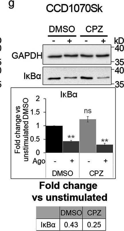
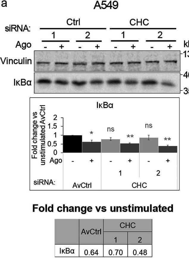
Detection of Lymphotoxin beta R/TNFRSF3 by Western Blot Blocking clathrin-dependent endocytosis enhances activation of canonical NF-kappa B signaling by LT beta R. A549 (a), HEK293T (b, d) and CCD1070Sk (c, d) cells were transfected with siRNAs targeting clathrin (CHC) (two oligonucleotides) along with control, non-targeting siRNAs (two oligonucleotides) or treated with chlorpromazine (CPZ, e-g) along with DMSO, and stimulated or not with Ago for 1 h. Lysates of cells were analyzed by Western blotting with antibodies against the indicated proteins. Graphs show densitometric analysis of abundance of I kappa B alpha, normalized to loading controls (GAPDH or vinculin). Values are presented as a fold change vs unstimulated non-targeting controls – averaged non-targeting controls (AvCtrl) or DMSO, set as 1. Data represent the means ± SEM, n = 3 (a, b, f, g), n = 4 (c, e); ns - P > 0.05; *P ≤ 0.05; **P ≤ 0.01; ***P ≤ 0.001 by one sample t test. Tables present the fold change of I kappa B alpha abundance in stimulated vs unstimulated cells (means, n ≥ 3). d HEK293T and CCD1070Sk cells were analyzed with respect to the efficiency of clathrin knock-down. Representative blots are shown. The blots of GAPDH shown in panels b and c are also shown in panel d Image collected and cropped by CiteAb from the following open publication (https://pubmed.ncbi.nlm.nih.gov/33148272), licensed under a CC-BY license. Not internally tested by R&D Systems.
 View Larger
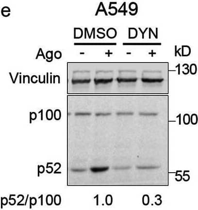
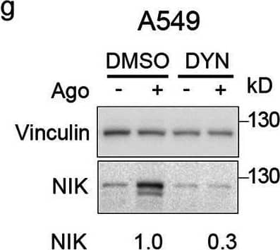
Detection of Lymphotoxin beta R/TNFRSF3 by Western Blot Clathrin and dynamin deficiency reduces activation of the non-canonical NF-kappa B pathway. a-c A549, HEK293T and CCD1070Sk cells were transfected with siRNAs targeting clathrin (CHC) (two oligonucleotides) along with control, non-targeting siRNAs (two oligonucleotides) and stimulated or not with Ago for 1 h. d, f A549 cells were transfected with siRNAs targeting dynamin-1/2 (three combinations of oligonucleotides targeting dynamin-1 and dynamin-2, see Methods) along with non-targeting control (Ctrl) siRNAs (two combinations of oligonucleotides, see Methods) and stimulated or not with Ago for 1 h. e, g A549 cells were treated with dynasore (DYN) or DMSO and stimulated or not with Ago for 1 h. Lysates of cells were analyzed by Western blotting with antibodies against the indicated proteins. Representative blots are shown. Values presented below blots represent the averaged p52/p100/loading control (a-e) or NIK/loading control ratio (f-g) from at least three experiments (normalized to the selected control, set as 1) in cells stimulated with Ago Image collected and cropped by CiteAb from the following open publication (https://pubmed.ncbi.nlm.nih.gov/33148272), licensed under a CC-BY license. Not internally tested by R&D Systems.
 View Larger
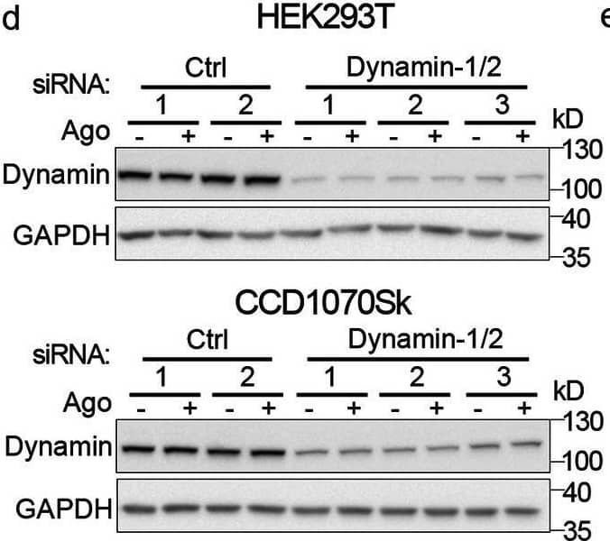
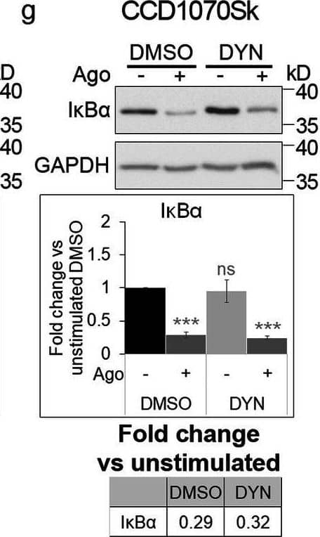
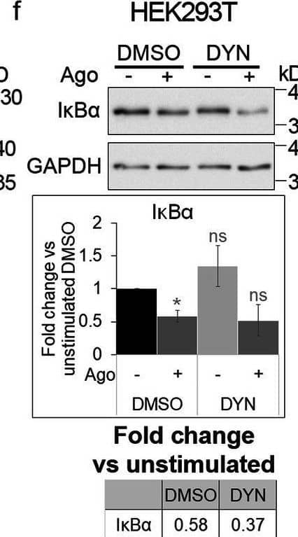
Detection of Lymphotoxin beta R/TNFRSF3 by Western Blot Blocking dynamin-dependent endocytosis enhances activation of canonical NF-kappa B signaling by LT beta R. A549 (a), HEK293T (b, d) and CCD1070Sk (c, d) cells were transfected with siRNAs targeting dynamin-1/2 (three combinations of oligonucleotides targeting dynamin-1 and dynamin-2, see Methods) along with non-targeting control (Ctrl) siRNAs (two combinations of oligonucleotides, see Methods) or treated with dynasore (DYN, e-g) along with DMSO, and stimulated or not with Ago for 1 h. Lysates of cells were analyzed by Western blotting with antibodies against the indicated proteins. Graphs show densitometric analysis of abundance of I kappa B alpha, normalized to loading controls (GAPDH or vinculin). Values are presented as a fold change vs unstimulated non-targeting controls – averaged non-targeting controls (AvCtrl) or DMSO, set as 1. Data represent the means ± SEM, n = 3 (b, c, f), n = 4 (a, e, g); ns - P > 0.05; *P ≤ 0.05; **P ≤ 0.01; ***P ≤ 0.001 by one sample t test. Tables present the fold change of I kappa B alpha abundance in stimulated vs unstimulated cells (means, n ≥ 3). d HEK293T and CCD1070Sk cells were analyzed with respect to the efficiency of dynamin knock-down. Representative blots are shown Image collected and cropped by CiteAb from the following open publication (https://pubmed.ncbi.nlm.nih.gov/33148272), licensed under a CC-BY license. Not internally tested by R&D Systems.
 View Larger
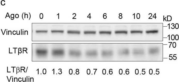
Detection of Lymphotoxin beta R/TNFRSF3 by Western Blot LT beta R is internalized and trafficked towards degradation upon ligand binding. A549 cells were stimulated with Ago for the indicated time periods and immunostained for LT beta R, EEA1 and LAMP1 (a) or trans- (TGN46) and cis-Golgi (GM130) (b). Insets show magnified views of boxed regions in the main images. Scale bars, 20 μm. Graphs represent the analysis of colocalization between LT beta R and EEA1 or LAMP1, and integral intensity of LT beta R (a) and colocalization between LT beta R and GM130 or TGN46 (b). Data represent the means ± SEM, n ≥ 5 (a), n = 3 (b); ns - P > 0.05; *P ≤ 0.05; **P ≤ 0.01; ***P ≤ 0.001 by one sample t test. c Lysates of A549 cells stimulated with Ago for different time periods were analyzed by Western blotting with antibodies against LT beta R and vinculin (used as a loading control). Representative blots are shown. Values below blots represent the averaged LT beta R/vinculin ratio (n = 5) in cells stimulated with Ago for the indicated time periods. Values are normalized to unstimulated control (time 0) set as 1.d Lysates of A549, HEK293T, CCD1070Sk and HeLa cells pretreated or not for 20 h (A549, HEK293T, CCD1070Sk) or 16 h (HeLa) with lysosomal degradation inhibitor, chloroquine (CQ), stimulated or not with Ago for the next 4 h were analyzed by Western blotting with antibodies against LT beta R and GAPDH (used as a loading control). Representative blots are shown. Table presents the fold change of LT beta R abundance in stimulated vs unstimulated cells (means, n ≥ 3) Image collected and cropped by CiteAb from the following open publication (https://pubmed.ncbi.nlm.nih.gov/33148272), licensed under a CC-BY license. Not internally tested by R&D Systems.
 View Larger
Detection of Lymphotoxin beta R/TNFRSF3 by Western Blot Clathrin and dynamin deficiency reduces activation of the non-canonical NF-kappa B pathway. a-c A549, HEK293T and CCD1070Sk cells were transfected with siRNAs targeting clathrin (CHC) (two oligonucleotides) along with control, non-targeting siRNAs (two oligonucleotides) and stimulated or not with Ago for 1 h. d, f A549 cells were transfected with siRNAs targeting dynamin-1/2 (three combinations of oligonucleotides targeting dynamin-1 and dynamin-2, see Methods) along with non-targeting control (Ctrl) siRNAs (two combinations of oligonucleotides, see Methods) and stimulated or not with Ago for 1 h. e, g A549 cells were treated with dynasore (DYN) or DMSO and stimulated or not with Ago for 1 h. Lysates of cells were analyzed by Western blotting with antibodies against the indicated proteins. Representative blots are shown. Values presented below blots represent the averaged p52/p100/loading control (a-e) or NIK/loading control ratio (f-g) from at least three experiments (normalized to the selected control, set as 1) in cells stimulated with Ago Image collected and cropped by CiteAb from the following open publication (https://pubmed.ncbi.nlm.nih.gov/33148272), licensed under a CC-BY license. Not internally tested by R&D Systems.
 View Larger
Detection of Lymphotoxin beta R/TNFRSF3 by Western Blot Blocking dynamin-dependent endocytosis enhances activation of canonical NF-kappa B signaling by LT beta R. A549 (a), HEK293T (b, d) and CCD1070Sk (c, d) cells were transfected with siRNAs targeting dynamin-1/2 (three combinations of oligonucleotides targeting dynamin-1 and dynamin-2, see Methods) along with non-targeting control (Ctrl) siRNAs (two combinations of oligonucleotides, see Methods) or treated with dynasore (DYN, e-g) along with DMSO, and stimulated or not with Ago for 1 h. Lysates of cells were analyzed by Western blotting with antibodies against the indicated proteins. Graphs show densitometric analysis of abundance of I kappa B alpha, normalized to loading controls (GAPDH or vinculin). Values are presented as a fold change vs unstimulated non-targeting controls – averaged non-targeting controls (AvCtrl) or DMSO, set as 1. Data represent the means ± SEM, n = 3 (b, c, f), n = 4 (a, e, g); ns - P > 0.05; *P ≤ 0.05; **P ≤ 0.01; ***P ≤ 0.001 by one sample t test. Tables present the fold change of I kappa B alpha abundance in stimulated vs unstimulated cells (means, n ≥ 3). d HEK293T and CCD1070Sk cells were analyzed with respect to the efficiency of dynamin knock-down. Representative blots are shown Image collected and cropped by CiteAb from the following open publication (https://pubmed.ncbi.nlm.nih.gov/33148272), licensed under a CC-BY license. Not internally tested by R&D Systems.
 View Larger
Detection of Lymphotoxin beta R/TNFRSF3 by Western Blot Blocking clathrin-dependent endocytosis enhances activation of canonical NF-kappa B signaling by LT beta R. A549 (a), HEK293T (b, d) and CCD1070Sk (c, d) cells were transfected with siRNAs targeting clathrin (CHC) (two oligonucleotides) along with control, non-targeting siRNAs (two oligonucleotides) or treated with chlorpromazine (CPZ, e-g) along with DMSO, and stimulated or not with Ago for 1 h. Lysates of cells were analyzed by Western blotting with antibodies against the indicated proteins. Graphs show densitometric analysis of abundance of I kappa B alpha, normalized to loading controls (GAPDH or vinculin). Values are presented as a fold change vs unstimulated non-targeting controls – averaged non-targeting controls (AvCtrl) or DMSO, set as 1. Data represent the means ± SEM, n = 3 (a, b, f, g), n = 4 (c, e); ns - P > 0.05; *P ≤ 0.05; **P ≤ 0.01; ***P ≤ 0.001 by one sample t test. Tables present the fold change of I kappa B alpha abundance in stimulated vs unstimulated cells (means, n ≥ 3). d HEK293T and CCD1070Sk cells were analyzed with respect to the efficiency of clathrin knock-down. Representative blots are shown. The blots of GAPDH shown in panels b and c are also shown in panel d Image collected and cropped by CiteAb from the following open publication (https://pubmed.ncbi.nlm.nih.gov/33148272), licensed under a CC-BY license. Not internally tested by R&D Systems.
 View Larger
Detection of Lymphotoxin beta R/TNFRSF3 by Western Blot Blocking dynamin-dependent endocytosis enhances activation of canonical NF-kappa B signaling by LT beta R. A549 (a), HEK293T (b, d) and CCD1070Sk (c, d) cells were transfected with siRNAs targeting dynamin-1/2 (three combinations of oligonucleotides targeting dynamin-1 and dynamin-2, see Methods) along with non-targeting control (Ctrl) siRNAs (two combinations of oligonucleotides, see Methods) or treated with dynasore (DYN, e-g) along with DMSO, and stimulated or not with Ago for 1 h. Lysates of cells were analyzed by Western blotting with antibodies against the indicated proteins. Graphs show densitometric analysis of abundance of I kappa B alpha, normalized to loading controls (GAPDH or vinculin). Values are presented as a fold change vs unstimulated non-targeting controls – averaged non-targeting controls (AvCtrl) or DMSO, set as 1. Data represent the means ± SEM, n = 3 (b, c, f), n = 4 (a, e, g); ns - P > 0.05; *P ≤ 0.05; **P ≤ 0.01; ***P ≤ 0.001 by one sample t test. Tables present the fold change of I kappa B alpha abundance in stimulated vs unstimulated cells (means, n ≥ 3). d HEK293T and CCD1070Sk cells were analyzed with respect to the efficiency of dynamin knock-down. Representative blots are shown Image collected and cropped by CiteAb from the following open publication (https://pubmed.ncbi.nlm.nih.gov/33148272), licensed under a CC-BY license. Not internally tested by R&D Systems.
 View Larger
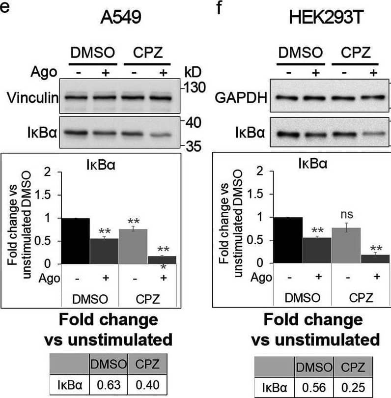
Detection of Lymphotoxin beta R/TNFRSF3 by Western Blot Blocking clathrin-dependent endocytosis enhances activation of canonical NF-kappa B signaling by LT beta R. A549 (a), HEK293T (b, d) and CCD1070Sk (c, d) cells were transfected with siRNAs targeting clathrin (CHC) (two oligonucleotides) along with control, non-targeting siRNAs (two oligonucleotides) or treated with chlorpromazine (CPZ, e-g) along with DMSO, and stimulated or not with Ago for 1 h. Lysates of cells were analyzed by Western blotting with antibodies against the indicated proteins. Graphs show densitometric analysis of abundance of I kappa B alpha, normalized to loading controls (GAPDH or vinculin). Values are presented as a fold change vs unstimulated non-targeting controls – averaged non-targeting controls (AvCtrl) or DMSO, set as 1. Data represent the means ± SEM, n = 3 (a, b, f, g), n = 4 (c, e); ns - P > 0.05; *P ≤ 0.05; **P ≤ 0.01; ***P ≤ 0.001 by one sample t test. Tables present the fold change of I kappa B alpha abundance in stimulated vs unstimulated cells (means, n ≥ 3). d HEK293T and CCD1070Sk cells were analyzed with respect to the efficiency of clathrin knock-down. Representative blots are shown. The blots of GAPDH shown in panels b and c are also shown in panel d Image collected and cropped by CiteAb from the following open publication (https://pubmed.ncbi.nlm.nih.gov/33148272), licensed under a CC-BY license. Not internally tested by R&D Systems.
 View Larger
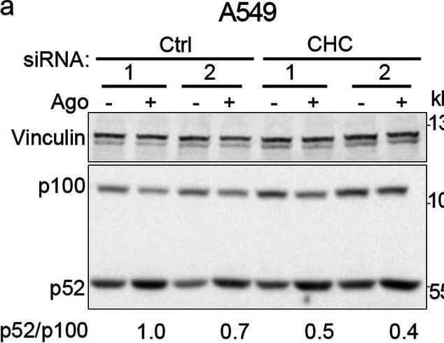
Detection of Lymphotoxin beta R/TNFRSF3 by Western Blot Clathrin and dynamin deficiency reduces activation of the non-canonical NF-kappa B pathway. a-c A549, HEK293T and CCD1070Sk cells were transfected with siRNAs targeting clathrin (CHC) (two oligonucleotides) along with control, non-targeting siRNAs (two oligonucleotides) and stimulated or not with Ago for 1 h. d, f A549 cells were transfected with siRNAs targeting dynamin-1/2 (three combinations of oligonucleotides targeting dynamin-1 and dynamin-2, see Methods) along with non-targeting control (Ctrl) siRNAs (two combinations of oligonucleotides, see Methods) and stimulated or not with Ago for 1 h. e, g A549 cells were treated with dynasore (DYN) or DMSO and stimulated or not with Ago for 1 h. Lysates of cells were analyzed by Western blotting with antibodies against the indicated proteins. Representative blots are shown. Values presented below blots represent the averaged p52/p100/loading control (a-e) or NIK/loading control ratio (f-g) from at least three experiments (normalized to the selected control, set as 1) in cells stimulated with Ago Image collected and cropped by CiteAb from the following open publication (https://pubmed.ncbi.nlm.nih.gov/33148272), licensed under a CC-BY license. Not internally tested by R&D Systems.
 View Larger
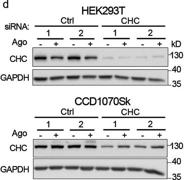
Detection of Lymphotoxin beta R/TNFRSF3 by Western Blot Blocking clathrin-dependent endocytosis enhances activation of canonical NF-kappa B signaling by LT beta R. A549 (a), HEK293T (b, d) and CCD1070Sk (c, d) cells were transfected with siRNAs targeting clathrin (CHC) (two oligonucleotides) along with control, non-targeting siRNAs (two oligonucleotides) or treated with chlorpromazine (CPZ, e-g) along with DMSO, and stimulated or not with Ago for 1 h. Lysates of cells were analyzed by Western blotting with antibodies against the indicated proteins. Graphs show densitometric analysis of abundance of I kappa B alpha, normalized to loading controls (GAPDH or vinculin). Values are presented as a fold change vs unstimulated non-targeting controls – averaged non-targeting controls (AvCtrl) or DMSO, set as 1. Data represent the means ± SEM, n = 3 (a, b, f, g), n = 4 (c, e); ns - P > 0.05; *P ≤ 0.05; **P ≤ 0.01; ***P ≤ 0.001 by one sample t test. Tables present the fold change of I kappa B alpha abundance in stimulated vs unstimulated cells (means, n ≥ 3). d HEK293T and CCD1070Sk cells were analyzed with respect to the efficiency of clathrin knock-down. Representative blots are shown. The blots of GAPDH shown in panels b and c are also shown in panel d Image collected and cropped by CiteAb from the following open publication (https://pubmed.ncbi.nlm.nih.gov/33148272), licensed under a CC-BY license. Not internally tested by R&D Systems.
 View Larger
Detection of Lymphotoxin beta R/TNFRSF3 by Western Blot LT beta R is internalized and trafficked towards degradation upon ligand binding. A549 cells were stimulated with Ago for the indicated time periods and immunostained for LT beta R, EEA1 and LAMP1 (a) or trans- (TGN46) and cis-Golgi (GM130) (b). Insets show magnified views of boxed regions in the main images. Scale bars, 20 μm. Graphs represent the analysis of colocalization between LT beta R and EEA1 or LAMP1, and integral intensity of LT beta R (a) and colocalization between LT beta R and GM130 or TGN46 (b). Data represent the means ± SEM, n ≥ 5 (a), n = 3 (b); ns - P > 0.05; *P ≤ 0.05; **P ≤ 0.01; ***P ≤ 0.001 by one sample t test. c Lysates of A549 cells stimulated with Ago for different time periods were analyzed by Western blotting with antibodies against LT beta R and vinculin (used as a loading control). Representative blots are shown. Values below blots represent the averaged LT beta R/vinculin ratio (n = 5) in cells stimulated with Ago for the indicated time periods. Values are normalized to unstimulated control (time 0) set as 1.d Lysates of A549, HEK293T, CCD1070Sk and HeLa cells pretreated or not for 20 h (A549, HEK293T, CCD1070Sk) or 16 h (HeLa) with lysosomal degradation inhibitor, chloroquine (CQ), stimulated or not with Ago for the next 4 h were analyzed by Western blotting with antibodies against LT beta R and GAPDH (used as a loading control). Representative blots are shown. Table presents the fold change of LT beta R abundance in stimulated vs unstimulated cells (means, n ≥ 3) Image collected and cropped by CiteAb from the following open publication (https://pubmed.ncbi.nlm.nih.gov/33148272), licensed under a CC-BY license. Not internally tested by R&D Systems.
 View Larger
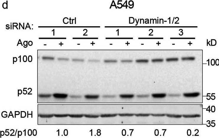
Detection of Lymphotoxin beta R/TNFRSF3 by Western Blot Clathrin and dynamin deficiency reduces activation of the non-canonical NF-kappa B pathway. a-c A549, HEK293T and CCD1070Sk cells were transfected with siRNAs targeting clathrin (CHC) (two oligonucleotides) along with control, non-targeting siRNAs (two oligonucleotides) and stimulated or not with Ago for 1 h. d, f A549 cells were transfected with siRNAs targeting dynamin-1/2 (three combinations of oligonucleotides targeting dynamin-1 and dynamin-2, see Methods) along with non-targeting control (Ctrl) siRNAs (two combinations of oligonucleotides, see Methods) and stimulated or not with Ago for 1 h. e, g A549 cells were treated with dynasore (DYN) or DMSO and stimulated or not with Ago for 1 h. Lysates of cells were analyzed by Western blotting with antibodies against the indicated proteins. Representative blots are shown. Values presented below blots represent the averaged p52/p100/loading control (a-e) or NIK/loading control ratio (f-g) from at least three experiments (normalized to the selected control, set as 1) in cells stimulated with Ago Image collected and cropped by CiteAb from the following open publication (https://pubmed.ncbi.nlm.nih.gov/33148272), licensed under a CC-BY license. Not internally tested by R&D Systems.
 View Larger
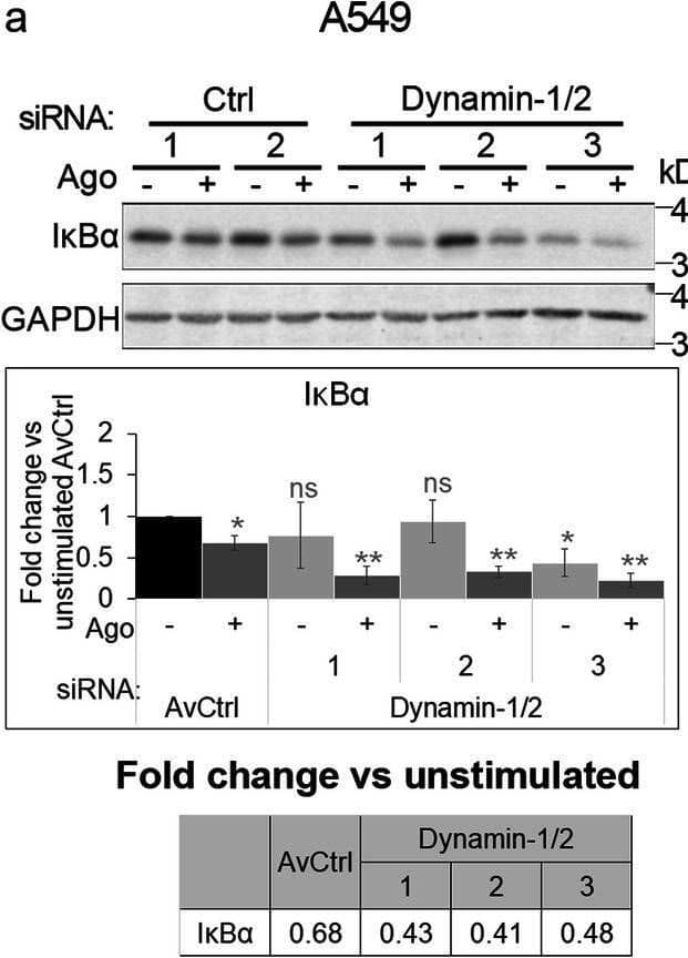
Detection of Lymphotoxin beta R/TNFRSF3 by Western Blot Blocking dynamin-dependent endocytosis enhances activation of canonical NF-kappa B signaling by LT beta R. A549 (a), HEK293T (b, d) and CCD1070Sk (c, d) cells were transfected with siRNAs targeting dynamin-1/2 (three combinations of oligonucleotides targeting dynamin-1 and dynamin-2, see Methods) along with non-targeting control (Ctrl) siRNAs (two combinations of oligonucleotides, see Methods) or treated with dynasore (DYN, e-g) along with DMSO, and stimulated or not with Ago for 1 h. Lysates of cells were analyzed by Western blotting with antibodies against the indicated proteins. Graphs show densitometric analysis of abundance of I kappa B alpha, normalized to loading controls (GAPDH or vinculin). Values are presented as a fold change vs unstimulated non-targeting controls – averaged non-targeting controls (AvCtrl) or DMSO, set as 1. Data represent the means ± SEM, n = 3 (b, c, f), n = 4 (a, e, g); ns - P > 0.05; *P ≤ 0.05; **P ≤ 0.01; ***P ≤ 0.001 by one sample t test. Tables present the fold change of I kappa B alpha abundance in stimulated vs unstimulated cells (means, n ≥ 3). d HEK293T and CCD1070Sk cells were analyzed with respect to the efficiency of dynamin knock-down. Representative blots are shown Image collected and cropped by CiteAb from the following open publication (https://pubmed.ncbi.nlm.nih.gov/33148272), licensed under a CC-BY license. Not internally tested by R&D Systems.
 View Larger
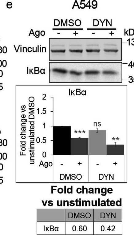
Detection of Lymphotoxin beta R/TNFRSF3 by Western Blot Blocking dynamin-dependent endocytosis enhances activation of canonical NF-kappa B signaling by LT beta R. A549 (a), HEK293T (b, d) and CCD1070Sk (c, d) cells were transfected with siRNAs targeting dynamin-1/2 (three combinations of oligonucleotides targeting dynamin-1 and dynamin-2, see Methods) along with non-targeting control (Ctrl) siRNAs (two combinations of oligonucleotides, see Methods) or treated with dynasore (DYN, e-g) along with DMSO, and stimulated or not with Ago for 1 h. Lysates of cells were analyzed by Western blotting with antibodies against the indicated proteins. Graphs show densitometric analysis of abundance of I kappa B alpha, normalized to loading controls (GAPDH or vinculin). Values are presented as a fold change vs unstimulated non-targeting controls – averaged non-targeting controls (AvCtrl) or DMSO, set as 1. Data represent the means ± SEM, n = 3 (b, c, f), n = 4 (a, e, g); ns - P > 0.05; *P ≤ 0.05; **P ≤ 0.01; ***P ≤ 0.001 by one sample t test. Tables present the fold change of I kappa B alpha abundance in stimulated vs unstimulated cells (means, n ≥ 3). d HEK293T and CCD1070Sk cells were analyzed with respect to the efficiency of dynamin knock-down. Representative blots are shown Image collected and cropped by CiteAb from the following open publication (https://pubmed.ncbi.nlm.nih.gov/33148272), licensed under a CC-BY license. Not internally tested by R&D Systems.
Reconstitution Calculator
Preparation and Storage
- 12 months from date of receipt, -20 to -70 °C as supplied.
- 1 month, 2 to 8 °C under sterile conditions after reconstitution.
- 6 months, -20 to -70 °C under sterile conditions after reconstitution.
Background: Lymphotoxin beta R/TNFRSF3
Lymphotoxin beta receptor (LT beta R), also known as TNF RIII and TNF R-related protein (TNF Rrp), was originally identified as a transcribed sequence on human chromosome 12p with homology to the TNF receptor superfamily. In the new TNF nomenclature, LT beta R is referred to as TNFRSF3. Human LT beta R cDNA encodes a 435 amino acid (aa) residue type I membrane protein with a putative 30 aa residue signal peptide, a 193 aa residue extracellular domain and a 171 aa residue cytoplasmic domain. The extracellular domain of LT beta R contains four cysteine-rich motif characteristic of the TNF receptor superfamily. The cytoplasmic region of LT beta R share little sequence similarity with other TNF receptor family members, suggesting that different signaling mechanisms may be utilized. LT beta R is expressed in a variety of tissues including visceral and lymphoid tissues. LT beta R is also expressed by cell lines of monocytic, epithelial, and fibroblastic origins but not by T and B lymphocytes. The human and mouse LT beta R share 76% aa sequence homology. The TNF family ligands that have been shown to bind and activate LT beta R include LIGHT (also a ligand for HVEM) and the heterotrimeric lymphotoxin LT alpha 1/ beta 2 or LT alpha 2/ beta 1. Depending on the cell type, activation of LT beta R has been shown to induce NF kappa B activation, chemokine production, growth arrest, and apoptosis. In vivo, LT beta R has been shown to play a critical role in controlling cellular immune functions and lymphoid organogenesis.
- Zhai, Y. et al. (1998) J. Clin. Invest. 102:1142.
- Rennert, P.D. et al. (1998) Immunity 9:71.
- Degli-Esposti, M.A. et al. (1997) J. Immunol 158:1756.
- Mackay, F. et al. (1996) J. Biol. Chem. 271:8618.
- Crowe, P.D. et al. (1994) Science 264:707.
Product Datasheets
Citations for Human Lymphotoxin beta R/TNFRSF3 Antibody
R&D Systems personnel manually curate a database that contains references using R&D Systems products. The data collected includes not only links to publications in PubMed, but also provides information about sample types, species, and experimental conditions.
10
Citations: Showing 1 - 10
Filter your results:
Filter by:
-
Retinoic Acid and Lymphotoxin Signaling Promote Differentiation of Human Intestinal M Cells
Authors: Siyuan Ding, Yanhua Song, Kevin F. Brulois, Junliang Pan, Julia Y. Co, Lili Ren et al.
Gastroenterology
-
Clathrin- and dynamin-dependent endocytosis limits canonical NF-&kappaB signaling triggered by lymphotoxin &beta receptor
Authors: M Maksymowic, M Mi?czy?ska, M Banach-Or?
Cell Commun Signal, 2020-11-04;18(1):176.
Species: Human
Sample Types: Cell Lysates
Applications: Western Blot -
TNF-alpha blockade induces IL-10 expression in human CD4+ T cells.
Authors: Evans H, Roostalu U, Walter G, Gullick N, Frederiksen K, Roberts C, Sumner J, Baeten D, Gerwien J, Cope A, Geissmann F, Kirkham B, Taams L
Nat Commun, 2014-01-01;5(0):3199.
Species: Human
Sample Types: Whole Cells
Applications: Neutralization -
Mitogenic signalling and the p16INK4a-Rb pathway cooperate to enforce irreversible cellular senescence.
Authors: Takahashi A, Ohtani N, Yamakoshi K, Iida S, Tahara H, Nakayama K, Nakayama KI, Ide T, Saya H, Hara E
Nat. Cell Biol., 2006-10-08;8(11):1291-7.
Species: Human
Sample Types: Cell Lysates
Applications: Western Blot -
Hepatitis C virus NS5A-regulated gene expression and signaling revealed via microarray and comparative promoter analyses.
Authors: Girard S, Vossman E, Misek DE, Podevin P, Hanash S, Brechot C, Beretta L
Hepatology, 2004-09-01;40(3):708-18.
Species: Human
Sample Types: Cell Lysates
Applications: Western Blot -
LIGHT, a member of the tumor necrosis factor ligand superfamily, prevents tumor necrosis factor-alpha-mediated human primary hepatocyte apoptosis, but not Fas-mediated apoptosis.
Authors: Hikichi Y, Tsuji I, Shintani Y
J. Biol. Chem., 2002-10-18;277(51):50054-61.
Species: Human
Sample Types: Whole Cells
Applications: Flow Cytometry -
Cholesterol restricts lymphotoxin beta receptor-triggered NF-kappa B signaling
Authors: Magdalena Banach-Orłowska, Renata Wyszyńska, Beata Pyrzyńska, Małgorzata Maksymowicz, Jakub Gołąb, Marta Miączyńska
Cell Communication and Signaling
-
The topology of lymphotoxin ? receptor accumulated upon endolysosomal dysfunction dictates the NF-?B signaling outcome
Authors: M Banach-Or?, K Jastrz?bsk, J Cendrowski, M Maksymowic, K Wojciechow, M Korosty?sk, D Moreau, J Gruenberg, M Miaczynska
J. Cell. Sci., 2018-11-21;0(0):.
-
Induction of the Alternative NF-kappa B Pathway by Lymphotoxin alpha beta (LT alpha beta ) Relies on Internalization of LT beta Receptor
Authors: Corinne Ganeff, Caroline Remouchamps, Layla Boutaffala, Cécile Benezech, Géraldine Galopin, Sarah Vandepaer et al.
Molecular and Cellular Biology
-
Non-canonical NF-kappa B signalling and ETS1/2 cooperatively drive C250T mutant TERT promoter activation
Authors: Yinghui Li, Qi-Ling Zhou, Wenjie Sun, Prashant Chandrasekharan, Hui Shan Cheng, Zhe Ying et al.
Nature Cell Biology
FAQs
-
Is the bioassay for Catalog # AF629 indicative of agonistic or antagonistic function of the antibody?
The assay is indicative of agonistic function.
Reviews for Human Lymphotoxin beta R/TNFRSF3 Antibody
There are currently no reviews for this product. Be the first to review Human Lymphotoxin beta R/TNFRSF3 Antibody and earn rewards!
Have you used Human Lymphotoxin beta R/TNFRSF3 Antibody?
Submit a review and receive an Amazon gift card.
$25/€18/£15/$25CAN/¥75 Yuan/¥2500 Yen for a review with an image
$10/€7/£6/$10 CAD/¥70 Yuan/¥1110 Yen for a review without an image

