
Neurofascin 155 (NF155) is a type I transmembrane glycoprotein that belongs to the L1CAM family of cell adhesion proteins (1, 2). The rat NF155 cDNA encodes a 1240 amino acid (aa) precursor that contains a 24 aa signal sequence, a 1086 aa extracellular domain (ECD), a 21 aa transmembrane segment, and a 109 aa cytoplasmic domain. The ECD consists of six Ig-like domains and four fibronectin type III repeats, the second of which has an RGD motif. A splice variant of Neurofascin, NF186, lacks the RGD-containing fibronectin type III domain but instead has a mucin-like domain and an additional non-RGD fibronectin type III domain (3). Within shared regions of the ECD, rat NF155 shares 45% and 39% aa sequence identity with rat Nr-CAM and L1CAM, respectively, and 98% aa sequence identity with human and mouse NF155. NF155 is transiently expressed by oligodendrocytes at the onset of axon myelination, whereas NF186 is neuronally expressed in nodes of Ranvier (4‑6). Clustering of NF155 in paranodal oligodengroglia lipid raft domains is stabilized by dimerization of its cytoplasmic domains and association with intracellular ankyrin (6‑9). NF155 interacts with axonal contactin and plays a role in node of Ranvier formation and the establishment of saltatory conduction (5, 9‑12). The ECD of NF155 is cleaved from oligodengroglia membranes by metalloproteases, a process which is required for NF155 transport from the glial cell body to the axoglial junction (13). In addition to distinct expression patterns, Neurofascin isoforms have different functional properties. NF155 promotes neuronal adhesion and neurite outgrowth, whereas NF186 inhibits neuronal adhesion (4, 7, 13).
Neurofascin Antibody (727030)
R&D Systems | Catalog # MAB3235

Scientific images may be resized, cropped, or adjusted for brightness or contrast for presentation purposes. These updates are limited to presentation and do not affect the underlying scientific interpretation of the data. Where available, additional source data may be provided upon request.
Key Product Details
Species Reactivity
Validated:
Cited:
Applications
Validated:
Cited:
Label
Antibody Source
Product Specifications
Immunogen
Ile25-Ala1031
Accession # NP_446361
Specificity
Clonality
Host
Isotype
Scientific Data Images for Neurofascin Antibody (727030)
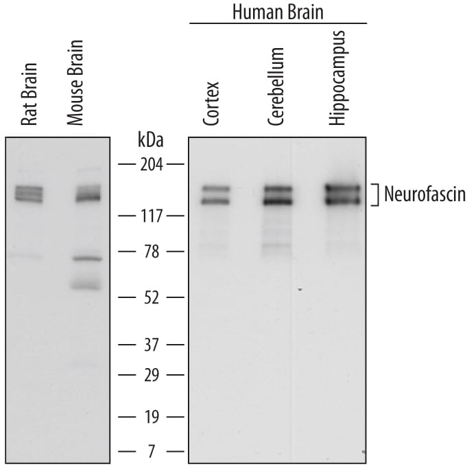
Detection of Human, Mouse, and Rat Neurofascin by Western Blot.
Western blot shows lysates of rat brain tissue, mouse brain tissue, human brain (cortex) tissue, human brain (cerebellum) tissue, and human brain (hippocampus) tissue. PVDF membrane was probed with 0.5 µg/mL of Mouse Anti-Human/Mouse/Rat Neurofascin Monoclonal Antibody (Catalog # MAB3235) followed by HRP-conjugated Anti-Mouse IgG Secondary Antibody (Catalog # HAF007). Specific bands were detected for Neurofascin at approximately 140 to 155 kDa (as indicated). This experiment was conducted under reducing conditions and using Immunoblot Buffer Group 1.Applications for Neurofascin Antibody (727030)
Western Blot
Sample: Rat brain tissue, mouse brain tissue, human brain (cortex) tissue, human brain (cerebellum) tissue, and human brain (hippocampus) tissue
Formulation, Preparation, and Storage
Purification
Reconstitution
Sterile PBS to a final concentration of 0.5 mg/mL. For liquid material, refer to CoA for concentration.
Formulation
Shipping
Stability & Storage
- 12 months from date of receipt, -20 to -70 °C as supplied.
- 1 month, 2 to 8 °C under sterile conditions after reconstitution.
- 6 months, -20 to -70 °C under sterile conditions after reconstitution.
Calculators
Background: Neurofascin
References
- Sherman, D.L. and P.J. Brophy (2005) Nat. Rev. Neurosci. 6:683.
- Coman, I. et al. (2005) J. Neurol. Sci. 233:67.
- Volkmer, H. et al. (1992) J. Cell Biol. 118:149.
- Koticha, D. et al. (2005) Mol. Cell. Neurosci. 30:137.
- Collinson, J.M. et al. (1998) Glia 23:11.
- Schafer, D.P. et al. (2004) J. Neurosci. 24:3176.
- Maier, O. et al. (2005) Mol. Cell. Neurosci. 28:390.
- Zhang, X. et al. (1998) J. Biol. Chem. 273:30785.
- Sherman, D.L. et al. (2005) Neuron 48:737.
- Gollan, L. et al. (2003) J. Cell Biol. 163:1213.
- Charles, P. et al. (2002) Curr. Biol. 12:217.
- Volkmer, H. et al. (1996) J. Cell Biol. 135:1059.
- Maier, O. et al. (2006) Exp. Cell Res. 312:500.
Alternate Names
Gene Symbol
UniProt
Additional Neurofascin Products
Product Documents for Neurofascin Antibody (727030)
Certificate of Analysis
To download a Certificate of Analysis, please enter a lot or batch number in the search box below.
Note: Certificate of Analysis not available for kit components.
Product Specific Notices for Neurofascin Antibody (727030)
For research use only
Related Research Areas
Citations for Neurofascin Antibody (727030)
 Powered by Bioz
Customer Reviews for Neurofascin Antibody (727030)
There are currently no reviews for this product. Be the first to review Neurofascin Antibody (727030) and earn rewards!
Have you used Neurofascin Antibody (727030)?
Submit a review and receive an Amazon gift card!
$25/€18/£15/$25CAN/¥2500 for a review with an image
$10/€7/£6/$10CAN/¥1110 for a review without an image
Submit a review
Protocols
Find general support by application which include: protocols, troubleshooting, illustrated assays, videos and webinars.
- Cellular Response to Hypoxia Protocols
- R&D Systems Quality Control Western Blot Protocol
- Troubleshooting Guide: Western Blot Figures
- Western Blot Conditions
- Western Blot Protocol
- Western Blot Protocol for Cell Lysates
- Western Blot Troubleshooting
- Western Blot Troubleshooting Guide
- View all Protocols, Troubleshooting, Illustrated assays and Webinars