Placenta growth factor (PlGF) is a member of the vascular endothelial growth factor (VEGF) family of growth factors. As a result of alternative splicing, at least two PlGF mRNAs encoding monomeric PlGF precursors containing 149 and 170 amino acid residues have been described. The expression of PlGF is not widespread but has been detected in human umbilical vein endothelial cells, placenta, choriocarcinoma cell lines, and in renal cell carcinoma associated with angiogenesis. The PlGF proteins bind with high-affinity to VEGF R1/Flt-1 but not to VEGF R2/Flk-1.
Human PlGF Antibody (37203)
R&D Systems | Catalog # MAB264

Key Product Details
Validated by
Biological Validation
Species Reactivity
Validated:
Human
Cited:
Human, Mouse
Applications
Validated:
ELISA Capture (Matched Antibody Pair)
Cited:
Immunohistochemistry, Neutralization, In vivo assay
Label
Unconjugated
Antibody Source
Monoclonal Mouse IgG1 Clone # 37203
Loading...
Product Specifications
Immunogen
E. coli-derived recombinant human PlGF
Ala21-Arg149
Accession # P49763
Ala21-Arg149
Accession # P49763
Specificity
Detects human PlGF in ELISAs. In ELISAs, this antibody does not cross-react with recombinant human (rh) VEGF, rhPDGF‑AA, rhPDGF‑AB, or rhPDGF‑BB.
Clonality
Monoclonal
Host
Mouse
Isotype
IgG1
Scientific Data Images for Human PlGF Antibody (37203)
Detection of Human PLGF by Block/Neutralize
Anti-PlGF neutralising antibody blocks DMOG-induced inhibition of ECFC tube formation. ECFCs were stained with calcein (Invitrogen) and grown on Matrigel®. Cells were treated with conditioned medium harvested from ECFCs treated with a DMSO (vehicle) plus isotype control IgG, b DMOG plus isotype control IgG, c DMSO plus anti-PlGF neutralising antibody or d DMOG plus anti-PlGF neutralising antibody. e At 48 h, total tube area (μm2) was quantified using NIS Elements software (Nikon). Data plotted as mean ± SD and are representative of three independent experiments. **p < 0.01; ***p < 0.001. Scale bars: 200 μm (n = 3). DMOG dimethyloxalylglycine, PlGF placental growth factor Image collected and cropped by CiteAb from the following publication (https://pubmed.ncbi.nlm.nih.gov/27899144), licensed under a CC-BY license. Not internally tested by R&D Systems.
Human PlGF ELISA Standard Curve
Recombinant Human PlGF (Catalog # 264-PGB) was serially diluted and captured by Mouse Anti-Human PlGF Monoclonal Antibody (Catalog # MAB264) coated on a Clear Polystyrene Microplate (Catalog # DY990). Goat Anti-Human PlGF Antigen Affinity-purified Polyclonal Antibody (Catalog # AF-264-PB) was biotinylated and incubated with the protein captured on the plate. Detection of the standard curve was achieved by incubating Streptavidin-HRP (Catalog # DY998)Applications for Human PlGF Antibody (37203)
Human PlGF Sandwich Immunoassay
Please Note: Optimal dilutions of this antibody should be experimentally determined.
Reviewed Applications
Read 2 reviews rated 5 using MAB264 in the following applications:
Formulation, Preparation, and Storage
Purification
Protein A or G purified from hybridoma culture supernatant
Reconstitution
Reconstitute at 0.5 mg/mL in sterile PBS. For liquid material, refer to CoA for concentration.
Loading...
Formulation
Lyophilized from a 0.2 μm filtered solution in PBS with Trehalose. *Small pack size (SP) is supplied either lyophilized or as a 0.2 µm filtered solution in PBS.
Shipping
Lyophilized product is shipped at ambient temperature. Liquid small pack size (-SP) is shipped with polar packs. Upon receipt, store immediately at the temperature recommended below.
Stability & Storage
Use a manual defrost freezer and avoid repeated freeze-thaw cycles.
- 12 months from date of receipt, -20 to -70 °C as supplied.
- 1 month, 2 to 8 °C under sterile conditions after reconstitution.
- 6 months, -20 to -70 °C under sterile conditions after reconstitution.
Calculators
Background: PlGF
Long Name
Placenta Growth Factor
Alternate Names
PGF, PGFL
Gene Symbol
PGF
UniProt
Additional PlGF Products
Product Documents for Human PlGF Antibody (37203)
Certificate of Analysis
To download a Certificate of Analysis, please enter a lot or batch number in the search box below.
Note: Certificate of Analysis not available for kit components.
Product Specific Notices for Human PlGF Antibody (37203)
For research use only
Related Research Areas
Citations for Human PlGF Antibody (37203)
 Powered by Bioz
Customer Reviews for Human PlGF Antibody (37203) (2)
5 out of 5
2 Customer Ratings
Have you used Human PlGF Antibody (37203)?
Submit a review and receive an Amazon gift card!
$25/€18/£15/$25CAN/¥2500 Yen for a review with an image
$10/€7/£6/$10CAN/¥1110 Yen for a review without an image
Submit a review
Customer Images
Showing
1
-
2 of
2 reviews
Showing All
Filter By:
-
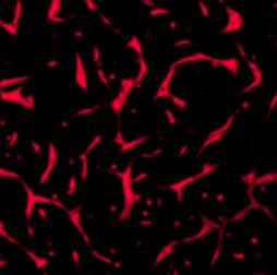
Application: Immunocytochemistry/ImmunofluorescenceSample Tested: Endothelial colony-forming cellsSpecies: MouseVerified Customer | Posted 02/17/2021Cells were fixed in 4% paraformaldehyde for 20 min at room temperature. Following PBS washes, cells were permeabilised with 0.1% Triton X-100 for 10 min at RT and then blocked in 1% bovine serum albumin for 2 h in PBS before overnight incubation in primary antibody at 4 °C. After washing with PBS, cells were incubated with appropriate secondary antibody for 1 h at RT and imaged

-
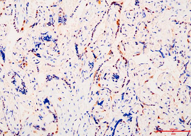
Application: ImmunohistochemistrySample Tested: Placental tissueSpecies: HumanVerified Customer | Posted 11/26/2020Human placenta were fixed in 4% paraformaldehyde. Tissue was embedded and 5 micron sections cut. Sections were permeabilized with PBS containing 0.1% Triton X-100, then blocked for 30 min in 3% BSA in PBS containing 0.1% Triton X-100. Goat Anti-Human PIGF Antigen was diluted in blocking buffer to 1:200 and applied to sections for 1 hour at RT. Sections were washed 3X with PBS containing 0.1% Triton X-100, then incubated with secondary antibody.
There are no reviews that match your criteria.
Loading...