Goat anti-Human IgG Fc Secondary Antibody (Pre-adsorbed)
Novus Biologicals | Catalog # NBP2-60678
Key Product Details
Species Reactivity
Human
Applications
Immunohistochemistry, Western Blot, ELISA, Dot Blot
Label
Unconjugated
Antibody Source
Polyclonal Goat IgG
Format
Pre-adsorbed
Loading...
Product Specifications
Immunogen
Human IgG F(c) fragment
Specificity
This antibody was pre-adsorbed against Mouse Serum Proteins. No reaction was observed against Human IgG F(ab) or Mouse Serum Proteins. Specificity was confirmed by ELISA minimal cross reactivity against Mouse IgG.
Clonality
Polyclonal
Host
Goat
Isotype
IgG
Description
This product was prepared from monospecific antiserum by immunoaffinity chromatography using Human IgG coupled to agarose beads followed by solid phase adsorption(s) to remove any unwanted reactivities. Assay by immunoelectrophoresis resulted in a single precipitin arc against anti-Goat Serum, Human IgG, Human IgG F(c) and Human Serum
Store vial at 4C prior to opening. This product is stable for several weeks at 4C as an undiluted liquid. Dilute only prior to immediate use. For extended storage aliquot contents and freeze at -20C or below. Avoid cycles of freezing and thawing.
Store vial at 4C prior to opening. This product is stable for several weeks at 4C as an undiluted liquid. Dilute only prior to immediate use. For extended storage aliquot contents and freeze at -20C or below. Avoid cycles of freezing and thawing.
Scientific Data Images for Goat anti-Human IgG Fc Secondary Antibody (Pre-adsorbed)
![ELISA: Goat anti-Human IgG Fc Secondary Antibody (Pre-adsorbed) [NBP2-60678]](https://resources.rndsystems.com/images/products/Goat-anti-Human-IgG-Fc-Secondary-Antibody-Pre-adsorbed-ELISA-NBP2-60678-img0002.jpg "ELISA: Goat anti-Human IgG Fc Secondary Antibody (Pre-adsorbed) [NBP2-60678]")
ELISA: Goat anti-Human IgG Fc Secondary Antibody (Pre-adsorbed) [NBP2-60678]
ELISA: Goat anti-Human IgG Fc Secondary Antibody (Pre-adsorbed) [NBP2-60678] - ELISA results of purified Goat anti-Human IgG F(c) Antibody Biotin conjugated tested against purified Human IgG F(c). Each well was coated in duplicate with 1.0 ug of Human IgG F(c). The starting dilution of antibody was 5 ug/ml and the X-axis represents the Log10 of a 3-fold dilution. This titration is a 4-parameter curve fit where the IC50 is defined as the titer of the antibody.Assay performed using 3percent fish gelatin as blocking buffer, Streptavidin Peroxidase Conjugated and TMB substrate. Image using the Biotin form of this antibody.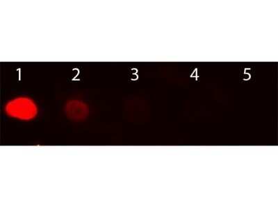
Dot Blot: Goat anti-Human IgG Fc Secondary Antibody (Pre-adsorbed) [NBP2-60678] - Load: Lane 1 - 100 ng Lane 2 - 33.3 ng Lane 3 - 11.1 ng Lane 4 - 3.70 ng Lane 5 - 1.23 ng. Primary antibody: n/a. Secondary antibody: Goat anti-Human IgG Fc Antibody Rhodamine Conjugated at 1:1,000 for 60 min at RT. Blocked with blocking buffer for 60 min at RT. Image from the Rhodamine version of this antibody.
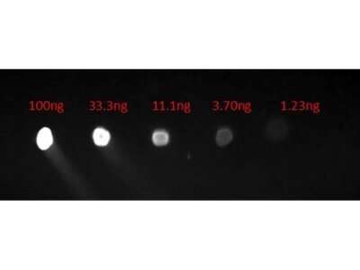
Dot Blot: Goat anti-Human IgG Fc Secondary Antibody (Pre-adsorbed) [NBP2-60678] - Lane 1: 100ng. Lane 2: 33.3ng. Lane 3: 11.1ng. Lane 4: 3.7ng. Lane 5: 1.23ng. Secondary Antibody: Goat Anti-Human F(c) FITC 1 ug/mL. Blocked with blocking buffer for 30 min at RT. Image from the FITC version of this antibody.
![ELISA: Goat anti-Human IgG Fc Secondary Antibody (Pre-adsorbed) [NBP2-60678]](https://resources.rndsystems.com/images/products/Goat-anti-Human-IgG-Fc-Secondary-Antibody-Pre-adsorbed-ELISA-NBP2-60678-img0007.jpg "ELISA: Goat anti-Human IgG Fc Secondary Antibody (Pre-adsorbed) [NBP2-60678]")
ELISA: Goat anti-Human IgG Fc Secondary Antibody (Pre-adsorbed) [NBP2-60678]
ELISA: Goat anti-Human IgG Fc Secondary Antibody (Pre-adsorbed) [NBP2-60678] - ELISA results of purified Goat anti-Human IgG Fc Secondary antibody (Pre-adsorbed) (min x Mouse serum proteins) tested against purified HumanIgG Fc. Each well was coated in duplicate with 1.0 ug of HumanIgG Fc as well as Mouse IgG. The starting dilution of antibody was 5ug/ml and the X-axis represents the Log10 of a 3-fold dilution. This titration is a 4-parameter curve fit where the IC50 is defined as the titer of the antibody. Assay performed using 3% fish gelatin as blocking buffer, Donkey anti-Goat IgG Antibody Peroxidase Conjugated (Min X Ch GP Ham Hs Ms Rb & Rt Serum Proteins) and TMB substrate.Applications for Goat anti-Human IgG Fc Secondary Antibody (Pre-adsorbed)
Application
Recommended Usage
ELISA
1:15000-1:1:60000
Immunohistochemistry
1:1000-1:3000
Western Blot
1:1000-1:5000
Application Notes
This product has been tested by dot blot and ELISA. This antibody is suitable for ELISA, western blot, and immunohistochemistry, as well as other assays requiring lot-to-lot consistency.
Formulation, Preparation, and Storage
Purification
Multi-step
Formulation
0.02 M Potassium Phosphate, 0.15 M Sodium Chloride, pH 7.2
Format
Pre-adsorbed
Preservative
0.01% Sodium Azide
Concentration
Please see the vial label for concentration. If unlisted please contact technical services.
Shipping
The product is shipped with polar packs. Upon receipt, store it immediately at the temperature recommended below.
Stability & Storage
Store at 4C short term. Aliquot and store at -20C long term. Avoid freeze-thaw cycles.
Product Documents for Goat anti-Human IgG Fc Secondary Antibody (Pre-adsorbed)
Certificate of Analysis
To download a Certificate of Analysis, please enter a lot or batch number in the search box below.
Product Specific Notices for Goat anti-Human IgG Fc Secondary Antibody (Pre-adsorbed)
This product is for research use only and is not approved for use in humans or in clinical diagnosis. Secondary Antibodies are guaranteed for 1 year from date of receipt.
Customer Reviews for Goat anti-Human IgG Fc Secondary Antibody (Pre-adsorbed)
There are currently no reviews for this product. Be the first to review Goat anti-Human IgG Fc Secondary Antibody (Pre-adsorbed) and earn rewards!
Have you used Goat anti-Human IgG Fc Secondary Antibody (Pre-adsorbed)?
Submit a review and receive an Amazon gift card!
$25/€18/£15/$25CAN/¥2500 Yen for a review with an image
$10/€7/£6/$10CAN/¥1110 Yen for a review without an image
Submit a review
Protocols
Find general support by application which include: protocols, troubleshooting, illustrated assays, videos and webinars.
- Antigen Retrieval Protocol (PIER)
- Antigen Retrieval for Frozen Sections Protocol
- Appropriate Fixation of IHC/ICC Samples
- Cellular Response to Hypoxia Protocols
- Chromogenic IHC Staining of Formalin-Fixed Paraffin-Embedded (FFPE) Tissue Protocol
- Chromogenic Immunohistochemistry Staining of Frozen Tissue
- ClariTSA™ Fluorophore Kits
- Detection & Visualization of Antibody Binding
- ELISA Sample Preparation & Collection Guide
- ELISA Troubleshooting Guide
- Fluorescent IHC Staining of Frozen Tissue Protocol
- Graphic Protocol for Heat-induced Epitope Retrieval
- Graphic Protocol for the Preparation and Fluorescent IHC Staining of Frozen Tissue Sections
- Graphic Protocol for the Preparation and Fluorescent IHC Staining of Paraffin-embedded Tissue Sections
- Graphic Protocol for the Preparation of Gelatin-coated Slides for Histological Tissue Sections
- How to Run an R&D Systems DuoSet ELISA
- How to Run an R&D Systems Quantikine ELISA
- How to Run an R&D Systems Quantikine™ QuicKit™ ELISA
- IHC Sample Preparation (Frozen sections vs Paraffin)
- Immunofluorescent IHC Staining of Formalin-Fixed Paraffin-Embedded (FFPE) Tissue Protocol
- Immunohistochemistry (IHC) and Immunocytochemistry (ICC) Protocols
- Immunohistochemistry Frozen Troubleshooting
- Immunohistochemistry Paraffin Troubleshooting
- Preparing Samples for IHC/ICC Experiments
- Preventing Non-Specific Staining (Non-Specific Binding)
- Primary Antibody Selection & Optimization
- Protocol for Heat-Induced Epitope Retrieval (HIER)
- Protocol for Making a 4% Formaldehyde Solution in PBS
- Protocol for VisUCyte™ HRP Polymer Detection Reagent
- Protocol for the Preparation & Fixation of Cells on Coverslips
- Protocol for the Preparation and Chromogenic IHC Staining of Frozen Tissue Sections
- Protocol for the Preparation and Chromogenic IHC Staining of Frozen Tissue Sections - Graphic
- Protocol for the Preparation and Chromogenic IHC Staining of Paraffin-embedded Tissue Sections
- Protocol for the Preparation and Chromogenic IHC Staining of Paraffin-embedded Tissue Sections - Graphic
- Protocol for the Preparation and Fluorescent IHC Staining of Frozen Tissue Sections
- Protocol for the Preparation and Fluorescent IHC Staining of Paraffin-embedded Tissue Sections
- Protocol for the Preparation of Gelatin-coated Slides for Histological Tissue Sections
- Quantikine HS ELISA Kit Assay Principle, Alkaline Phosphatase
- Quantikine HS ELISA Kit Principle, Streptavidin-HRP Polymer
- R&D Systems Quality Control Western Blot Protocol
- Sandwich ELISA (Colorimetric) – Biotin/Streptavidin Detection Protocol
- Sandwich ELISA (Colorimetric) – Direct Detection Protocol
- TUNEL and Active Caspase-3 Detection by IHC/ICC Protocol
- The Importance of IHC/ICC Controls
- Troubleshooting Guide: ELISA
- Troubleshooting Guide: Immunohistochemistry
- Troubleshooting Guide: Western Blot Figures
- Western Blot Conditions
- Western Blot Protocol
- Western Blot Protocol for Cell Lysates
- Western Blot Troubleshooting
- Western Blot Troubleshooting Guide
- View all Protocols, Troubleshooting, Illustrated assays and Webinars
Loading...