Rabbit anti-Mouse IgG (H+L) Secondary Antibody [Biotin]
Novus Biologicals | Catalog # NB720-B
Key Product Details
Species Reactivity
Mouse
Applications
Immunohistochemistry, Western Blot, ELISA
Label
Biotin
Antibody Source
Polyclonal Rabbit IgG
Loading...
Product Specifications
Immunogen
Mouse IgG whole molecule
Clonality
Polyclonal
Host
Rabbit
Isotype
IgG
Description
For extended storage aliquot contents and freeze at -20C or below. Avoid cycles of freezing and thawing. Centrifuge product if not completely clear after standing at room
This product was prepared from monospecific antiserum by immunoaffinity chromatography using Mouse IgG coupled to agarose beads followed by solid phase adsorption(s) to remove any unwanted reactivities. Assay by immunoelectrophoresis resulted in a single precipitin arc against anti-biotin, anti-Rabbit Serum, Mouse IgG and Mouse Serum.
This product was prepared from monospecific antiserum by immunoaffinity chromatography using Mouse IgG coupled to agarose beads followed by solid phase adsorption(s) to remove any unwanted reactivities. Assay by immunoelectrophoresis resulted in a single precipitin arc against anti-biotin, anti-Rabbit Serum, Mouse IgG and Mouse Serum.
Scientific Data Images for Rabbit anti-Mouse IgG (H+L) Secondary Antibody [Biotin]
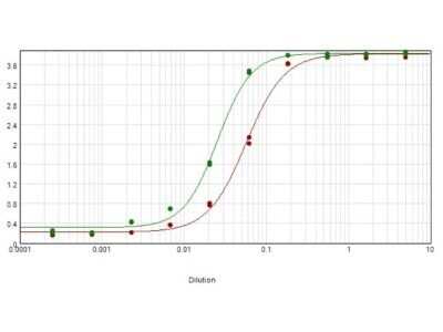
ELISA: Rabbit anti-Mouse IgG (H+L) Secondary Antibody [Biotin] [NB720-B] - ELISA results of purified Rabbit anti-Mouse IgG (H+L) Secondary Antibody [Biotin] tested against purified Mouse IgG. Each well was coated in duplicate with 1.0 ug of Mouse IgG (green line). The starting dilution of antibody was 5ug/ml and the X-axis represents the Log10 of a 3-fold dilution. This titration is a 4-parameter curve fit where the IC50 is defined as the titer of the antibody. Assay performed using blocking buffer, Streptavidin HRP conjugate 1:10000, and TMB substrate.
![Rabbit anti-Mouse IgG (H+L) Secondary Antibody [Biotin]](https://resources.rndsystems.com/images/products/nb720-b_rabbit-polyclonal-rabbit-anti-mouse-igg-h-l-secondary-antibody-biotin-310202416171481.jpg "Western Blot: Rabbit anti-Mouse IgG (H+L) Secondary Antibody [Biotin] [NB720-B] -")
Western Blot: Rabbit anti-Mouse IgG (H+L) Secondary Antibody [Biotin] [NB720-B] -
Western Blot: Rabbit anti-Mouse IgG (H+L) Secondary Antibody [Biotin] [NB720-B] - (A) Confocal microscopy of NCTC liver cells before & after exposure to amifostine (100 μg/ml), for double PDH(red)/phosphoPDH(green), phosphoPDH(red), LDH5 (red) & HIF1 alpha (red) expression. Western blot bands following exposure to 100 & 500 μg/ml of amifostine at 30 min is also shown. (B) Western blot expression of phosphoPDH, PDK1, LDH5 & HIF1 alpha in hepatoma HepG2 cells, at 0, 30 & 60 min following exposure to 100 μg/ml of amifostine. (C) Confocal microscopy of NCTC liver cells before & after exposure to amifostine (100 μg/ml), for HIF1 alpha (green), LDH5 (red), PDK1 (green) & phosphoPDH(red) expression with & without silencing of the HIF1 alpha gene. (D) Acetyl-CoA levels (pmol) in NCTC cells at 0, 30 & 60 min following exposure to 100 μg/ml of amifostine. (E) ATP levels (pmol) in NCTC cells at 0, 30 & 60 min following exposure to 100 μg/ml of amifostine. (F) Time course recording of NCTC & HepG2 cell mitochondrial membrane potentials as assessed with the JC1 method & confocal imaging (0–20 minutes), showed a rapid transient reduction of green (monomer) & red (aggregate) forms of the dye that was subsequently restored to normal levels. (G) Mitochondrial ROS (mtROS) production by NCTC & HepG2 cells, after exposure to 18Gy of ionizing radiation with & without pre-incubation with amifostine, showing a strong effect of amifostine in normal NCTC cells. mtROS were low in hepatoma HepG2 cells compared to NCTC hepatocytes & were increased only in dividing neoplastic cells. Image collected & cropped by CiteAb from the following publication (https://pubmed.ncbi.nlm.nih.gov/27507219), licensed under a CC-BY license. Not internally tested by Novus Biologicals.![Rabbit anti-Mouse IgG (H+L) Secondary Antibody [Biotin]](https://resources.rndsystems.com/images/products/nb720-b_rabbit-polyclonal-rabbit-anti-mouse-igg-h-l-secondary-antibody-biotin-310202416163717.jpg "Western Blot: Rabbit anti-Mouse IgG (H+L) Secondary Antibody [Biotin] [NB720-B] -")
Western Blot: Rabbit anti-Mouse IgG (H+L) Secondary Antibody [Biotin] [NB720-B] -
Western Blot: Rabbit anti-Mouse IgG (H+L) Secondary Antibody [Biotin] [NB720-B] - (A) Western blot images & band densitometry analysis of levels of proteins involved in anaerobic metabolism, as assessed in mouse liver before & after administration of amifostine. Bars show standard deviation & asterisks refer to p-values (*p < 0.05, **p < 0.001). (B) Confocal immunofluorescent microscopy after staining with anti-PDK1/red antibody showing increased cytoplasmic expression in mouse hepatocytes from 0 (i) to 30 min (ii) & regression thereafter (iii). Double immunostaining with PDH/red & phosphorylated pPDH/green (iv), showed an intensification of the expression of the inactive pPDH form of the enzyme 30 min (v) following amifostine injection & trend for restoration of normal PDH levels at 60 min (vi). Confocal immunofluorescent microscopy after staining for LDH1/red showed stable levels of expression in mouse hepatocytes (i,ii,iii). In contrast, LDH5/red expression was sharply induced 30 min following amifostine injection & decreased thereafter (iv, v, vi). Similar patterns were noted for GLUT2 expression (i,ii,iii) & for HIF1 alpha expression (iv,v,iv). (C) Analysis of the fluorescence intensity of confocal microscopy images (from five representative tissue areas for each staining). Bars show standard deviation & asterisks refer to p-values (*p < 0.05, **p < 0.001). (D) mRNA expression levels of LDHA, PDK1 & GLUT2 (three mice for each time point) following exposure to amifostine, as measured with quantitative RT-PCR. Bars show standard deviation & asterisks refer to p-values (**p < 0.001). Image collected & cropped by CiteAb from the following publication (https://pubmed.ncbi.nlm.nih.gov/27507219), licensed under a CC-BY license. Not internally tested by Novus Biologicals.Applications for Rabbit anti-Mouse IgG (H+L) Secondary Antibody [Biotin]
Application
Recommended Usage
ELISA
1:300000
Immunohistochemistry
1:1000 - 1:5000
Western Blot
1:20000 - 1:10000
Application Notes
This product is available in a variety of formats. Anti-Mouse IgG Biotin Antibody has been tested by ELISA and is suitable for western blot, ELISA and immunohistochemistry as well as other antibody based assays requiring lot-to-lot consistency.
Formulation, Preparation, and Storage
Purification
Multi-step
Formulation
0.02 M Potassium Phosphate, 0.15 M Sodium Chloride, pH 7.2, 10 mg/mL Bovine Serum Albumin (BSA) - Immunoglobulin and Protease free
Preservative
0.01% Sodium Azide
Concentration
Please see the vial label for concentration. If unlisted please contact technical services.
Shipping
The product is shipped with polar packs. Upon receipt, store it immediately at the temperature recommended below.
Stability & Storage
Store at 4C short term. Aliquot and store at -20C long term. Avoid freeze-thaw cycles.
Background: IgG (H+L)
The 4 IgG subclasses, sharing 95% amino acid identity, include IgG1, IgG2, IgG3, and IgG4 for humans and IgG1, IgG2a, IgG2b, and IgG3 for mice. The relative abundance of each human subclass is 60% for IgG1, 32% for IgG2, 4% for IgG3, and 4% for IgG4. In an IgG deficiency, there may be a shortage of one or more subclasses (4).
References
1. Painter RH. (1998) Encyclopedia of Immunology (Second Edition). Elsevier. 1208-1211
2. Chapter 9 - Antibodies. (2012) Immunology for Pharmacy. Mosby 70-78
3. Schroeder H, Cavacini, L. (2010) Structure and Function of Immunoglobulins. J Allergy Clin Immunol. 125(2 0 2): S41-S52. PMID: 20176268
4. Vidarsson G, Dekkers G, Rispens T. (2014) IgG subclasses and allotypes: from structure to effector functions. Front Immunol. 5:520. PMID: 25368619
Additional IgG (H+L) Products
Product Documents for Rabbit anti-Mouse IgG (H+L) Secondary Antibody [Biotin]
Certificate of Analysis
To download a Certificate of Analysis, please enter a lot or batch number in the search box below.
Product Specific Notices for Rabbit anti-Mouse IgG (H+L) Secondary Antibody [Biotin]
This product is for research use only and is not approved for use in humans or in clinical diagnosis. Secondary Antibodies are guaranteed for 1 year from date of receipt.
Citations for Rabbit anti-Mouse IgG (H+L) Secondary Antibody [Biotin]
 Powered by Bioz
Customer Reviews for Rabbit anti-Mouse IgG (H+L) Secondary Antibody [Biotin]
There are currently no reviews for this product. Be the first to review Rabbit anti-Mouse IgG (H+L) Secondary Antibody [Biotin] and earn rewards!
Have you used Rabbit anti-Mouse IgG (H+L) Secondary Antibody [Biotin]?
Submit a review and receive an Amazon gift card!
$25/€18/£15/$25CAN/¥2500 Yen for a review with an image
$10/€7/£6/$10CAN/¥1110 Yen for a review without an image
Submit a review
Protocols
Find general support by application which include: protocols, troubleshooting, illustrated assays, videos and webinars.
- Antigen Retrieval Protocol (PIER)
- Antigen Retrieval for Frozen Sections Protocol
- Appropriate Fixation of IHC/ICC Samples
- Cellular Response to Hypoxia Protocols
- Chromogenic IHC Staining of Formalin-Fixed Paraffin-Embedded (FFPE) Tissue Protocol
- Chromogenic Immunohistochemistry Staining of Frozen Tissue
- ClariTSA™ Fluorophore Kits
- Detection & Visualization of Antibody Binding
- ELISA Sample Preparation & Collection Guide
- ELISA Troubleshooting Guide
- Fluorescent IHC Staining of Frozen Tissue Protocol
- Graphic Protocol for Heat-induced Epitope Retrieval
- Graphic Protocol for the Preparation and Fluorescent IHC Staining of Frozen Tissue Sections
- Graphic Protocol for the Preparation and Fluorescent IHC Staining of Paraffin-embedded Tissue Sections
- Graphic Protocol for the Preparation of Gelatin-coated Slides for Histological Tissue Sections
- How to Run an R&D Systems DuoSet ELISA
- How to Run an R&D Systems Quantikine ELISA
- How to Run an R&D Systems Quantikine™ QuicKit™ ELISA
- IHC Sample Preparation (Frozen sections vs Paraffin)
- Immunofluorescent IHC Staining of Formalin-Fixed Paraffin-Embedded (FFPE) Tissue Protocol
- Immunohistochemistry (IHC) and Immunocytochemistry (ICC) Protocols
- Immunohistochemistry Frozen Troubleshooting
- Immunohistochemistry Paraffin Troubleshooting
- Preparing Samples for IHC/ICC Experiments
- Preventing Non-Specific Staining (Non-Specific Binding)
- Primary Antibody Selection & Optimization
- Protocol for Heat-Induced Epitope Retrieval (HIER)
- Protocol for Making a 4% Formaldehyde Solution in PBS
- Protocol for VisUCyte™ HRP Polymer Detection Reagent
- Protocol for the Preparation & Fixation of Cells on Coverslips
- Protocol for the Preparation and Chromogenic IHC Staining of Frozen Tissue Sections
- Protocol for the Preparation and Chromogenic IHC Staining of Frozen Tissue Sections - Graphic
- Protocol for the Preparation and Chromogenic IHC Staining of Paraffin-embedded Tissue Sections
- Protocol for the Preparation and Chromogenic IHC Staining of Paraffin-embedded Tissue Sections - Graphic
- Protocol for the Preparation and Fluorescent IHC Staining of Frozen Tissue Sections
- Protocol for the Preparation and Fluorescent IHC Staining of Paraffin-embedded Tissue Sections
- Protocol for the Preparation of Gelatin-coated Slides for Histological Tissue Sections
- Quantikine HS ELISA Kit Assay Principle, Alkaline Phosphatase
- Quantikine HS ELISA Kit Principle, Streptavidin-HRP Polymer
- R&D Systems Quality Control Western Blot Protocol
- Sandwich ELISA (Colorimetric) – Biotin/Streptavidin Detection Protocol
- Sandwich ELISA (Colorimetric) – Direct Detection Protocol
- TUNEL and Active Caspase-3 Detection by IHC/ICC Protocol
- The Importance of IHC/ICC Controls
- Troubleshooting Guide: ELISA
- Troubleshooting Guide: Immunohistochemistry
- Troubleshooting Guide: Western Blot Figures
- Western Blot Conditions
- Western Blot Protocol
- Western Blot Protocol for Cell Lysates
- Western Blot Troubleshooting
- Western Blot Troubleshooting Guide
- View all Protocols, Troubleshooting, Illustrated assays and Webinars
Loading...