Key Product Details
Species Reactivity
Validated:
Human
Cited:
Human
Applications
Immunohistochemistry, Immunohistochemistry-Paraffin, Western Blot
Label
Unconjugated
Antibody Source
Monoclonal Mouse IgG1 kappa Clone # IM260
Loading...
Product Specifications
Immunogen
Recombinant heavy chain of human IgM
Localization
Cytoplasm, Cell Surface and Secreted
Marker
B-Cell Marker
Specificity
Recognizes a protein of 75kDa, identified as mu heavy chain of human immunoglobulins. It does not cross-react with alpha (IgA), gamma (IgG), epsilon (IgE), or delta (IgD), heavy chains, T-cells, monocytes, granulocytes, or erythrocytes. This monoclonal antibody is useful in the identification of leukemias, plasmacytomas, and certain non-Hodgkin s lymphomas. The most common feature of these malignancies is the restricted expression of a single heavy chain class. Demonstration of clonality in lymphoid infiltrates indicates that the infiltrate is clonal and therefore malignant.
Clonality
Monoclonal
Host
Mouse
Isotype
IgG1 kappa
Description
200ug/ml of antibody purified from Bioreactor Concentrate by Protein A or G. Prepared in 10 mM PBS with 0.05% BSA & 0.05% azide. Also available WITHOUT BSA & azide at 1.0 mg/ml. (NBP2-34649)
Antibody with azide - store at 2 to 8C. Antibody without azide - store at -20 to -80C.
Antibody with azide - store at 2 to 8C. Antibody without azide - store at -20 to -80C.
Scientific Data Images for IgM Antibody (IM260)
![Western Blot: IgM Antibody (IM260) [NBP2-34253]](https://resources.rndsystems.com/images/products/IgM-Antibody-IM260-Western-Blot-NBP2-34253-img0005.jpg "Western Blot: IgM Antibody (IM260) [NBP2-34253]")
Western Blot: IgM Antibody (IM260) [NBP2-34253]
Western Blot: IgM Antibody (IM260) [NBP2-34253] - Western Blot Analysis of Raji cell lysate using IgM Antibody (IM260)![Immunohistochemistry-Paraffin: IgM Antibody (IM260) [NBP2-34253]](https://resources.rndsystems.com/images/products/IgM-Antibody-IM260-Immunohistochemistry-Paraffin-NBP2-34253-img0004.jpg "Immunohistochemistry-Paraffin: IgM Antibody (IM260) [NBP2-34253]")
Immunohistochemistry-Paraffin: IgM Antibody (IM260) [NBP2-34253]
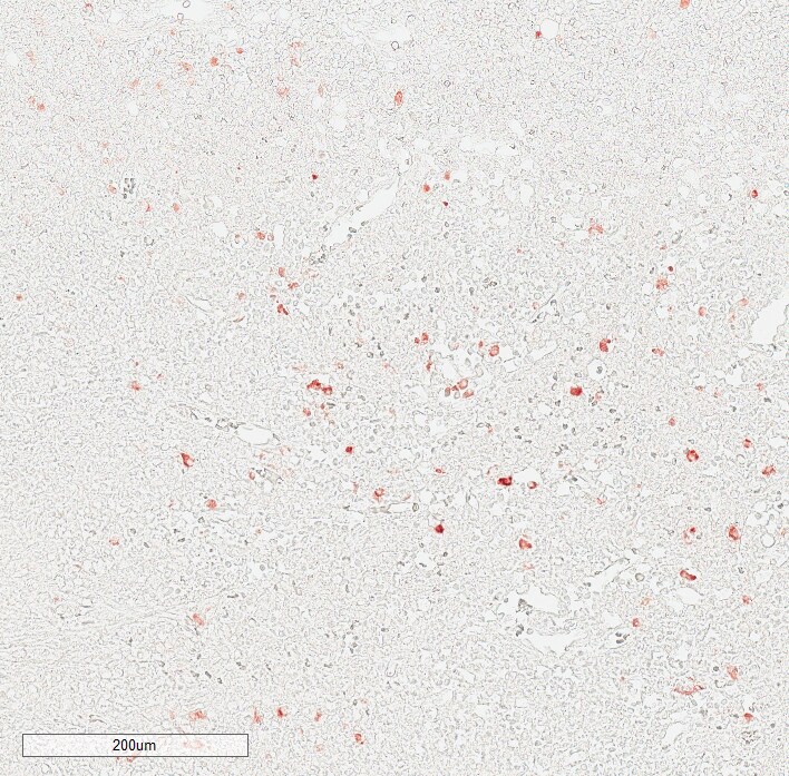
Immunohistochemistry-Paraffin: IgM Antibody (IM260) [NBP2-34253] - Formalin-fixed, paraffin-embedded human tonsil stained with IgM Antibody (IM260)![Immunohistochemistry-Paraffin: IgM Antibody (IM260) [NBP2-34253]](https://resources.rndsystems.com/images/products/IgM-Antibody-IM260-Immunohistochemistry-Paraffin-NBP2-34253-img0003.jpg "Immunohistochemistry-Paraffin: IgM Antibody (IM260) [NBP2-34253]")
Immunohistochemistry-Paraffin: IgM Antibody (IM260) [NBP2-34253]
Immunohistochemistry-Paraffin: IgM Antibody (IM260) [NBP2-34253] - Human lymph node. IgM antibody at 1:200 at RT for 1 hr. IHC-P image submitted by a verified customer review.Applications for IgM Antibody (IM260)
Application
Recommended Usage
Immunohistochemistry-Paraffin
1-2 ug/ml
Western Blot
1-2 ug/ml
Application Notes
Immunohistochemistry (Formalin-fixed): 1-2ug/ml for 30 minutes at RT. Staining of formalin-fixed tissues requires heating tissue sections in 10mM Tris with 1mM EDTA, pH 9.0, for 45 min at 95C followed by cooling at RT for 20 minutes.
Optimal dilution for a specific application should be determined.
Optimal dilution for a specific application should be determined.
Reviewed Applications
Read 1 review rated 5 using NBP2-34253 in the following applications:
Formulation, Preparation, and Storage
Purification
Protein A or G purified
Formulation
10 mM PBS with 0.05% BSA
Preservative
0.05% Sodium Azide
Concentration
0.2 mg/ml
Shipping
The product is shipped with polar packs. Upon receipt, store it immediately at the temperature recommended below.
Stability & Storage
Store at 4C.
Background: IgM
Long Name
Immunoglobulin M
Alternate Names
AGM1, DKFZp686I15196, DKFZp686I15212, FLJ00385, immunoglobulin heavy constant mu, MGC104996, MGC52291, MU, VH
Gene Symbol
IGHM
UniProt
Additional IgM Products
Product Documents for IgM Antibody (IM260)
Certificate of Analysis
To download a Certificate of Analysis, please enter a lot or batch number in the search box below.
Product Specific Notices for IgM Antibody (IM260)
This product is for research use only and is not approved for use in humans or in clinical diagnosis. Primary Antibodies are guaranteed for 1 year from date of receipt.
Citations for IgM Antibody (IM260)
 Powered by Bioz
Customer Reviews for IgM Antibody (IM260) (1)
5 out of 5
1 Customer Rating
Have you used IgM Antibody (IM260)?
Submit a review and receive an Amazon gift card!
$25/€18/£15/$25CAN/¥2500 Yen for a review with an image
$10/€7/£6/$10CAN/¥1110 Yen for a review without an image
Submit a review
Customer Images
Showing
1
-
1 of
1 review
Showing All
Filter By:
-
Application: Immunohistochemistry-ParaffinSample Tested: Human lymph nodeSpecies: HumanVerified Customer | Posted 09/17/20191:200 @ room temperature for 1 hour

There are no reviews that match your criteria.
Protocols
Find general support by application which include: protocols, troubleshooting, illustrated assays, videos and webinars.
- Antigen Retrieval Protocol (PIER)
- Antigen Retrieval for Frozen Sections Protocol
- Appropriate Fixation of IHC/ICC Samples
- Cellular Response to Hypoxia Protocols
- Chromogenic IHC Staining of Formalin-Fixed Paraffin-Embedded (FFPE) Tissue Protocol
- Chromogenic Immunohistochemistry Staining of Frozen Tissue
- ClariTSA™ Fluorophore Kits
- Detection & Visualization of Antibody Binding
- Fluorescent IHC Staining of Frozen Tissue Protocol
- Graphic Protocol for Heat-induced Epitope Retrieval
- Graphic Protocol for the Preparation and Fluorescent IHC Staining of Frozen Tissue Sections
- Graphic Protocol for the Preparation and Fluorescent IHC Staining of Paraffin-embedded Tissue Sections
- Graphic Protocol for the Preparation of Gelatin-coated Slides for Histological Tissue Sections
- IHC Sample Preparation (Frozen sections vs Paraffin)
- Immunofluorescent IHC Staining of Formalin-Fixed Paraffin-Embedded (FFPE) Tissue Protocol
- Immunohistochemistry (IHC) and Immunocytochemistry (ICC) Protocols
- Immunohistochemistry Frozen Troubleshooting
- Immunohistochemistry Paraffin Troubleshooting
- Preparing Samples for IHC/ICC Experiments
- Preventing Non-Specific Staining (Non-Specific Binding)
- Primary Antibody Selection & Optimization
- Protocol for Heat-Induced Epitope Retrieval (HIER)
- Protocol for Making a 4% Formaldehyde Solution in PBS
- Protocol for VisUCyte™ HRP Polymer Detection Reagent
- Protocol for the Preparation & Fixation of Cells on Coverslips
- Protocol for the Preparation and Chromogenic IHC Staining of Frozen Tissue Sections
- Protocol for the Preparation and Chromogenic IHC Staining of Frozen Tissue Sections - Graphic
- Protocol for the Preparation and Chromogenic IHC Staining of Paraffin-embedded Tissue Sections
- Protocol for the Preparation and Chromogenic IHC Staining of Paraffin-embedded Tissue Sections - Graphic
- Protocol for the Preparation and Fluorescent IHC Staining of Frozen Tissue Sections
- Protocol for the Preparation and Fluorescent IHC Staining of Paraffin-embedded Tissue Sections
- Protocol for the Preparation of Gelatin-coated Slides for Histological Tissue Sections
- R&D Systems Quality Control Western Blot Protocol
- TUNEL and Active Caspase-3 Detection by IHC/ICC Protocol
- The Importance of IHC/ICC Controls
- Troubleshooting Guide: Immunohistochemistry
- Troubleshooting Guide: Western Blot Figures
- Western Blot Conditions
- Western Blot Protocol
- Western Blot Protocol for Cell Lysates
- Western Blot Troubleshooting
- Western Blot Troubleshooting Guide
- View all Protocols, Troubleshooting, Illustrated assays and Webinars
Loading...