iNOS Antibody - BSA Free
Novus Biologicals | Catalog # NB300-605
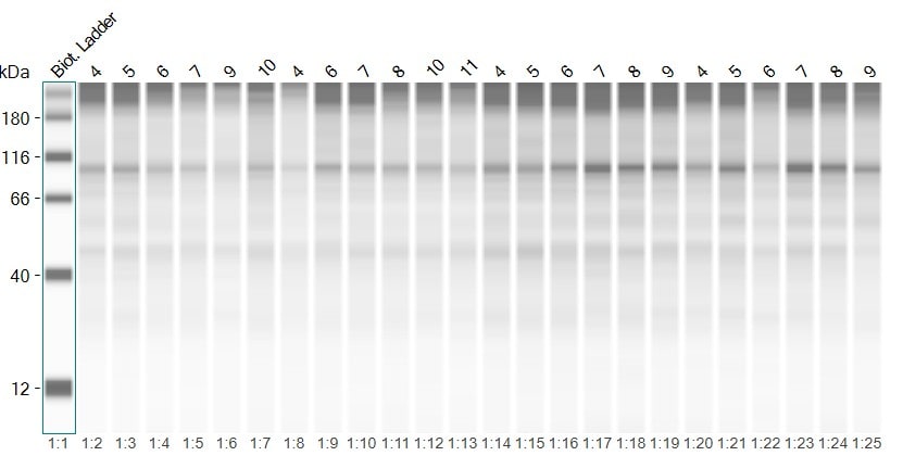
![Western Blot: iNOS AntibodyBSA Free [NB300-605]](https://resources.rndsystems.com/images/products/iNOS-Antibody-Western-Blot-NB300-605-img0015.jpg "Western Blot: iNOS AntibodyBSA Free [NB300-605]")
Key Product Details
Validated by
Biological Validation
Species Reactivity
Validated:
Human, Mouse, Rat, Porcine
Cited:
Human, Mouse, Rat, Porcine
Applications
Validated:
Immunohistochemistry, Immunohistochemistry-Paraffin, Immunohistochemistry-Frozen, Western Blot, Immunoblotting, Flow Cytometry, Immunocytochemistry/ Immunofluorescence, In vitro assay
Cited:
Immunohistochemistry-Paraffin, Immunohistochemistry-Frozen, Western Blot, Immunoblotting, Flow Cytometry, Immunocytochemistry/ Immunofluorescence, Immunoprecipitation, IF/IHC
Label
Unconjugated
Antibody Source
Polyclonal Rabbit IgG
Format
BSA Free
Loading...
Product Specifications
Immunogen
Sythetic peptide made to an internal portion of mouse iNOS (between amino acids 12-48) [UniProt P29477].
Reactivity Notes
Porcine reactivity reported in scientific literature (PMID: 31292486).
Specificity
This antibody detects iNOS. It does not detect other NOS isoforms.
Clonality
Polyclonal
Host
Rabbit
Isotype
IgG
Theoretical MW
131 kDa.
Disclaimer note: The observed molecular weight of the protein may vary from the listed predicted molecular weight due to post translational modifications, post translation cleavages, relative charges, and other experimental factors.
Disclaimer note: The observed molecular weight of the protein may vary from the listed predicted molecular weight due to post translational modifications, post translation cleavages, relative charges, and other experimental factors.
Scientific Data Images for iNOS Antibody - BSA Free
Western Blot: iNOS AntibodyBSA Free [NB300-605]
Western Blot: iNOS Antibody [NB300-605] - Analysis of iNOS was performed by loading 20 ug of RAW264 whole cell lysate untreated (left lane) or stimulated with LPS at 1 ug/mL for 16 hours (right lane) and 10 uL of PageRuler Plus Prestained Protein Ladder onto a 4-20% Tris-Glycine polyacrylamide gel. Proteins were transferred to a nitrocellulose membrane and blocked with 5% Milk in TBST for at least 1 hour. The membrane was probed with an iNOS Rabbit polyclonal antibody at a dilution of 1:1000 overnight at 4C on a rocking platform, washed in TBST, and probed with a Goat anti-Rabbit IgG (H+L) Secondary Antibody, HRP conjugate at a dilution of 1:1000 for 1 hour. Chemiluminescent detection was performed using SuperSignal West Pico.![Immunohistochemistry-Paraffin: iNOS Antibody - BSA Free [NB300-605]](https://resources.rndsystems.com/images/products/iNOS-Antibody-Immunohistochemistry-Paraffin-NB300-605-img0010.jpg "Immunohistochemistry-Paraffin: iNOS Antibody - BSA Free [NB300-605]")
Immunohistochemistry-Paraffin: iNOS Antibody - BSA Free [NB300-605]
Immunohistochemistry-Paraffin: iNOS Antibody [NB300-605] - Immunohistochemistry was performed on normal deparaffinized human Lung tissue.![Immunohistochemistry-Paraffin: iNOS Antibody - BSA Free [NB300-605]](https://resources.rndsystems.com/images/products/iNOS-Antibody-Immunohistochemistry-Paraffin-NB300-605-img0016.jpg "Immunohistochemistry-Paraffin: iNOS Antibody - BSA Free [NB300-605]")
Immunohistochemistry-Paraffin: iNOS Antibody - BSA Free [NB300-605]
Immunohistochemistry-Paraffin: iNOS Antibody [NB300-605] - Immunohistochemistry was performed on normal deparaffinized human Heart tissue.![Immunocytochemistry/ Immunofluorescence: iNOS Antibody - BSA Free [NB300-605]](https://resources.rndsystems.com/images/products/iNOS-Antibody-Immunocytochemistry-Immunofluorescence-NB300-605-img0012.jpg "Immunocytochemistry/ Immunofluorescence: iNOS Antibody - BSA Free [NB300-605]")
Immunocytochemistry/ Immunofluorescence: iNOS Antibody - BSA Free [NB300-605]
Immunocytochemistry/Immunofluorescence: iNOS Antibody [NB300-605] - Analysis of iNOS in A549 Cells. Cells were grown on chamber slides and fixed with formaldehyde prior to staining. Cells were probed without (control) or with a iNOS polyclonal antibody at a dilution of 1:20 overnight at 4C, washed with PBS and incubated with a DyLight 488 conjugated secondary antibody. iNOS staining (green), F-Actin staining with Phalloidin (red) and nuclei with DAPI (blue) is shown.![Western Blot: iNOS AntibodyBSA Free [NB300-605]](https://resources.rndsystems.com/images/products/iNOS-Antibody-Western-Blot-NB300-605-img0005.jpg "Western Blot: iNOS AntibodyBSA Free [NB300-605]")
![Western Blot: iNOS AntibodyBSA Free [NB300-605]](https://resources.rndsystems.com/images/products/iNOS-Antibody-Western-Blot-NB300-605-img0017.jpg "Western Blot: iNOS AntibodyBSA Free [NB300-605]")
![Immunocytochemistry/ Immunofluorescence: iNOS Antibody - BSA Free [NB300-605]](https://resources.rndsystems.com/images/products/iNOS-Antibody-Immunocytochemistry-Immunofluorescence-NB300-605-img0013.jpg "Immunocytochemistry/ Immunofluorescence: iNOS Antibody - BSA Free [NB300-605]")
Immunocytochemistry/ Immunofluorescence: iNOS Antibody - BSA Free [NB300-605]
Immunocytochemistry/Immunofluorescence: iNOS Antibody [NB300-605] - Analysis of iNOS in NIH-3T3 Cells. Cells were grown on chamber slides and fixed with formaldehyde prior to staining. Cells were probed without (control) or with a iNOS polyclonal antibody at a dilution of 1:20 overnight at 4C, washed with PBS and incubated with a DyLight 488 conjugated secondary antibody. iNOS staining (green), F-Actin staining with Phalloidin (red) and nuclei with DAPI (blue) is shown.
Western Blot: iNOS Antibody - BSA Free [NB300-605] -
Western Blot: iNOS Antibody - BSA Free [NB300-605] - Stimulation of IMA 2.1 cells had no effect on relative survival after viral infection. IMA 2.1 cells were stimulated with LPS (10 µg/ml), IFN-gamma, (10 ng/ml) LPS + IFN-gamma, IL-4 (10 ng/ml), FGF (5 ng/ml) or FGF + LPS for 24 h in medium with 2% FBS. Polarization of cells was analyzed by Griess assay (a), Western blot (b) & FACS analysis by detection of MHCII+ cells (c). d MTT assay was performed to detect the relative percentage of VACV-mediated cell death in the presence of stimulating factors. e Viral replication was analyzed by standard plaque assay in the presence of stimulating factors. All experiments were performed in triplicate. Image collected & cropped by CiteAb from the following publication (http://www.translational-medicine.com/content/13/1/216), licensed under a CC-BY license. Not internally tested by Novus Biologicals.
Immunocytochemistry/ Immunofluorescence: iNOS Antibody - BSA Free [NB300-605] -
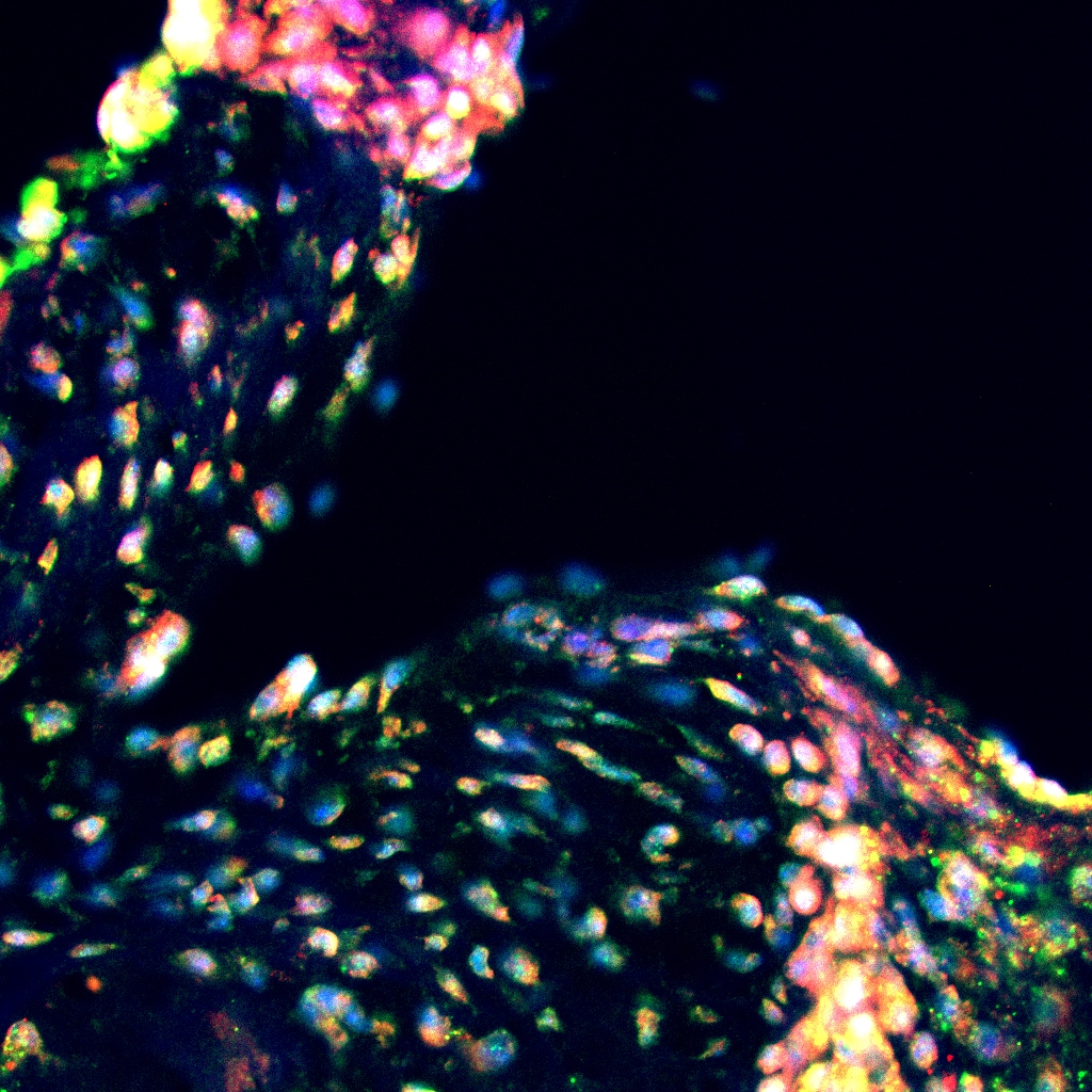
Immunocytochemistry/ Immunofluorescence: iNOS Antibody - BSA Free [NB300-605] - Nfix is mainly expressed by anti-inflammatory MPs. (a) Percentage of F4/80+ MPs positive for Nfix in Tibialis Anterior muscles (TA) of WT mice injected by CTX at D2, D4 & D7, post-injury. Immunostaining for F4/80 (green), Nfix (red) & DAPI (blue) at D4 & D7 after CTX injection; (b) Percentage of Ly6C+ & Ly6C- sorted MPs positive for Nfix in TA muscles of WT mice injected by CTX at D2, D4 & D7 post-injury; (c) Percentage of Nfix+ MPs after M1 & M2c polarization (with IFN gamma & IL10, respectively). * p < 0.05; *** p < 0.001; for (b) * p < 0.05 Ly6C+ vs. Ly6C+ at D4 & D7; # p < 0.05 Ly6C− D7 vs. D2. Results are means ± SEM of at least three independent experiments. Scale bar = 50 μm. Image collected & cropped by CiteAb from the following publication (https://pubmed.ncbi.nlm.nih.gov/32183151), licensed under a CC-BY license. Not internally tested by Novus Biologicals.
Western Blot: iNOS Antibody - BSA Free [NB300-605] -
Western Blot: iNOS Antibody - BSA Free [NB300-605] - Indolepropionic acid (IPA) induced oxidative stress, cellular energy stress, & decreased the proportions of cancer stem cells. 500,000 cells/well 4T1 cells were treated with IPA in the concentrations indicated for 24 h; then, (A) lipid peroxidation was measured by TBARS assay, & (B) 4HNE expression was assessed by Western blotting (representative figure, n = 3). In the same cells (C), the protein expression of NRF2 (at 68 kDa) & iNOS were determined by Western blotting (n = 3), while (D) the mRNA expression of catalase (cat) was determined by RT-qPCR (n = 3). (E) The expression of the indicated proteins (pACC, ACC, FOXO1, & PGC-1 beta ) were determined by Western blotting (n = 3, except for PGC-1 beta, where n = 2). (F) 100,000 cells/well 4T1 cells were treated with the indicated concentration of IPA for 24 h; then, the proportions of aldehyde dehydrogenase-positive cells were determined in Aldefluor assays using flow cytometry (n = 3). For Western blots, a typical experiment was displayed. Fold data were log2 transformed to achieve normal distribution. Statistical significance was determined using the ANOVA test followed by Dunnett’s post-hoc test, except for panel F, where Student’s t-test was used. * & *** indicate statistically significant difference between control & treated samples at p < 0.05 & p < 0.001, respectively. Image collected & cropped by CiteAb from the following publication (https://pubmed.ncbi.nlm.nih.gov/32854297), licensed under a CC-BY license. Not internally tested by Novus Biologicals.
Western Blot: iNOS Antibody - BSA Free [NB300-605] -
Western Blot: iNOS Antibody - BSA Free [NB300-605] - NRF2 activation modulated LCA-induced oxidative stress responses in 4T1 breast cancer cells. The 4T1 cells were treated with 0.3 μm LCA & the NRF2 activator RA839 or tBHQ in the concentrations indicated for 48 h. Lipid peroxidation was determined by measuring (A) TBARS (n = 4) & (B,C) 4-HNE levels using western blotting (n = 3). (D) The 4T1 cells were treated with LCA (0.3 μm) and/or GSH & NAC (both at 5 mm) antioxidants for 48 h, then total protein concentration was determined using the sulforhodamine B assay (n = 3). For statistical analysis ANOVA test was used followed by the Dunnett post-hoc test, where all groups were compared to the LCA-treated cohort. Data are plotted as mean ± SEM. ** p < 0.01, LCA vs. LCA/NRF2-activator-treated groups (GSH, reduced glutathione; LCA, lithocholic acid; NAC, N-acetylcysteine; TBARS, thiobarbituric acid reactive substances; 4HNE, 4-hydroxynonenal). Image collected & cropped by CiteAb from the following publication (https://pubmed.ncbi.nlm.nih.gov/31461945), licensed under a CC-BY license. Not internally tested by Novus Biologicals.
Western Blot: iNOS Antibody - BSA Free [NB300-605] -
Western Blot: iNOS Antibody - BSA Free [NB300-605] - Impaired amount of virus progeny after stimulation of BV-2 cells with IFN-gamma or IFN-gamma & LPS. BV-2 cells were stimulated with LPS (10 µg/ml), IFN-gamma, (10 ng/ml), LPS + IFN-gamma or IL-4 (10 ng/ml) for 24 h in medium with 2% FBS. Polarization of cells was analyzed by Griess assay (a), Western blot (b) & by detection of the percentage of MHCII+ cells by FACS analysis (c). The amount of virus progeny (pfu/ml) was analyzed by standard plaque assay in the presence of stimulating factors & in normal growth medium in cells & supernatants (d). Statistical analysis was performed related to infection medium. To analyze the recovery of viral replication, normal growth medium was applied after infection of pre-stimulated cells. Standard plaque assay was performed to determine viral progeny in cells & supernatant after 24, 48, 72 & 96 hpi (e). To analyze the relative survival of the cells in the presence of stimulating factors a MTT assay was performed (f). The cellular density of stimulated cells relative to unstimulated (w/o) cells was detected via optical density measurement after 24 & 48 h of cultivation (g). Two-sided t test with unequal variances was used for statistics *p < 0.05, **p < 0.01, ***p < 0.001. Active replication was defined as virus titer above the virus titer of the infection medium (red line). All experiments were performed in triplicate & repeated in an independent experiment. Image collected & cropped by CiteAb from the following publication (http://www.translational-medicine.com/content/13/1/216), licensed under a CC-BY license. Not internally tested by Novus Biologicals.
Western Blot: iNOS Antibody - BSA Free [NB300-605] -
Western Blot: iNOS Antibody - BSA Free [NB300-605] - Inhibitory effect of MMPP on amyloidogenesis & STAT3 translocation in astrocytes & microglia cells. The expression of APP, BACE1 & C99 was detected by Western blotting using specific antibodies in astrocytes (a) & microglia cells (b). Each blot is representative of three experiments. The activity of beta -secretase was investigated using assay kit in astrocytes (c) & microglia cells (d). Values are presented as mean ± S.D. of the three independent experiments performed in triplicate. #p < 0.05 compared to control, *p < 0.05 compared to LPS. Iba-1, COX-2, & iNOS proteins were detected by Western blotting using specific antibodies in astrocytes (e) & microglia cells (f). NO level was measured in astrocytes (g) & microglia cells (h). Activation of STAT3 was investigated using EMSA in astrocytes (i) microglial cells (j) were determined & the expression of STAT3 & phopho-STAT3 was also detected by Western blotting using specific antibodies (k), (l). For the cropped images, samples were run in the same gels under same experimental conditions & processed in parallel. Each band is representative for three experiments Image collected & cropped by CiteAb from the following publication (http://link.springer.com/10.1007/s12017-017-8469-3), licensed under a CC-BY license. Not internally tested by Novus Biologicals.
Immunohistochemistry: iNOS Antibody - BSA Free [NB300-605] -
TSA, PXD, or MGCD restore osteoimmune balance and reduce osteolytic activity. A Histological examination by H&E staining revealed differences in calvarial bone morphology between groups. Immunohistochemical staining demonstrated that treatment with TSA, PXD, or MGCD reduced osteoclast activity, as shown by decreased CTSK expression (B), suppressed M1 macrophage polarization indicated by reduced iNOS expression (D), enhanced M2 macrophage polarization indicated by increased ARG1 expression (F), and promoted osteogenesis as reflected by elevated RUNX2 expression (H). Quantification of CTSK- (C), iNOS- (E), ARG1- (G), and RUNX2- (I) positive cells. Data are presented as mean +/- SD (n = 6 per group). *P < 0.05, **P < 0.01, and ***P < 0.001. Scale bar = 100 μm Image collected and cropped by CiteAb from the following open publication (https://pubmed.ncbi.nlm.nih.gov/41174647), licensed under a CC-BY license. Not internally tested by Novus Biologicals.Applications for iNOS Antibody - BSA Free
Application
Recommended Usage
Flow Cytometry
reported in scientific literature (PMID 31536479)
Immunocytochemistry/ Immunofluorescence
1:50
Immunohistochemistry
1:10 - 1:500
Immunohistochemistry-Frozen
reported in scientific literature (PMID 35005642)
Immunohistochemistry-Paraffin
1:20
In vitro assay
reported in scientific literature (PMID 27998907)
Western Blot
1:200 - 1:800
Application Notes
WB: Detects an approx. 135 kDa protein representing recombinant human iNOS and human iNOS from cytokine stimulated A549 cells. Also detects purified recombinant mouse iNOS, mouse iNOS from cytokine stimulated RAW 264.7 cells and cytokine stimulated rat fibroblast iNOS. However, the signals are not as strong as those seen with the human samples.
Reviewed Applications
Read 5 reviews rated 4.2 using NB300-605 in the following applications:
Flow Cytometry Panel Builder
Bio-Techne Knows Flow Cytometry
Save time and reduce costly mistakes by quickly finding compatible reagents using the Panel Builder Tool.
Advanced Features
- Spectra Viewer - Custom analysis of spectra from multiple fluorochromes
- Spillover Popups - Visualize the spectra of individual fluorochromes
- Antigen Density Selector - Match fluorochrome brightness with antigen density

Formulation, Preparation, and Storage
Purification
Affinity purified
Formulation
PBS
Format
BSA Free
Preservative
0.02% Sodium Azide
Concentration
0.5 mg/ml
Shipping
The product is shipped with polar packs. Upon receipt, store it immediately at the temperature recommended below.
Stability & Storage
Store at -20C. Avoid freeze-thaw cycles.
Background: iNOS
The 131 kDa enzyme, iNOS, is found in a variety of cell types including macrophages, hepatocytes, synoviocytes, and smooth muscle cells. While constitutively expressed in kidneys, in other tissues iNOS is induced by bacterial lipopolysaccharides (LPS), growth factors, and cytokines such as IFN-gamma, TNF, IL-1 and IL-2. iNOS is not regulated by the level of intracellular Ca2+ and is constantly active as a dimer when expressed. iNOS activity is elevated in a variety of diseases including atherosclerosis, heart failure, sepsis, solid tumors, and type 2 diabetes. Acting as a critical mediator of inflammation and apoptosis, iNOS inhibitors have been shown to alleviate obesity and stress inducted insulin resistance in mouse models (2,3).
References
1. Forstermann U, and Sessa WC. (2012) Nitric oxide synthases: regulation and function. Eur Heart J. 33(7): 829-837. PMID: 21890489
2. Aktan F. (2004) iNOS-mediated nitric oxide production and its regulation. Life Sci. 75(6):639-53. PMID: 15172174
3. Cinelli MA, Do HT, Miley GP, Silverman RB. (2020) Inducible nitric oxide synthase: Regulation, structure, and inhibition. Med Res Rev. 40(1):158-189. PMID: 31192483
Long Name
Inducible Nitic Oxide Synthase
Alternate Names
NOS2, NOS2A
Gene Symbol
NOS2
UniProt
Additional iNOS Products
Product Documents for iNOS Antibody - BSA Free
Certificate of Analysis
To download a Certificate of Analysis, please enter a lot or batch number in the search box below.
Product Specific Notices for iNOS Antibody - BSA Free
This product is for research use only and is not approved for use in humans or in clinical diagnosis. Primary Antibodies are guaranteed for 1 year from date of receipt.
Citations for iNOS Antibody - BSA Free
 Powered by Bioz
Customer Reviews for iNOS Antibody - BSA Free (5)
4.2 out of 5
5 Customer Ratings
Have you used iNOS Antibody - BSA Free?
Submit a review and receive an Amazon gift card!
$25/€18/£15/$25CAN/¥2500 Yen for a review with an image
$10/€7/£6/$10CAN/¥1110 Yen for a review without an image
Submit a review
Customer Images
_248b4ab4-9a39-47b1-8c2c-740a51a00d93.jpg)

-(02-ml)_NB300-605_7041.jpg)
Showing
1
-
5 of
5 reviews
Showing All
Filter By:
-
Application: Simple WesternSample Tested: lung homogenateSpecies: MouseVerified Customer | Posted 09/08/2025Lung homogenate protein expression of iNOS in treated and untreated miceI tested the protein concentration in my samples using JESS simple western
 Bio-Techne ResponseThis review reflects a new species or application tested on a primary antibody.
 -
Application: Western BlotSample Tested: mouse lung homogenateSpecies: mouse lung homogenateVerified Customer | Posted 09/08/2025iNOS protein expression in mouse lung homogenateI ran it on my mouse homogenate samples to test the inos response after smoke exposure
-
Application: Western BlotSample Tested: Liver tissueSpecies: MouseVerified Customer | Posted 09/15/2020n/a
-
Application: ImmunofluorescenceSample Tested: Mouse aortic rootSpecies: MouseVerified Customer | Posted 11/13/2015Immunofluorescence in mouse paraffin section (RED)
-
Application: Western BlotSample Tested: human astrocyte lysateSpecies: HumanVerified Customer | Posted 04/28/2014iNOS in stimulated astrocytes
There are no reviews that match your criteria.
Protocols
View specific protocols for iNOS Antibody - BSA Free (NB300-605):
Immunohistochemistry-Paraffin Embedded Sections
Antigen Unmasking:
Bring slides to a boil in 10 mM sodium citrate buffer (pH 6.0) then maintain at a sub-boiling temperature for 10 minutes. Cool slides on bench-top for 30 minutes (keep slides in the sodium citrate buffer at all times).
Staining:
1. Wash sections in deionized water three times for 5 minutes each.
2. Wash sections in PBS for 5 minutes.
3. Block each section with 100-400 ul blocking solution (1% BSA in PBS) for 1 hour at room temperature.
4. Remove blocking solution and add 100-400 ul diluted primary antibody. Incubate overnight at 4 C.
5. Remove antibody solution and wash sections in wash buffer three times for 5 minutes each.
6. Add 100-400 ul HRP polymer conjugated secondary antibody. Incubate 30 minutes at room temperature.
7. Wash sections three times in wash buffer for 5 minutes each.
8. Add 100-400 ul DAB substrate to each section and monitor staining closely.
9. As soon as the sections develop, immerse slides in deionized water.
10. Counterstain sections in hematoxylin.
11. Wash sections in deionized water two times for 5 minutes each.
12. Dehydrate sections.
13. Mount coverslips.
Western Blot Protocol
1. Perform SDS-PAGE on samples to be analyzed, loading 10-25 ug of total protein per lane.
2. Transfer proteins to PVDF membrane according to the instructions provided by the manufacturer of the membrane and transfer apparatus.
3. Stain the membrane with Ponceau S (or similar product) to assess transfer success, and mark molecular weight standards where appropriate.
4. Rinse the blot TBS -0.05% Tween 20 (TBST).
5. Block the membrane in 5% Non-fat milk in TBST (blocking buffer) for at least 1 hour.
6. Wash the membrane in TBST three times for 10 minutes each.
7. Dilute primary antibody in blocking buffer and incubate overnight at 4C with gentle rocking.
8. Wash the membrane in TBST three times for 10 minutes each.
9. Incubate the membrane in diluted HRP conjugated secondary antibody in blocking buffer (as per manufacturer's instructions) for 1 hour at room temperature.
10. Wash the blot in TBST three times for 10 minutes each (this step can be repeated as required to reduce background).
11. Apply the detection reagent of choice in accordance with the manufacturer's instructions.
Find general support by application which include: protocols, troubleshooting, illustrated assays, videos and webinars.
- 7-Amino Actinomycin D (7-AAD) Cell Viability Flow Cytometry Protocol
- Antigen Retrieval Protocol (PIER)
- Antigen Retrieval for Frozen Sections Protocol
- Appropriate Fixation of IHC/ICC Samples
- Cellular Response to Hypoxia Protocols
- Chromogenic IHC Staining of Formalin-Fixed Paraffin-Embedded (FFPE) Tissue Protocol
- Chromogenic Immunohistochemistry Staining of Frozen Tissue
- ClariTSA™ Fluorophore Kits
- Detection & Visualization of Antibody Binding
- Extracellular Membrane Flow Cytometry Protocol
- Flow Cytometry Protocol for Cell Surface Markers
- Flow Cytometry Protocol for Staining Membrane Associated Proteins
- Flow Cytometry Staining Protocols
- Flow Cytometry Troubleshooting Guide
- Fluorescent IHC Staining of Frozen Tissue Protocol
- Graphic Protocol for Heat-induced Epitope Retrieval
- Graphic Protocol for the Preparation and Fluorescent IHC Staining of Frozen Tissue Sections
- Graphic Protocol for the Preparation and Fluorescent IHC Staining of Paraffin-embedded Tissue Sections
- Graphic Protocol for the Preparation of Gelatin-coated Slides for Histological Tissue Sections
- ICC Cell Smear Protocol for Suspension Cells
- ICC Immunocytochemistry Protocol Videos
- ICC for Adherent Cells
- IHC Sample Preparation (Frozen sections vs Paraffin)
- Immunocytochemistry (ICC) Protocol
- Immunocytochemistry Troubleshooting
- Immunofluorescence of Organoids Embedded in Cultrex Basement Membrane Extract
- Immunofluorescent IHC Staining of Formalin-Fixed Paraffin-Embedded (FFPE) Tissue Protocol
- Immunohistochemistry (IHC) and Immunocytochemistry (ICC) Protocols
- Immunohistochemistry Frozen Troubleshooting
- Immunohistochemistry Paraffin Troubleshooting
- Intracellular Flow Cytometry Protocol Using Alcohol (Methanol)
- Intracellular Flow Cytometry Protocol Using Detergents
- Intracellular Nuclear Staining Flow Cytometry Protocol Using Detergents
- Intracellular Staining Flow Cytometry Protocol Using Alcohol Permeabilization
- Intracellular Staining Flow Cytometry Protocol Using Detergents to Permeabilize Cells
- Preparing Samples for IHC/ICC Experiments
- Preventing Non-Specific Staining (Non-Specific Binding)
- Primary Antibody Selection & Optimization
- Propidium Iodide Cell Viability Flow Cytometry Protocol
- Protocol for Heat-Induced Epitope Retrieval (HIER)
- Protocol for Liperfluo
- Protocol for Making a 4% Formaldehyde Solution in PBS
- Protocol for VisUCyte™ HRP Polymer Detection Reagent
- Protocol for the Characterization of Human Th22 Cells
- Protocol for the Characterization of Human Th9 Cells
- Protocol for the Fluorescent ICC Staining of Cell Smears - Graphic
- Protocol for the Fluorescent ICC Staining of Cultured Cells on Coverslips - Graphic
- Protocol for the Preparation & Fixation of Cells on Coverslips
- Protocol for the Preparation and Chromogenic IHC Staining of Frozen Tissue Sections
- Protocol for the Preparation and Chromogenic IHC Staining of Frozen Tissue Sections - Graphic
- Protocol for the Preparation and Chromogenic IHC Staining of Paraffin-embedded Tissue Sections
- Protocol for the Preparation and Chromogenic IHC Staining of Paraffin-embedded Tissue Sections - Graphic
- Protocol for the Preparation and Fluorescent ICC Staining of Cells on Coverslips
- Protocol for the Preparation and Fluorescent ICC Staining of Non-adherent Cells
- Protocol for the Preparation and Fluorescent ICC Staining of Stem Cells on Coverslips
- Protocol for the Preparation and Fluorescent IHC Staining of Frozen Tissue Sections
- Protocol for the Preparation and Fluorescent IHC Staining of Paraffin-embedded Tissue Sections
- Protocol for the Preparation of Gelatin-coated Slides for Histological Tissue Sections
- Protocol for the Preparation of a Cell Smear for Non-adherent Cell ICC - Graphic
- Protocol: Annexin V and PI Staining by Flow Cytometry
- Protocol: Annexin V and PI Staining for Apoptosis by Flow Cytometry
- R&D Systems Quality Control Western Blot Protocol
- TUNEL and Active Caspase-3 Detection by IHC/ICC Protocol
- The Importance of IHC/ICC Controls
- Troubleshooting Guide: Fluorokine Flow Cytometry Kits
- Troubleshooting Guide: Immunohistochemistry
- Troubleshooting Guide: Western Blot Figures
- Western Blot Conditions
- Western Blot Protocol
- Western Blot Protocol for Cell Lysates
- Western Blot Troubleshooting
- Western Blot Troubleshooting Guide
- View all Protocols, Troubleshooting, Illustrated assays and Webinars
FAQs for iNOS Antibody - BSA Free
Showing
1
-
2 of
2 FAQs
Showing All
-
Q: We need to evaluate the level of iNOS in Rat sciatic nerve by Western Blot. I see that you have few antibodies suitable for that, could you please recommend the best what you have for our purposes?
A:
We have 4 iNOS antibodies that are suitable for your experiment. The best choice for you will depend on your specific preferences, in this case the main difference will be the immunogen location. If the immunogen is not important to you, then I would recommend choosing NB300-605 as it gives you the best value and we are able to provide the exact immunogen.
-
Q: Would there be any interference from other iNOS isoforms on a Western blot or any other method for NB300-605?
A: NB300-605 will only recognize the iNOS isoform, so there should be no interference from eNOS, nNOS or bNOS.
-
Q: We need to evaluate the level of iNOS in Rat sciatic nerve by Western Blot. I see that you have few antibodies suitable for that, could you please recommend the best what you have for our purposes?
A:
We have 4 iNOS antibodies that are suitable for your experiment. The best choice for you will depend on your specific preferences, in this case the main difference will be the immunogen location. If the immunogen is not important to you, then I would recommend choosing NB300-605 as it gives you the best value and we are able to provide the exact immunogen.
-
Q: Would there be any interference from other iNOS isoforms on a Western blot or any other method for NB300-605?
A: NB300-605 will only recognize the iNOS isoform, so there should be no interference from eNOS, nNOS or bNOS.
Loading...