INSIG-1 Antibody - BSA Free
Novus Biologicals | Catalog # NB110-55244
![Western Blot: INSIG-1 AntibodyBSA Free [NB110-55244]](https://resources.rndsystems.com/images/products/INSIG-1-Antibody-Western-Blot-NB110-55244-img0002.jpg "Western Blot: INSIG-1 AntibodyBSA Free [NB110-55244]")
Key Product Details
Species Reactivity
Validated:
Human, Rat
Cited:
Human, Rat
Applications
Validated:
Western Blot, Immunocytochemistry/ Immunofluorescence
Cited:
Western Blot
Label
Unconjugated
Antibody Source
Polyclonal Rabbit IgG
Format
BSA Free
Loading...
Product Specifications
Immunogen
A synthetic peptide representing an internal region of the human INSIG-1 protein (between residues 1-100) according to Swiss-Prot# O15503.
Localization
ER Membrane
Specificity
This is specific for Insig1 but not Insig2.
Clonality
Polyclonal
Host
Rabbit
Isotype
IgG
Theoretical MW
30 kDa.
Disclaimer note: The observed molecular weight of the protein may vary from the listed predicted molecular weight due to post translational modifications, post translation cleavages, relative charges, and other experimental factors.
Disclaimer note: The observed molecular weight of the protein may vary from the listed predicted molecular weight due to post translational modifications, post translation cleavages, relative charges, and other experimental factors.
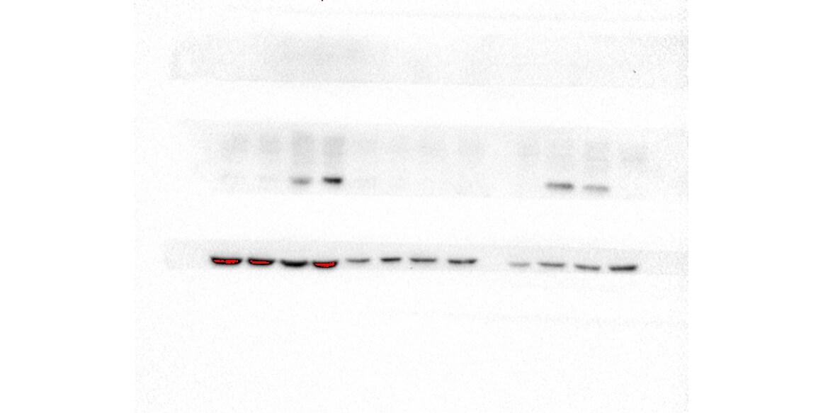
Scientific Data Images for INSIG-1 Antibody - BSA Free
Western Blot: INSIG-1 AntibodyBSA Free [NB110-55244]
Western Blot: INSIG-1 Antibody [NB110-55244] - Detection of Insig 1 in HepG2 lyaste using NB110-55244.![Immunocytochemistry/ Immunofluorescence: INSIG-1 Antibody - BSA Free [NB110-55244]](https://resources.rndsystems.com/images/products/INSIG-1-Antibody-Immunocytochemistry-Immunofluorescence-NB110-55244-img0003.jpg "Immunocytochemistry/ Immunofluorescence: INSIG-1 Antibody - BSA Free [NB110-55244]")
Immunocytochemistry/ Immunofluorescence: INSIG-1 Antibody - BSA Free [NB110-55244]
Immunocytochemistry/Immunofluorescence: INSIG-1 Antibody [NB110-55244] - INSIG-1 antibody was tested in HepG2 cells with Dylight 488 (green). Nuclei and alpha-tubulin were counterstained with DAPI (blue) and Dylight 550 (red).
Western Blot: INSIG-1 Antibody - BSA Free [NB110-55244] -
Validation of interaction between TDP-43 and SREBP2. (A) Post-translational modification of SREBP2. Anchor protein SCAP induces SREBP2 to translocate from endoplasmic reticulum (ER) to Golgi apparatus. SREBP2 is cleaved by S1P/S2P protease, and N-terminal SREBP2 is translocated to bind the SRE transcriptional domain of DNA for expression of target genes. (B) Immunoblots of proteins controlling SREBP processing pathway in TDP-43 abundant condition (Dox+) in comparison with cholesterol depleted condition. S: cholesterol-sufficient condition, D: cholesterol-depleted condition. The panels were cropped from original blots presented in Supplementary Fig. 3. (C) Quantification of the protein levels of cholesterol-related factors relative to beta -actin between Dox− and Dox+ condition; *p < 0.05, t-test, error bars = s.d. n = 3. (D) HEK293T cells were transfected with or without pcDNA3.1-TDP-43 and pFLAG-NSREBP2 (1–481) and the lysates were immunoprecipitated with FLAG antibody for the following immunoblots using TDP-43 antibody. Image collected and cropped by CiteAb from the following open publication (https://pubmed.ncbi.nlm.nih.gov/35568729), licensed under a CC-BY license. Not internally tested by Novus Biologicals.Applications for INSIG-1 Antibody - BSA Free
Application
Recommended Usage
Immunocytochemistry/ Immunofluorescence
1:100-1:1000
Western Blot
2 ug/ml
Application Notes
This INSIG-1 antibody is useful for Immunocytochemistry/Immunofluorescence and Western Blot, where a band is seen at ~30 kDa representing the Insig1 protein. In ICC/IF positive staining was localized to the ER membrane in HepG2 cells. The observed molecular weight of the protein may vary from the listed predicted molecular weight due to post translational modifications, post translation cleavages, relative charges, and other experimental factors.
Reviewed Applications
Read 1 review rated 5 using NB110-55244 in the following applications:
Formulation, Preparation, and Storage
Purification
Immunogen affinity purified
Formulation
PBS and 30% Glycerol
Format
BSA Free
Preservative
0.1% Sodium Azide
Concentration
1.02 mg/ml
Shipping
The product is shipped with polar packs. Upon receipt, store it immediately at the temperature recommended below.
Stability & Storage
Aliquot and store at -20C or -80C. Avoid freeze-thaw cycles.
Background: INSIG-1
Alternate Names
CL6, CL-6, INSIG-1, INSIG-1 membrane protein, insulin induced gene 1, insulin-induced gene 1 protein, MGC1405
Gene Symbol
INSIG1
Additional INSIG-1 Products
Product Documents for INSIG-1 Antibody - BSA Free
Certificate of Analysis
To download a Certificate of Analysis, please enter a lot or batch number in the search box below.
Product Specific Notices for INSIG-1 Antibody - BSA Free
This product is for research use only and is not approved for use in humans or in clinical diagnosis. Primary Antibodies are guaranteed for 1 year from date of receipt.
Citations for INSIG-1 Antibody - BSA Free
 Powered by Bioz
Customer Reviews for INSIG-1 Antibody - BSA Free (1)
5 out of 5
1 Customer Rating
Have you used INSIG-1 Antibody - BSA Free?
Submit a review and receive an Amazon gift card!
$25/€18/£15/$25CAN/¥2500 Yen for a review with an image
$10/€7/£6/$10CAN/¥1110 Yen for a review without an image
Submit a review
Customer Images
Showing
1
-
1 of
1 review
Showing All
Filter By:
-
Application: Western BlotSample Tested: U251 glioma cell line; whole cell lysate and U87 whole cell lysatesSpecies: HumanVerified Customer | Posted 07/15/2018There are three PVDF membranes in the picture, B-actin at the bottom and insig1 in the middle. It can be seen that in the human glioma cell line U87, the molecular weight of the insig1 protein in the U251 lysate is about 25-26kD。the molecular weight of the insig1 protein in the U251 lysate is about 25-26kD。(not the 30kD indicated on the manual). The strips are clearly visible and meet the submission requirements. The dilution ratio is (2ug/ml)

There are no reviews that match your criteria.
Protocols
View specific protocols for INSIG-1 Antibody - BSA Free (NB110-55244):
INSIG-1 Antibody:
Immunocytochemistry Protocol
Culture cells to appropriate density in 35 mm culture dishes or 6-well plates.
1. Remove culture medium and add 10% formalin to the dish. Fix at room temperature for 30 minutes.
2. Remove the formalin and add ice cold methanol. Incubate for 5-10 minutes.
3. Remove methanol and add washing solution (i.e. PBS). Be sure to not let the specimen dry out. Wash three times for 10 minutes.
4. To block nonspecific antibody binding incubate in 10% normal goat serum from 1 hour to overnight at room temperature.
5. Add primary antibody at appropriate dilution and incubate at room temperature from 2 hours to overnight at room temperature.
6. Remove primary antibody and replace with washing solution. Wash three times for 10 minutes.
7. Add secondary antibody at appropriate dilution. Incubate for 1 hour at room temperature.
8. Remove antibody and replace with wash solution, then wash for 10 minutes. Add Hoechst 33258 to wash solution at 1:25,0000 and incubate for 10 minutes. Wash a third time for 10 minutes.
9. Cells can be viewed directly after washing. The plates can also be stored in PBS containing Azide covered in Parafilm (TM). Cells can also be cover-slipped using Fluoromount, with appropriate sealing.
*The above information is only intended as a guide. The researcher should determine what protocol best meets their needs. Please follow safe laboratory procedures.
INSIG-1 Antibody:
Western Blot Protocol
1. Perform SDS-PAGE (4-12% MOPS) on samples to be analyzed, loading 30 ug of total protein per lane.
2. Transfer proteins to Nitrocellulose according to the instructions provided by the manufacturer of the transfer
apparatus.
3. Rinse membrane with dH2O and then stain the blot using Ponceau S for 1-2 minutes to access the transfer of proteins onto the nitrocellulose membrane. Rinse the blot in water to remove excess stain and mark the lane locations and locations of molecular weight markers using a pencil.
4. Rinse the blot in TBS for approximately 5 minutes.
5. Block the membrane using 5% non-fat dry milk + 1% BSA in TBS, 1 hour at room temperature.
6. Rinse the membrane in dH2O and then wash the membrane in wash buffer [TBS + 0.1% Tween] 3 times for 10 minutes each.
7. Dilute the rabbit anti-Insig1 primary antibody (NB 110-55244) in blocking buffer and incubate 2 hours at room temperature.
8. Rinse the membrane in dH2O and then wash the membrane in wash buffer [TBS + 0.1% Tween] 3 times for 10 minutes each.
9. Apply the diluted rabbit-IgG HRP-conjugated secondary antibody in blocking buffer (as per manufacturers
instructions) and incubate 1 hour at room temperature.
10. Wash the blot in wash buffer [TBS + 0.1% Tween] 3 times for 10 minutes each (this step can be repeated as required to reduce background).
11. Apply the detection reagent of choice in accordance with the manufacturers instructions (Pierce, ECL).
Note: Tween-20 can be added to the blocking or antibody diultion buffer at a final concentration of 0.05-0.2%, provided it does not interfere with antibody-antigen binding.
Find general support by application which include: protocols, troubleshooting, illustrated assays, videos and webinars.
- Appropriate Fixation of IHC/ICC Samples
- Cellular Response to Hypoxia Protocols
- ClariTSA™ Fluorophore Kits
- Detection & Visualization of Antibody Binding
- ICC Cell Smear Protocol for Suspension Cells
- ICC Immunocytochemistry Protocol Videos
- ICC for Adherent Cells
- Immunocytochemistry (ICC) Protocol
- Immunocytochemistry Troubleshooting
- Immunofluorescence of Organoids Embedded in Cultrex Basement Membrane Extract
- Immunohistochemistry (IHC) and Immunocytochemistry (ICC) Protocols
- Preparing Samples for IHC/ICC Experiments
- Preventing Non-Specific Staining (Non-Specific Binding)
- Primary Antibody Selection & Optimization
- Protocol for VisUCyte™ HRP Polymer Detection Reagent
- Protocol for the Fluorescent ICC Staining of Cell Smears - Graphic
- Protocol for the Fluorescent ICC Staining of Cultured Cells on Coverslips - Graphic
- Protocol for the Preparation and Fluorescent ICC Staining of Cells on Coverslips
- Protocol for the Preparation and Fluorescent ICC Staining of Non-adherent Cells
- Protocol for the Preparation and Fluorescent ICC Staining of Stem Cells on Coverslips
- Protocol for the Preparation of a Cell Smear for Non-adherent Cell ICC - Graphic
- R&D Systems Quality Control Western Blot Protocol
- TUNEL and Active Caspase-3 Detection by IHC/ICC Protocol
- The Importance of IHC/ICC Controls
- Troubleshooting Guide: Western Blot Figures
- Western Blot Conditions
- Western Blot Protocol
- Western Blot Protocol for Cell Lysates
- Western Blot Troubleshooting
- Western Blot Troubleshooting Guide
- View all Protocols, Troubleshooting, Illustrated assays and Webinars
Loading...