![Western Blot: IP3R1 Antibody [NBP1-21398]](https://resources.rndsystems.com/images/products/IP3R1-Antibody-Western-Blot-NBP1-21398-img0002.jpg "Western Blot: IP3R1 Antibody [NBP1-21398]")
Loading...
Key Product Details
Validated by
Independent Antibodies
Species Reactivity
Validated:
Human, Mouse
Cited:
Mouse
Predicted:
Rat (100%). Backed by our 100% Guarantee.
Applications
Validated:
Immunohistochemistry, Immunohistochemistry-Paraffin, Western Blot, Immunocytochemistry/ Immunofluorescence, Immunoprecipitation
Cited:
Western Blot
Label
Unconjugated
Antibody Source
Polyclonal Rabbit IgG
Loading...
Product Specifications
Immunogen
The immunogen recognized by this antibody maps to a region between residue 2708 and 2758 of human inositol 1,4,5-trisphosphate receptor type 1 using the numbering given in entry Q14643.2 (GeneID 3708).
Clonality
Polyclonal
Host
Rabbit
Isotype
IgG
Scientific Data Images for IP3R1 Antibody
![Immunohistochemistry-Paraffin: IP3R1 Antibody [NBP1-21398]](https://resources.rndsystems.com/images/products/IP3R1-Antibody-Immunohistochemistry-NBP1-21398-img0006.jpg "Immunohistochemistry-Paraffin: IP3R1 Antibody [NBP1-21398]")
Immunohistochemistry-Paraffin: IP3R1 Antibody [NBP1-21398]
Immunohistochemistry-Paraffin: IP3R1 Antibody [NBP1-21398] - Section of mouse cerebellum. Antibody: Affinity purified rabbit anti- IP3R1 used at a dilution of 1:100 (2ug/ml). Detection: Red-fluorescent Goat anti-Rabbit IgG-heavy and light chain cross-adsorbed Antibody DyLight 594 Conjugated used ata dilution of 1:100. Counterstain: DAPI (blue).![Immunohistochemistry-Paraffin: IP3R1 Antibody [NBP1-21398]](https://resources.rndsystems.com/images/products/IP3R1-Antibody-Immunohistochemistry-NBP1-21398-img0005.jpg "Immunohistochemistry-Paraffin: IP3R1 Antibody [NBP1-21398]")
Immunohistochemistry-Paraffin: IP3R1 Antibody [NBP1-21398]
Immunohistochemistry-Paraffin: IP3R1 Antibody [NBP1-21398] - Section of mouse cerebellum. Antibody: Affinity purified rabbit anti- IP3R1 used at a dilution of 1:200 (1ug/ml). Detection: DAB. Counterstain: IHC Hematoxylin (blue).Applications for IP3R1 Antibody
Application
Recommended Usage
Immunocytochemistry/ Immunofluorescence
1:50-1:250
Immunohistochemistry-Paraffin
1:100 - 1:500
Immunoprecipitation
2-5 ug/mg lysate
Western Blot
1:2000-1:10000
Application Notes
Epitope retrieval with Tris-EDTA pH9.0 is recommended for FFPE tissue sections.
Reviewed Applications
Read 1 review rated 5 using NBP1-21398 in the following applications:
Formulation, Preparation, and Storage
Purification
Immunogen affinity purified
Formulation
TBS and 0.1% BSA
Preservative
0.09% Sodium Azide
Concentration
0.2 mg/ml
Shipping
The product is shipped with polar packs. Upon receipt, store it immediately at the temperature recommended below.
Stability & Storage
Store at 4C. Do not freeze.
Background: IP3R1
Long Name
Inositol 1,4,5-trisphosphate receptor type 1
Alternate Names
InsP3R1, IP3R 1, ITPR1
Entrez Gene IDs
3708 (Human)
Gene Symbol
ITPR1
UniProt
Additional IP3R1 Products
Product Documents for IP3R1 Antibody
Certificate of Analysis
To download a Certificate of Analysis, please enter a lot or batch number in the search box below.
Product Specific Notices for IP3R1 Antibody
This product is for research use only and is not approved for use in humans or in clinical diagnosis. Primary Antibodies are guaranteed for 1 year from date of receipt.
Citations for IP3R1 Antibody
 Powered by Bioz
Customer Reviews for IP3R1 Antibody (1)
5 out of 5
1 Customer Rating
Have you used IP3R1 Antibody?
Submit a review and receive an Amazon gift card!
$25/€18/£15/$25CAN/¥2500 Yen for a review with an image
$10/€7/£6/$10CAN/¥1110 Yen for a review without an image
Submit a review
Customer Images
Showing
1
-
1 of
1 review
Showing All
Filter By:
-
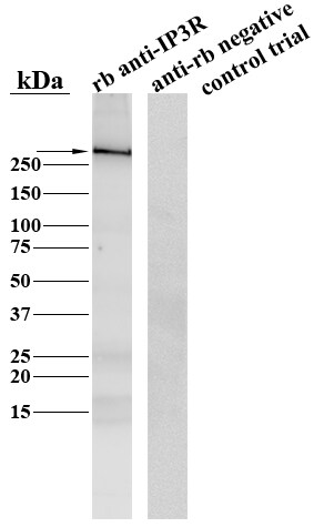
Application: Western BlotSample Tested: HindbrainSpecies: OtherVerified Customer | Posted 04/20/2016Western Blot showing an IP3R immunoreactive band around 270kDa

There are no reviews that match your criteria.
Protocols
Find general support by application which include: protocols, troubleshooting, illustrated assays, videos and webinars.
- Antigen Retrieval Protocol (PIER)
- Antigen Retrieval for Frozen Sections Protocol
- Appropriate Fixation of IHC/ICC Samples
- Cellular Response to Hypoxia Protocols
- Chromogenic IHC Staining of Formalin-Fixed Paraffin-Embedded (FFPE) Tissue Protocol
- Chromogenic Immunohistochemistry Staining of Frozen Tissue
- ClariTSA™ Fluorophore Kits
- Detection & Visualization of Antibody Binding
- Fluorescent IHC Staining of Frozen Tissue Protocol
- Graphic Protocol for Heat-induced Epitope Retrieval
- Graphic Protocol for the Preparation and Fluorescent IHC Staining of Frozen Tissue Sections
- Graphic Protocol for the Preparation and Fluorescent IHC Staining of Paraffin-embedded Tissue Sections
- Graphic Protocol for the Preparation of Gelatin-coated Slides for Histological Tissue Sections
- ICC Cell Smear Protocol for Suspension Cells
- ICC Immunocytochemistry Protocol Videos
- ICC for Adherent Cells
- IHC Sample Preparation (Frozen sections vs Paraffin)
- Immunocytochemistry (ICC) Protocol
- Immunocytochemistry Troubleshooting
- Immunofluorescence of Organoids Embedded in Cultrex Basement Membrane Extract
- Immunofluorescent IHC Staining of Formalin-Fixed Paraffin-Embedded (FFPE) Tissue Protocol
- Immunohistochemistry (IHC) and Immunocytochemistry (ICC) Protocols
- Immunohistochemistry Frozen Troubleshooting
- Immunohistochemistry Paraffin Troubleshooting
- Immunoprecipitation Protocol
- Preparing Samples for IHC/ICC Experiments
- Preventing Non-Specific Staining (Non-Specific Binding)
- Primary Antibody Selection & Optimization
- Protocol for Heat-Induced Epitope Retrieval (HIER)
- Protocol for Making a 4% Formaldehyde Solution in PBS
- Protocol for VisUCyte™ HRP Polymer Detection Reagent
- Protocol for the Fluorescent ICC Staining of Cell Smears - Graphic
- Protocol for the Fluorescent ICC Staining of Cultured Cells on Coverslips - Graphic
- Protocol for the Preparation & Fixation of Cells on Coverslips
- Protocol for the Preparation and Chromogenic IHC Staining of Frozen Tissue Sections
- Protocol for the Preparation and Chromogenic IHC Staining of Frozen Tissue Sections - Graphic
- Protocol for the Preparation and Chromogenic IHC Staining of Paraffin-embedded Tissue Sections
- Protocol for the Preparation and Chromogenic IHC Staining of Paraffin-embedded Tissue Sections - Graphic
- Protocol for the Preparation and Fluorescent ICC Staining of Cells on Coverslips
- Protocol for the Preparation and Fluorescent ICC Staining of Non-adherent Cells
- Protocol for the Preparation and Fluorescent ICC Staining of Stem Cells on Coverslips
- Protocol for the Preparation and Fluorescent IHC Staining of Frozen Tissue Sections
- Protocol for the Preparation and Fluorescent IHC Staining of Paraffin-embedded Tissue Sections
- Protocol for the Preparation of Gelatin-coated Slides for Histological Tissue Sections
- Protocol for the Preparation of a Cell Smear for Non-adherent Cell ICC - Graphic
- R&D Systems Quality Control Western Blot Protocol
- TUNEL and Active Caspase-3 Detection by IHC/ICC Protocol
- The Importance of IHC/ICC Controls
- Troubleshooting Guide: Immunohistochemistry
- Troubleshooting Guide: Western Blot Figures
- Western Blot Conditions
- Western Blot Protocol
- Western Blot Protocol for Cell Lysates
- Western Blot Troubleshooting
- Western Blot Troubleshooting Guide
- View all Protocols, Troubleshooting, Illustrated assays and Webinars
Loading...