Key Product Details
Species Reactivity
Human, Chicken (Negative), Mouse (Negative), Primate, Rat (Negative)
Applications
Flow Cytometry, Flow (Intracellular), Immunocytochemistry/ Immunofluorescence
Label
Unconjugated
Antibody Source
Monoclonal Mouse IgG1 kappa Clone # KU729
Loading...
Product Specifications
Immunogen
Nuclear extract of human HL-60 cells
Reactivity Notes
Does not react with Mouse, Rat and Chicken.
Localization
Nuclear
Marker
Nuclear Marker
Specificity
Recognizes a dimer of two proteins of 70kDa and ~80kDa, identified as two subunits of Ku. This monoclonal antibody recognizes a conformational epitope of p70/p80 dimer, which is destroyed during Western blotting. The p70/p80 dimer is important for function of a 460kDa DNA-dependent protein kinase. Ku protein plays a role in cell signaling, proliferation, DNA repair, replication, transcriptional activation, and apoptosis.
Clonality
Monoclonal
Host
Mouse
Isotype
IgG1 kappa
Description
200ug/ml of antibody purified from Bioreactor Concentrate by Protein A or G. Prepared in 10 mM PBS with 0.05% BSA & 0.05% azide. Also available WITHOUT BSA & azide at 1.0 mg/ml. (NBP2-34663)
Antibody with azide - store at 2 to 8C. Antibody without azide - store at -20 to -80 C.
Antibody with azide - store at 2 to 8C. Antibody without azide - store at -20 to -80 C.
Scientific Data Images for Ku70/XRCC6 Antibody (KU729)
![Immunocytochemistry/ Immunofluorescence: Ku70/XRCC6 Antibody (KU729) [NBP2-34247]](https://resources.rndsystems.com/images/products/Ku70-XRCC6-Antibody-KU729-Immunocytochemistry-Immunofluorescence-NBP2-34247-img0008.jpg "Immunocytochemistry/ Immunofluorescence: Ku70/XRCC6 Antibody (KU729) [NBP2-34247]")
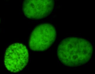
Immunocytochemistry/ Immunofluorescence: Ku70/XRCC6 Antibody (KU729) [NBP2-34247]
Immunocytochemistry/Immunofluorescence: Ku70/XRCC6 Antibody (KU729) [NBP2-34247] - Immunofluorescent analysis of HeLa cells using Ku70/XRCC6 antibody (KU729). Image from verified customer review.![Flow Cytometry: Ku70/XRCC6 Antibody (KU729) [NBP2-34247]](https://resources.rndsystems.com/images/products/Ku70-XRCC6-Antibody-KU729-Flow-Cytometry-NBP2-34247-img0003.jpg "Flow Cytometry: Ku70/XRCC6 Antibody (KU729) [NBP2-34247]")
Flow Cytometry: Ku70/XRCC6 Antibody (KU729) [NBP2-34247]
Flow Cytometry: Ku70/XRCC6 Antibody (KU729) [NBP2-34247] - Flow Cytometric Analysis of human Ku (p70/p80) on 293T cells. Black: cells alone; Grey: Isotype Control; Green: AF488-labeled Ku70/XRCC6 Antibody (KU729).![Immunocytochemistry/ Immunofluorescence: Ku70/XRCC6 Antibody (KU729) [NBP2-34247]](https://resources.rndsystems.com/images/products/Ku70-XRCC6-Antibody-KU729-Immunocytochemistry-Immunofluorescence-NBP2-34247-img0004.jpg "Immunocytochemistry/ Immunofluorescence: Ku70/XRCC6 Antibody (KU729) [NBP2-34247]")
Immunocytochemistry/ Immunofluorescence: Ku70/XRCC6 Antibody (KU729) [NBP2-34247]
Immunocytochemistry/Immunofluorescence: Ku70/XRCC6 Antibody (KU729) [NBP2-34247] - Immunofluorescent staining of paraformaldehyde-fixed HeLa cells using followed by goat anti-Mouse IgG-CF488 (Green). Phalloidin (Red) is used to label cell membrane.![Flow Cytometry: Ku70/XRCC6 Antibody (KU729) [NBP2-34247]](https://resources.rndsystems.com/images/products/Ku70-XRCC6-Antibody-KU729-Flow-Cytometry-NBP2-34247-img0002.jpg "Flow Cytometry: Ku70/XRCC6 Antibody (KU729) [NBP2-34247]")
Flow Cytometry: Ku70/XRCC6 Antibody (KU729) [NBP2-34247]
Flow Cytometry: Ku70/XRCC6 Antibody (KU729) [NBP2-34247] - Flow Cytometry of human Ku (p70/p80) on K562 cells. Black: cells alone; Green: Isotype Control; Red: PE-labeled Ku70/XRCC6 Antibody (KU729).: Ku70/XRCC6 Antibody (KU729) [NBP2-34247] -")
Flow (Intracellular): Ku70/XRCC6 Antibody (KU729) [NBP2-34247] -
Flow (Intracellular): Ku70/XRCC6 Antibody (KU729) [NBP2-34247] - An intracellular stain was performed on Hek293 cells with Ku70/XRCC6 Antibody (KU729) NBP2-34663AF647 (blue) and a matched isotype control (orange). Cells were fixed with 4% PFA and then permeabilized with 0.1% saponin. Cells were incubated in an antibody dilution of 2.5 ug/mL for 30 minutes at room temperature. Both antibodies were conjugated to Alexa Fluor 647.: Ku70/XRCC6 Antibody (KU729) [NBP2-34247] -")
Flow (Intracellular): Ku70/XRCC6 Antibody (KU729) [NBP2-34247] -
Flow (Intracellular): Ku70/XRCC6 Antibody (KU729) [NBP2-34247] - An intracellular stain was performed on Hek293 cells with Ku70/XRCC6 Antibody (KU729) NBP2-34663AF647 (blue) and a matched isotype control (orange). Cells were fixed with 4% PFA and then permeabilized with 0.1% saponin. Cells were incubated in an antibody dilution of 2.5 ug/mL for 30 minutes at room temperature. Both antibodies were conjugated to Alexa Fluor 647.Applications for Ku70/XRCC6 Antibody (KU729)
Application
Recommended Usage
Flow Cytometry
1-2 ug/million cells
Immunocytochemistry/ Immunofluorescence
1-2 ug/ml
Application Notes
Immunocytochemistry (Acetone-fixed cells): 0.5-1.0ug/ml for 30 minutes at RT. Optimal dilution for a specific application should be determined.
Optimal dilution for a specific application should be determined.
Optimal dilution for a specific application should be determined.
Reviewed Applications
Read 1 review rated 5 using NBP2-34247 in the following applications:
Flow Cytometry Panel Builder
Bio-Techne Knows Flow Cytometry
Save time and reduce costly mistakes by quickly finding compatible reagents using the Panel Builder Tool.
Advanced Features
- Spectra Viewer - Custom analysis of spectra from multiple fluorochromes
- Spillover Popups - Visualize the spectra of individual fluorochromes
- Antigen Density Selector - Match fluorochrome brightness with antigen density

Formulation, Preparation, and Storage
Purification
Protein A or G purified
Formulation
10 mM PBS with 0.05% BSA
Preservative
0.05% Sodium Azide
Concentration
0.2 mg/ml
Shipping
The product is shipped with polar packs. Upon receipt, store it immediately at the temperature recommended below.
Stability & Storage
Store at 4C.
Background: Ku70/XRCC6
Long Name
Lupus Ku Autoantigen Protein 70
Alternate Names
CTC75, CTCBF, G22P1, ML8, TLAA, XRCC6
Gene Symbol
XRCC6
Additional Ku70/XRCC6 Products
Product Documents for Ku70/XRCC6 Antibody (KU729)
Certificate of Analysis
To download a Certificate of Analysis, please enter a lot or batch number in the search box below.
Product Specific Notices for Ku70/XRCC6 Antibody (KU729)
This product is for research use only and is not approved for use in humans or in clinical diagnosis. Primary Antibodies are guaranteed for 1 year from date of receipt.
Customer Reviews for Ku70/XRCC6 Antibody (KU729) (1)
5 out of 5
1 Customer Rating
Have you used Ku70/XRCC6 Antibody (KU729)?
Submit a review and receive an Amazon gift card!
$25/€18/£15/$25CAN/¥2500 Yen for a review with an image
$10/€7/£6/$10CAN/¥1110 Yen for a review without an image
Submit a review
Customer Images
Showing
1
-
1 of
1 review
Showing All
Filter By:
-
Application: ImmunocytochemistrySample Tested: HeLa cellsSpecies: HumanVerified Customer | Posted 03/22/2022HeLa cells

There are no reviews that match your criteria.
Protocols
Find general support by application which include: protocols, troubleshooting, illustrated assays, videos and webinars.
- 7-Amino Actinomycin D (7-AAD) Cell Viability Flow Cytometry Protocol
- Appropriate Fixation of IHC/ICC Samples
- Cellular Response to Hypoxia Protocols
- ClariTSA™ Fluorophore Kits
- Detection & Visualization of Antibody Binding
- Extracellular Membrane Flow Cytometry Protocol
- Flow Cytometry Protocol for Cell Surface Markers
- Flow Cytometry Protocol for Staining Membrane Associated Proteins
- Flow Cytometry Staining Protocols
- Flow Cytometry Troubleshooting Guide
- ICC Cell Smear Protocol for Suspension Cells
- ICC Immunocytochemistry Protocol Videos
- ICC for Adherent Cells
- Immunocytochemistry (ICC) Protocol
- Immunocytochemistry Troubleshooting
- Immunofluorescence of Organoids Embedded in Cultrex Basement Membrane Extract
- Immunohistochemistry (IHC) and Immunocytochemistry (ICC) Protocols
- Intracellular Flow Cytometry Protocol Using Alcohol (Methanol)
- Intracellular Flow Cytometry Protocol Using Detergents
- Intracellular Nuclear Staining Flow Cytometry Protocol Using Detergents
- Intracellular Staining Flow Cytometry Protocol Using Alcohol Permeabilization
- Intracellular Staining Flow Cytometry Protocol Using Detergents to Permeabilize Cells
- Preparing Samples for IHC/ICC Experiments
- Preventing Non-Specific Staining (Non-Specific Binding)
- Primary Antibody Selection & Optimization
- Propidium Iodide Cell Viability Flow Cytometry Protocol
- Protocol for Liperfluo
- Protocol for VisUCyte™ HRP Polymer Detection Reagent
- Protocol for the Characterization of Human Th22 Cells
- Protocol for the Characterization of Human Th9 Cells
- Protocol for the Fluorescent ICC Staining of Cell Smears - Graphic
- Protocol for the Fluorescent ICC Staining of Cultured Cells on Coverslips - Graphic
- Protocol for the Preparation and Fluorescent ICC Staining of Cells on Coverslips
- Protocol for the Preparation and Fluorescent ICC Staining of Non-adherent Cells
- Protocol for the Preparation and Fluorescent ICC Staining of Stem Cells on Coverslips
- Protocol for the Preparation of a Cell Smear for Non-adherent Cell ICC - Graphic
- Protocol: Annexin V and PI Staining by Flow Cytometry
- Protocol: Annexin V and PI Staining for Apoptosis by Flow Cytometry
- TUNEL and Active Caspase-3 Detection by IHC/ICC Protocol
- The Importance of IHC/ICC Controls
- Troubleshooting Guide: Fluorokine Flow Cytometry Kits
- View all Protocols, Troubleshooting, Illustrated assays and Webinars
Loading...