Laminin gamma 1 Antibody (A5)
Novus Biologicals | Catalog # NBP2-44751
Key Product Details
Species Reactivity
Validated:
Human, Mouse, Bovine
Cited:
Mouse
Applications
Validated:
Immunohistochemistry, Immunohistochemistry-Paraffin, Immunohistochemistry-Frozen, Flow Cytometry, Immunofluorescence, Immunocytochemistry/ Immunofluorescence
Cited:
Immunohistochemistry-Frozen, Immunocytochemistry/ Immunofluorescence, IF/IHC
Label
Unconjugated
Antibody Source
Monoclonal Rat IgG2a Kappa Clone # A5
Loading...
Product Specifications
Immunogen
Murine EHS laminin preparation
Reactivity Notes
Bovine reported by a verified customer review.
Localization
Basement membrane
Specificity
Laminins are large hetero-trimeric, non-collagenous glycoproteins composed of alpha, beta, and gamma chains. This monoclonal antibody reacts with laminin B2/1 chain of ~210kDa and does not cross-react with other basement membrane components or fibronectin. Its specificity was established by immunoprecipitation and immunofluorescence of human skeletal muscle and kidney with laminin chain-specific monoclonal antibodys. Epithelial sheets in vivo are separated from the mesenchymal elements of the stroma by a thin layer of a specialized type of extracellular matrix termed the basement membrane (BM). This structure consists of individual components, some of which are ubiquitous in BMs and some are not. The ubiquitous ones comprise laminin (LN), entactin/nidogen (EN), collagen type IV (CIV), and large heparan sulfate proteoglycan (HSPG), which interact specifically with each other to form a continuous and regular BM. Alterations of BM integrity, from local discontinuities up to complete loss, are described in many types of human and animal epithelial neoplasms. This monoclonal antibody stains uniformly all human and murine basement membranes.
Clonality
Monoclonal
Host
Rat
Isotype
IgG2a Kappa
Theoretical MW
210 kDa.
Disclaimer note: The observed molecular weight of the protein may vary from the listed predicted molecular weight due to post translational modifications, post translation cleavages, relative charges, and other experimental factors.
Disclaimer note: The observed molecular weight of the protein may vary from the listed predicted molecular weight due to post translational modifications, post translation cleavages, relative charges, and other experimental factors.
Description
200ug/ml of antibody purified from Bioreactor Concentrate by Protein A or G. Prepared in 10 mM PBS with 0.05% BSA & 0.05% azide. Also available WITHOUT BSA at 1.0 mg/ml. (NBP2-47842)
Antibody with azide - store at 2 to 8C. Antibody without azide - store at -20 to -80 C.
Antibody with azide - store at 2 to 8C. Antibody without azide - store at -20 to -80 C.
Scientific Data Images for Laminin gamma 1 Antibody (A5)
![Immunohistochemistry-Paraffin: Laminin gamma 1 Antibody (A5) [NBP2-44751]](https://resources.rndsystems.com/images/products/Laminin-gamma-1-Antibody-A5-Immunohistochemistry-Paraffin-NBP2-44751-img0003.jpg "Immunohistochemistry-Paraffin: Laminin gamma 1 Antibody (A5) [NBP2-44751]")
Immunohistochemistry-Paraffin: Laminin gamma 1 Antibody (A5) [NBP2-44751]
Immunohistochemistry-Paraffin: Laminin gamma 1 Antibody (A5) [NBP2-44751] - Formalin-fixed, paraffin-embedded human Renal Cell Carcinoma stained with Laminin gamma 1 Antibody (A5).![Immunohistochemistry-Frozen: Laminin gamma 1 Antibody (A5) [NBP2-44751]](https://resources.rndsystems.com/images/products/Laminin-gamma-1-Antibody-A5-Immunohistochemistry-Frozen-NBP2-44751-img0002.jpg "Immunohistochemistry-Frozen: Laminin gamma 1 Antibody (A5) [NBP2-44751]")
Immunohistochemistry-Frozen: Laminin gamma 1 Antibody (A5) [NBP2-44751]
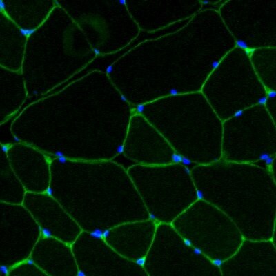
Immunohistochemistry-Frozen: Laminin gamma 1 Antibody (A5) [NBP2-44751] - Bovine frozen skeletal muscle section was fixed in 4% PFA for 5 min and stained with anti-laminin antibody (NBP2-44751) overnight at 4 C and then stained with a goat anti-rat-Alexa Fluor 488 secondary antibody (green) for 1 hour at room temperature. Section was then mounted in a mounting medium containing Dapi (blue). Image from verified customer review.![Immunohistochemistry-Paraffin: Laminin gamma 1 Antibody (A5) [NBP2-44751]](https://resources.rndsystems.com/images/products/Laminin-gamma-1-Antibody-A5-Immunohistochemistry-Paraffin-NBP2-44751-img0001.jpg "Immunohistochemistry-Paraffin: Laminin gamma 1 Antibody (A5) [NBP2-44751]")
Immunohistochemistry-Paraffin: Laminin gamma 1 Antibody (A5) [NBP2-44751]
Immunohistochemistry-Paraffin: Laminin gamma 1 Antibody (A5) [NBP2-44751] - Human Renal Cell Carcinoma stained with Laminin Monoclonal Antibody (A5).Applications for Laminin gamma 1 Antibody (A5)
Application
Recommended Usage
Flow Cytometry
0.5-1 ug/million cells
Immunocytochemistry/ Immunofluorescence
0.5-1.0 ug/ml
Immunofluorescence
1 - 2 ug/ml
Immunohistochemistry-Paraffin
1-2 ug/ml
Application Notes
Immunohistochemistry (Formalin-fixed): 1-2ug/ml for 30 minutes at RT. Staining of formalin-fixed tissues requires heating tissue sections in 10mM Tris with 1mM EDTA, pH 9.0, for 45 min at 95C followed by cooling at RT for 20 minutes.
Optimal dilution for a specific application should be determined.
Optimal dilution for a specific application should be determined.
Reviewed Applications
Read 1 review rated 5 using NBP2-44751 in the following applications:
Flow Cytometry Panel Builder
Bio-Techne Knows Flow Cytometry
Save time and reduce costly mistakes by quickly finding compatible reagents using the Panel Builder Tool.
Advanced Features
- Spectra Viewer - Custom analysis of spectra from multiple fluorochromes
- Spillover Popups - Visualize the spectra of individual fluorochromes
- Antigen Density Selector - Match fluorochrome brightness with antigen density

Formulation, Preparation, and Storage
Purification
Protein A or G purified
Formulation
10 mM PBS with 0.05% BSA
Preservative
0.05% Sodium Azide
Concentration
0.2 mg/ml
Shipping
The product is shipped with polar packs. Upon receipt, store it immediately at the temperature recommended below.
Stability & Storage
Store at 4C.
Background: Laminin gamma 1
Alternate Names
LAMC1, Laminin B2
Gene Symbol
LAMC1
UniProt
Additional Laminin gamma 1 Products
Product Documents for Laminin gamma 1 Antibody (A5)
Certificate of Analysis
To download a Certificate of Analysis, please enter a lot or batch number in the search box below.
Product Specific Notices for Laminin gamma 1 Antibody (A5)
This product is for research use only and is not approved for use in humans or in clinical diagnosis. Primary Antibodies are guaranteed for 1 year from date of receipt.
Related Research Areas
Citations for Laminin gamma 1 Antibody (A5)
 Powered by Bioz
Customer Reviews for Laminin gamma 1 Antibody (A5) (1)
5 out of 5
1 Customer Rating
Have you used Laminin gamma 1 Antibody (A5)?
Submit a review and receive an Amazon gift card!
$25/€18/£15/$25CAN/¥2500 Yen for a review with an image
$10/€7/£6/$10CAN/¥1110 Yen for a review without an image
Submit a review
Customer Images
Showing
1
-
1 of
1 review
Showing All
Filter By:
-
Application: Immunohistochemistry-FrozenSample Tested: Skeletal muscleSpecies: BovineVerified Customer | Posted 06/01/2018Bovine skeletal muscle. Laminin was stained with anti-laminin antibody (NBP2-44751) (green). Nuclei were stained with Dapi (blue).Bovine frozen skeletal muscle section was fixed in 4% PFA for 5 min and stained with anti-laminin antibody (NBP2-44751) overnight at 4 C and then stained with a goat anti-rat-Alexa Fluor 488 secondary antibody (green) for 1 hour at room temperature. Section was then mounted in a mounting medium containing Dapi (blue).

There are no reviews that match your criteria.
Protocols
Find general support by application which include: protocols, troubleshooting, illustrated assays, videos and webinars.
- 7-Amino Actinomycin D (7-AAD) Cell Viability Flow Cytometry Protocol
- Antigen Retrieval Protocol (PIER)
- Antigen Retrieval for Frozen Sections Protocol
- Appropriate Fixation of IHC/ICC Samples
- Cellular Response to Hypoxia Protocols
- Chromogenic IHC Staining of Formalin-Fixed Paraffin-Embedded (FFPE) Tissue Protocol
- Chromogenic Immunohistochemistry Staining of Frozen Tissue
- ClariTSA™ Fluorophore Kits
- Detection & Visualization of Antibody Binding
- Extracellular Membrane Flow Cytometry Protocol
- Flow Cytometry Protocol for Cell Surface Markers
- Flow Cytometry Protocol for Staining Membrane Associated Proteins
- Flow Cytometry Staining Protocols
- Flow Cytometry Troubleshooting Guide
- Fluorescent IHC Staining of Frozen Tissue Protocol
- Graphic Protocol for Heat-induced Epitope Retrieval
- Graphic Protocol for the Preparation and Fluorescent IHC Staining of Frozen Tissue Sections
- Graphic Protocol for the Preparation and Fluorescent IHC Staining of Paraffin-embedded Tissue Sections
- Graphic Protocol for the Preparation of Gelatin-coated Slides for Histological Tissue Sections
- ICC Cell Smear Protocol for Suspension Cells
- ICC Immunocytochemistry Protocol Videos
- ICC for Adherent Cells
- IHC Sample Preparation (Frozen sections vs Paraffin)
- Immunocytochemistry (ICC) Protocol
- Immunocytochemistry Troubleshooting
- Immunofluorescence of Organoids Embedded in Cultrex Basement Membrane Extract
- Immunofluorescent IHC Staining of Formalin-Fixed Paraffin-Embedded (FFPE) Tissue Protocol
- Immunohistochemistry (IHC) and Immunocytochemistry (ICC) Protocols
- Immunohistochemistry Frozen Troubleshooting
- Immunohistochemistry Paraffin Troubleshooting
- Intracellular Flow Cytometry Protocol Using Alcohol (Methanol)
- Intracellular Flow Cytometry Protocol Using Detergents
- Intracellular Nuclear Staining Flow Cytometry Protocol Using Detergents
- Intracellular Staining Flow Cytometry Protocol Using Alcohol Permeabilization
- Intracellular Staining Flow Cytometry Protocol Using Detergents to Permeabilize Cells
- Preparing Samples for IHC/ICC Experiments
- Preventing Non-Specific Staining (Non-Specific Binding)
- Primary Antibody Selection & Optimization
- Propidium Iodide Cell Viability Flow Cytometry Protocol
- Protocol for Heat-Induced Epitope Retrieval (HIER)
- Protocol for Liperfluo
- Protocol for Making a 4% Formaldehyde Solution in PBS
- Protocol for VisUCyte™ HRP Polymer Detection Reagent
- Protocol for the Characterization of Human Th22 Cells
- Protocol for the Characterization of Human Th9 Cells
- Protocol for the Fluorescent ICC Staining of Cell Smears - Graphic
- Protocol for the Fluorescent ICC Staining of Cultured Cells on Coverslips - Graphic
- Protocol for the Preparation & Fixation of Cells on Coverslips
- Protocol for the Preparation and Chromogenic IHC Staining of Frozen Tissue Sections
- Protocol for the Preparation and Chromogenic IHC Staining of Frozen Tissue Sections - Graphic
- Protocol for the Preparation and Chromogenic IHC Staining of Paraffin-embedded Tissue Sections
- Protocol for the Preparation and Chromogenic IHC Staining of Paraffin-embedded Tissue Sections - Graphic
- Protocol for the Preparation and Fluorescent ICC Staining of Cells on Coverslips
- Protocol for the Preparation and Fluorescent ICC Staining of Non-adherent Cells
- Protocol for the Preparation and Fluorescent ICC Staining of Stem Cells on Coverslips
- Protocol for the Preparation and Fluorescent IHC Staining of Frozen Tissue Sections
- Protocol for the Preparation and Fluorescent IHC Staining of Paraffin-embedded Tissue Sections
- Protocol for the Preparation of Gelatin-coated Slides for Histological Tissue Sections
- Protocol for the Preparation of a Cell Smear for Non-adherent Cell ICC - Graphic
- Protocol: Annexin V and PI Staining by Flow Cytometry
- Protocol: Annexin V and PI Staining for Apoptosis by Flow Cytometry
- TUNEL and Active Caspase-3 Detection by IHC/ICC Protocol
- The Importance of IHC/ICC Controls
- Troubleshooting Guide: Fluorokine Flow Cytometry Kits
- Troubleshooting Guide: Immunohistochemistry
- View all Protocols, Troubleshooting, Illustrated assays and Webinars
Loading...
Associated Pathways

