LAR/PTPRF Antibody (S165-38) - BSA Free
Novus Biologicals | Catalog # NBP2-42172
![Western Blot: LAR/PTPRF Antibody (S165-38) [NBP2-42172]](https://resources.rndsystems.com/images/products/LAR-PTPRF-Antibody-S165-38-Western-Blot-NBP2-42172-img0005.jpg "Western Blot: LAR/PTPRF Antibody (S165-38) [NBP2-42172]")
Key Product Details
Species Reactivity
Human, Mouse, Rat
Applications
Western Blot, Immunocytochemistry/ Immunofluorescence
Label
Unconjugated
Antibody Source
Monoclonal Mouse IgG2A Clone # S165-38
Format
BSA Free
Loading...
Product Specifications
Immunogen
Fusion protein amino acids 1315-1607 (cytoplasmic C-terminus) of human LAR. 97% identical in both rat and mouse. >80% identity with PTPRD and PTPRS. >50% identity with PTPRM and PTPRK.
Localization
Membrane
Specificity
Greater then 80% identity with PTPRD and PTPRS. Greater then 50% identity with PTPRM and PTPRK.Detects approx 85kDa (full length protein is 210 kDa -smaller due to proteolysis into P-subunit containing transmembrane and intracellular domains.
Clonality
Monoclonal
Host
Mouse
Isotype
IgG2A
Scientific Data Images for LAR/PTPRF Antibody (S165-38) - BSA Free
Western Blot: LAR/PTPRF Antibody (S165-38) [NBP2-42172]
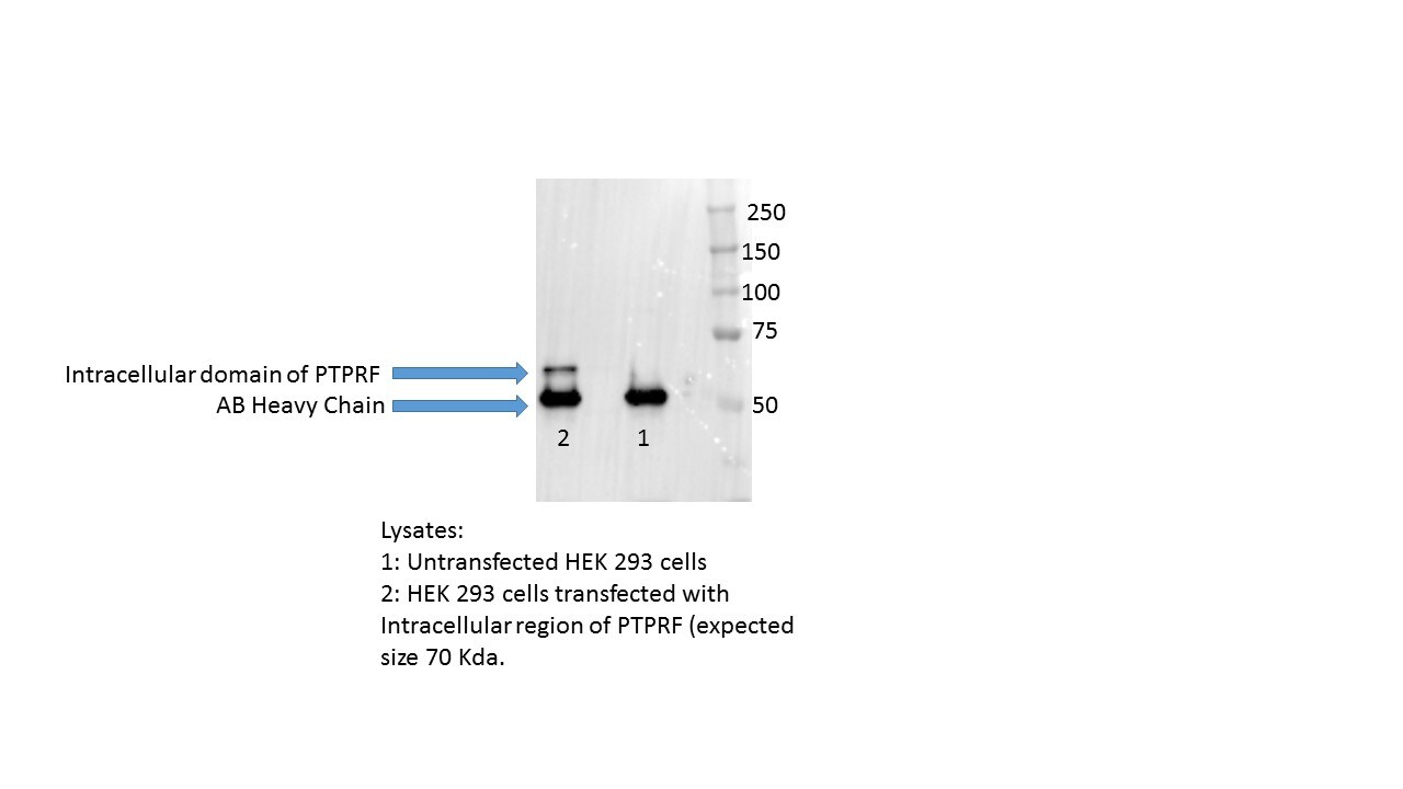
Western Blot: LAR/PTPRF Antibody (S165-38) [NBP2-42172] - Western Blot analysis of Rat Brain Membrane showing detection of LAR/PTPRF protein using Mouse Anti-LAR/PTPRF Monoclonal Antibody, Clone S165-38 (NBP2-42172). Primary Antibody: Mouse Anti-LAR/PTPRF Monoclonal Antibody (NBP2-42172) at 1:250.![Immunocytochemistry/ Immunofluorescence: LAR/PTPRF Antibody (S165-38) [NBP2-42172]](https://resources.rndsystems.com/images/products/LAR-PTPRF-Antibody-S165-38-Immunocytochemistry-Immunofluorescence-NBP2-42172-img0006.jpg "Immunocytochemistry/ Immunofluorescence: LAR/PTPRF Antibody (S165-38) [NBP2-42172]")
Immunocytochemistry/ Immunofluorescence: LAR/PTPRF Antibody (S165-38) [NBP2-42172]
Immunocytochemistry/Immunofluorescence: LAR/PTPRF Antibody (S165-38) [NBP2-42172] - Immunocytochemistry/Immunofluorescence analysis using Mouse Anti-LAR/PTPRF/PTPRF Monoclonal Antibody, Clone S165-38 (NBP2-42172). Tissue: Neuroblastoma cells (SH-SY5Y). Species: Human. Fixation: 4% PFA for 15 min. Primary Antibody: Mouse Anti-LAR/PTPRF/PTPRF Monoclonal Antibody (NBP2-42172) at 1:100 for overnight at 4C with slow rocking. Secondary Antibody: AlexaFluor 488 at 1:1000 for 1 hour at RT. Counterstain: Phalloidin-iFluor 647 (red) F-Actin stain; Hoechst (blue) nuclear stain at 1:800, 1.6mM for 20 min at RT. (A) Hoechst (blue) nuclear stain. (B) Phalloidin-iFluor 647 (red) F-Actin stain. (C) LAR/PTPRF/PTPRF Antibody (D) Composite.![Immunocytochemistry/ Immunofluorescence: LAR/PTPRF Antibody (S165-38) [NBP2-42172]](https://resources.rndsystems.com/images/products/LAR-PTPRF-Antibody-S165-38-Immunocytochemistry-Immunofluorescence-NBP2-42172-img0004.jpg "Immunocytochemistry/ Immunofluorescence: LAR/PTPRF Antibody (S165-38) [NBP2-42172]")
Immunocytochemistry/ Immunofluorescence: LAR/PTPRF Antibody (S165-38) [NBP2-42172]
Immunocytochemistry/Immunofluorescence: LAR/PTPRF Antibody (S165-38) [NBP2-42172] - Tissue: Neuroblastoma cell line SK-N-BE. Species: Human. Fixation: 4% Formaldehyde for 15 min at RT. Primary Antibody: Mouse Anti-LAR/PTPRF Monoclonal Antibody at 1:100 for 60 min at RT. Secondary Antibody: Goat Anti-Mouse ATTO 488 at 1:100 for 60 min at RT. Counterstain: Phalloidin Texas Red F-Actin stain; DAPI (blue) nuclear stain at 1:1000, 1:5000 for 60min RT, 5min RT. Localization: Membrane. Magnification: 60X.![Immunocytochemistry/ Immunofluorescence: LAR/PTPRF Antibody (S165-38) [NBP2-42172]](https://resources.rndsystems.com/images/products/LAR-PTPRF-Antibody-S165-38-Immunocytochemistry-Immunofluorescence-NBP2-42172-img0007.jpg "Immunocytochemistry/ Immunofluorescence: LAR/PTPRF Antibody (S165-38) [NBP2-42172]")
Immunocytochemistry/ Immunofluorescence: LAR/PTPRF Antibody (S165-38) [NBP2-42172]
Immunocytochemistry/Immunofluorescence: LAR/PTPRF Antibody (S165-38) [NBP2-42172] - Immunocytochemistry/Immunofluorescence analysis using Mouse Anti-LAR/PTPRF/PTPRF Monoclonal Antibody, Clone S165-38 (NBP2-42172). Tissue: Neuroblastoma cell line (SK-N-BE). Species: Human. Fixation: 4% Formaldehyde for 15 min at RT. Primary Antibody: Mouse Anti-LAR/PTPRF/PTPRF Monoclonal Antibody (NBP2-42172) at 1:100 for 60 min at RT. Secondary Antibody: Goat Anti-Mouse ATTO 488 at 1:100 for 60 min at RT. Counterstain: Phalloidin Texas Red F-Actin stain; DAPI (blue) nuclear stain at 1:1000, 1:5000 for 60min RT, 5min RT. Localization: Membrane. Magnification: 60X. (A) DAPI (blue) nuclear stain. (B) Phalloidin Texas Red F-Actin stain. (C) LAR/PTPRF/PTPRF Antibody. (D) Composite.Applications for LAR/PTPRF Antibody (S165-38) - BSA Free
Application
Recommended Usage
Western Blot
1:1000
Application Notes
1 ug/ml of LAR/PTPRF Antibody was sufficient for detection of LAR/PTPRF in 20 ug of rat brain lysate by colorimetric immunoblot analysis using Goat anti-mouse IgG:HRP as the secondary Antibody.
Reviewed Applications
Read 1 review rated 5 using NBP2-42172 in the following applications:
Formulation, Preparation, and Storage
Purification
Protein G purified
Formulation
PBS (pH 7.4), 50% Glycerol
Format
BSA Free
Preservative
0.1% Sodium Azide
Concentration
1 mg/ml
Shipping
The product is shipped with polar packs. Upon receipt, store it immediately at the temperature recommended below.
Stability & Storage
Store at 4C short term. Aliquot and store at -20C long term. Avoid freeze-thaw cycles.
Background: LAR
Long Name
Leukocyte Antigen-Related Tyrosine Phosphatase
Alternate Names
PTPRF
Gene Symbol
PTPRF
UniProt
Additional LAR Products
Product Documents for LAR/PTPRF Antibody (S165-38) - BSA Free
Certificate of Analysis
To download a Certificate of Analysis, please enter a lot or batch number in the search box below.
Product Specific Notices for LAR/PTPRF Antibody (S165-38) - BSA Free
This product is for research use only and is not approved for use in humans or in clinical diagnosis. Primary Antibodies are guaranteed for 1 year from date of receipt.
Related Research Areas
Customer Reviews for LAR/PTPRF Antibody (S165-38) - BSA Free (1)
5 out of 5
1 Customer Rating
Have you used LAR/PTPRF Antibody (S165-38) - BSA Free?
Submit a review and receive an Amazon gift card!
$25/€18/£15/$25CAN/¥2500 Yen for a review with an image
$10/€7/£6/$10CAN/¥1110 Yen for a review without an image
Submit a review
Customer Images
Showing
1
-
1 of
1 review
Showing All
Filter By:
-
Application: ImmunoprecipitationSample Tested: Sample 1: Untransfected HEK293 cellsSpecies: HumanVerified Customer | Posted 02/07/2017NSCS16

There are no reviews that match your criteria.
Protocols
Find general support by application which include: protocols, troubleshooting, illustrated assays, videos and webinars.
- Appropriate Fixation of IHC/ICC Samples
- Cellular Response to Hypoxia Protocols
- ClariTSA™ Fluorophore Kits
- Detection & Visualization of Antibody Binding
- ICC Cell Smear Protocol for Suspension Cells
- ICC Immunocytochemistry Protocol Videos
- ICC for Adherent Cells
- Immunocytochemistry (ICC) Protocol
- Immunocytochemistry Troubleshooting
- Immunofluorescence of Organoids Embedded in Cultrex Basement Membrane Extract
- Immunohistochemistry (IHC) and Immunocytochemistry (ICC) Protocols
- Preparing Samples for IHC/ICC Experiments
- Preventing Non-Specific Staining (Non-Specific Binding)
- Primary Antibody Selection & Optimization
- Protocol for VisUCyte™ HRP Polymer Detection Reagent
- Protocol for the Fluorescent ICC Staining of Cell Smears - Graphic
- Protocol for the Fluorescent ICC Staining of Cultured Cells on Coverslips - Graphic
- Protocol for the Preparation and Fluorescent ICC Staining of Cells on Coverslips
- Protocol for the Preparation and Fluorescent ICC Staining of Non-adherent Cells
- Protocol for the Preparation and Fluorescent ICC Staining of Stem Cells on Coverslips
- Protocol for the Preparation of a Cell Smear for Non-adherent Cell ICC - Graphic
- R&D Systems Quality Control Western Blot Protocol
- TUNEL and Active Caspase-3 Detection by IHC/ICC Protocol
- The Importance of IHC/ICC Controls
- Troubleshooting Guide: Western Blot Figures
- Western Blot Conditions
- Western Blot Protocol
- Western Blot Protocol for Cell Lysates
- Western Blot Troubleshooting
- Western Blot Troubleshooting Guide
- View all Protocols, Troubleshooting, Illustrated assays and Webinars
Loading...