Lyn Antibody (LYN-01) - BSA Free
Novus Biologicals | Catalog # NB500-519
![Western Blot: Lyn Antibody (LYN-01)BSA Free [NB500-519]](https://resources.rndsystems.com/images/products/Lyn-Antibody-LYN-01-Western-Blot-NB500-519-img0001.jpg "Western Blot: Lyn Antibody (LYN-01)BSA Free [NB500-519]")
Key Product Details
Species Reactivity
Validated:
Human, Mouse, Rat
Cited:
Human
Applications
Validated:
Western Blot, Immunocytochemistry/ Immunofluorescence, Immunoprecipitation
Cited:
Western Blot
Label
Unconjugated
Antibody Source
Monoclonal Mouse IgG1 Clone # LYN-01
Format
BSA Free
Loading...
Product Specifications
Immunogen
Bacterially expressed recombinant fragment of human Lyn, aa 8 - 238 (NM_001111097.2).
Reactivity Notes
Please note that this antibody is reactive to Mouse and derived from the same host, Mouse. Additional Mouse on Mouse blocking steps may be required for IHC and ICC experiments. Please contact Technical Support for more information.
Specificity
The LYN-01 reacts with Lyn (p56/p53), a non-receptor Src-family tyrosine kinase expressed in hematopoietic tissues.
Clonality
Monoclonal
Host
Mouse
Isotype
IgG1
Scientific Data Images for Lyn Antibody (LYN-01) - BSA Free
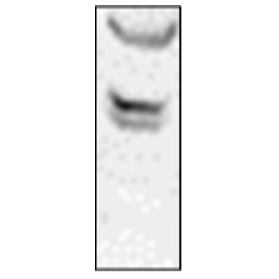
Western Blot: Lyn Antibody (LYN-01)BSA Free [NB500-519]
Western Blot: Lyn Antibody (LYN-01) [NB500-519] - Fig. 1. Western Blotting analysis (non-reducing conditions) of Lyn using anti-Lyn (LYN-01). Lane 1: negative control Lane 2: RBL rat basophilic leukemia cell line Lane 3: JURKAT human peripheral blood T cell leukemia cell line Lane 4: A-431 human epidermoid carcinoma cell line Lane 5: U-937 human histiocytic lymphoma cell line.![Lyn Antibody (LYN-01) - BSA Free Western Blot: Lyn Antibody (LYN-01) - BSA Free [NB500-519]](https://resources.rndsystems.com/images/products/nb500-519_mouse-monoclonal-lyn-antibody-lyn-01-13120241643337.jpg "Western Blot: Lyn Antibody (LYN-01) - BSA Free [NB500-519]")
Western Blot: Lyn Antibody (LYN-01) - BSA Free [NB500-519]
Analysis of human LYN using mouse monoclonal antibody LYN-01 on lysates of Raji cell line and Jurkat cell line (LYN non-expressing cell line; negative control) under non-reducing and reducing conditions. Nitrocellulose membrane was probed with 2 µg/ml of mouse anti-LYN monoclonal antibody followed by IRDye800-conjugated anti-mouse secondary antibody. LYN was detected around 55 kDa.Applications for Lyn Antibody (LYN-01) - BSA Free
Application
Recommended Usage
Immunocytochemistry/ Immunofluorescence
1:10-1:2000
Immunoprecipitation
1:50
Western Blot
1-2 ug/ml
Application Notes
For WB: Use non-reducing conditions. Purity is > 95% (by SDS-PAGE).
Reviewed Applications
Read 1 review rated 4 using NB500-519 in the following applications:
Formulation, Preparation, and Storage
Purification
Protein A purified
Formulation
Phosphate buffered saline (PBS), pH 7.4
Format
BSA Free
Preservative
15mM Sodium Azide
Concentration
1.0 mg/ml
Shipping
The product is shipped with polar packs. Upon receipt, store it immediately at the temperature recommended below.
Stability & Storage
Store at 4C. Do not freeze.
Background: Lyn
Long Name
V-Yes-1 Yamaguchi Sarcoma Viral Related Oncogene Homolog
Alternate Names
Hck-2, JTK8, v-yes-1
Entrez Gene IDs
4067 (Human)
Gene Symbol
LYN
UniProt
Additional Lyn Products
Product Documents for Lyn Antibody (LYN-01) - BSA Free
Certificate of Analysis
To download a Certificate of Analysis, please enter a lot or batch number in the search box below.
Product Specific Notices for Lyn Antibody (LYN-01) - BSA Free
This product is for research use only and is not approved for use in humans or in clinical diagnosis. Primary Antibodies are guaranteed for 1 year from date of receipt.
Related Research Areas
Citations for Lyn Antibody (LYN-01) - BSA Free
 Powered by Bioz
Customer Reviews for Lyn Antibody (LYN-01) - BSA Free (1)
4 out of 5
1 Customer Rating
Have you used Lyn Antibody (LYN-01) - BSA Free?
Submit a review and receive an Amazon gift card!
$25/€18/£15/$25CAN/¥2500 Yen for a review with an image
$10/€7/£6/$10CAN/¥1110 Yen for a review without an image
Submit a review
Customer Images
Showing
1
-
1 of
1 review
Showing All
Filter By:
-
Application: Western BlotSample Tested: Hela whole cell lysateSpecies: HumanVerified Customer | Posted 09/12/2012

There are no reviews that match your criteria.
Protocols
Find general support by application which include: protocols, troubleshooting, illustrated assays, videos and webinars.
- Appropriate Fixation of IHC/ICC Samples
- Cellular Response to Hypoxia Protocols
- ClariTSA™ Fluorophore Kits
- Detection & Visualization of Antibody Binding
- ICC Cell Smear Protocol for Suspension Cells
- ICC Immunocytochemistry Protocol Videos
- ICC for Adherent Cells
- Immunocytochemistry (ICC) Protocol
- Immunocytochemistry Troubleshooting
- Immunofluorescence of Organoids Embedded in Cultrex Basement Membrane Extract
- Immunohistochemistry (IHC) and Immunocytochemistry (ICC) Protocols
- Immunoprecipitation Protocol
- Preparing Samples for IHC/ICC Experiments
- Preventing Non-Specific Staining (Non-Specific Binding)
- Primary Antibody Selection & Optimization
- Protocol for VisUCyte™ HRP Polymer Detection Reagent
- Protocol for the Fluorescent ICC Staining of Cell Smears - Graphic
- Protocol for the Fluorescent ICC Staining of Cultured Cells on Coverslips - Graphic
- Protocol for the Preparation and Fluorescent ICC Staining of Cells on Coverslips
- Protocol for the Preparation and Fluorescent ICC Staining of Non-adherent Cells
- Protocol for the Preparation and Fluorescent ICC Staining of Stem Cells on Coverslips
- Protocol for the Preparation of a Cell Smear for Non-adherent Cell ICC - Graphic
- R&D Systems Quality Control Western Blot Protocol
- TUNEL and Active Caspase-3 Detection by IHC/ICC Protocol
- The Importance of IHC/ICC Controls
- Troubleshooting Guide: Western Blot Figures
- Western Blot Conditions
- Western Blot Protocol
- Western Blot Protocol for Cell Lysates
- Western Blot Troubleshooting
- Western Blot Troubleshooting Guide
- View all Protocols, Troubleshooting, Illustrated assays and Webinars
Loading...