
Lysine (K)-specific Demethylase 4A/KDM4A/JMJD2A Antibody
Novus Biologicals | Catalog # NB110-40585

![Western Blot: Lysine (K)-specific Demethylase 4A/KDM4A/JMJD2A Antibody [NB110-40585]](https://resources.rndsystems.com/images/products/Lysine-K-specific-Demethylase-4A-KDM4A-JMJD2A-Antibody-Western-Blot-NB110-40585-img0007.jpg "Western Blot: Lysine (K)-specific Demethylase 4A/KDM4A/JMJD2A Antibody [NB110-40585]")
Loading...
Key Product Details
Validated by
Independent Antibodies
Species Reactivity
Validated:
Human, Mouse
Cited:
Human
Applications
Validated:
Western Blot, Immunocytochemistry/ Immunofluorescence, Immunoprecipitation
Cited:
Western Blot, Immunocytochemistry/ Immunofluorescence, Immunoprecipitation
Label
Unconjugated
Antibody Source
Polyclonal Rabbit IgG
Loading...
Product Specifications
Immunogen
The immunogen recognized by this antibody maps to a region between residue 1010 and the C-terminus (residue 1064) of human Jumonji Domain Containing 2A using the numbering given in entry NP_055478.1 (GeneID 9682).
Clonality
Polyclonal
Host
Rabbit
Isotype
IgG
Scientific Data Images for Lysine (K)-specific Demethylase 4A/KDM4A/JMJD2A Antibody
Western Blot: Lysine (K)-specific Demethylase 4A/KDM4A/JMJD2A Antibody [NB110-40585]
Western Blot: Lysine (K)-specific Demethylase 4A/KDM4A/JMJD2A Antibody [NB110-40585] - Nuclear extract (50 ug) from HeLa, HEK293T, and mouse NIH 3T3 cells prepared using NETN lysis buffer. Antibody: Affinity purified rabbit anti-JMJD2A antibody used for WB at 0.1 ug/ml. Detection: Chemiluminescence with an exposure time of 3minutes.![Western Blot: Lysine (K)-specific Demethylase 4A/KDM4A/JMJD2A Antibody [NB110-40585]](https://resources.rndsystems.com/images/products/Lysine-K-specific-Demethylase-4A-KDM4A-JMJD2A-Antibody-Western-Blot-NB110-40585-img0006.jpg "Western Blot: Lysine (K)-specific Demethylase 4A/KDM4A/JMJD2A Antibody [NB110-40585]")
Western Blot: Lysine (K)-specific Demethylase 4A/KDM4A/JMJD2A Antibody [NB110-40585]
Lysine-K-specific-Demethylase-4A-KDM4A-JMJD2A-Antibody-Western-Blot-NB110-40585-img0006.jpg![Immunoprecipitation: Lysine (K)-specific Demethylase 4A/KDM4A/JMJD2A Antibody [NB110-40585]](https://resources.rndsystems.com/images/products/Lysine-K-specific-Demethylase-4A-KDM4A-JMJD2A-Antibody-Immunoprecipitation-NB110-40585-img0005.jpg "Immunoprecipitation: Lysine (K)-specific Demethylase 4A/KDM4A/JMJD2A Antibody [NB110-40585]")
Immunoprecipitation: Lysine (K)-specific Demethylase 4A/KDM4A/JMJD2A Antibody [NB110-40585]
Immunoprecipitation: Lysine (K)-specific Demethylase 4A/KDM4A/JMJD2A Antibody [NB110-40585] - Detection of human Lysine (K)-specific Demethylase 4A/KDM4A/JMJD2A by western blot of immunoprecipitates. Samples: Nuclear Extract (1 mg for IP; 20% of IP loaded) from HEK293T cells. Antibodies: Affinity purified rabbit anti-Lysine (K)-specific Demethylase 4A/KDM4A/JMJD2A antibody NB110-40585 used for IP at 6 ug per reaction. Lysine (K)-specific Demethylase 4A/KDM4A/JMJD2A was also immunoprecipitated by rabbit anti-Lysine (K)-specific Demethylase 4A/KDM4A/JMJD2A antibody NB110-40584. For blotting immunoprecipitated Lysine (K)-specific Demethylase 4A/KDM4A/JMJD2A, NB110-40585 was used at 1 ug/ml. Detection: Chemiluminescence with an exposure time of 30 seconds.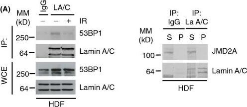
Western Blot: Lysine (K)-specific Demethylase 4A/KDM4A/JMJD2A Antibody [NB110-40585] -
Lamin A/C-53BP1 interaction is regulated in a DNA damage-dependent manner. (A) Left; HDF were subjected to IR (10 Gy) & allowed to recover for 1 h. Association between A-type lamins & 53BP1 was assessed by immunoprecipitation of endogenous lamin A/C followed by immunoblotting with 53BP1 & lamin A/C antibodies. WCE, whole cell extract, IP: immunoprecipitates. WCE represents 5% input. Right; endogenous lamin A/C was immunoprecipitated & supernatants or pellets were analysed by immunoblotting for interaction with JMJD2A. (B) U2OS/GFP-lamin A cells were pretreated with caffeine (20 mm) for 1 h before exposure to IR (10 Gy, 1 h recovery). Cell extracts were then subjected to immunoprecipitation using GFP-Trap beads, & bound complexes were then analysed by immunoblotting using 53BP1 & GFP antibodies. WCE represents 1% input. (C) As in (C) except cells were pretreated with 10 μm ATMi for 1 h before IR. WCE represents 1% input. (D) U2OS/GFP-lamin A cells were subjected to laser micro-irradiation, fixed 1 h later & immunostained with gamma -H2AX antibody. Scale bar, 10 μm. (E) U2OS cells were transfected with siCTRL or siLMNA & subjected to laser micro-irradiation, fixed 15 min later & then processed for immunofluorescence with gamma -H2AX & 53BP1 antibodies. Scale bar, 10 μm. Image collected & cropped by CiteAb from the following publication (https://pubmed.ncbi.nlm.nih.gov/25645366), licensed under a CC-BY license. Not internally tested by Novus Biologicals.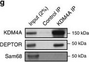
Western Blot: Lysine (K)-specific Demethylase 4A/KDM4A/JMJD2A Antibody [NB110-40585] -
Western Blot: Lysine (K)-specific Demethylase 4A/KDM4A/JMJD2A Antibody [NB110-40585] - KDM4A interacts with the mTORC1/2 complex.(a) Relative mRNA levels of negative & positive regulators of the PTEN/AKT/mTOR pathway following KDM4A depletion. Quantifications of mRNAs by RT–qPCR were normalized against beta -actin (ActB) mRNA. Asterisks denote a statistical difference between siKDM4A-treated cells & siGFP control cells, two-sided t-test P<0.05 (graph represents two independent experiences). Error bars represent standard deviation. (b) Co-immunoprecipitation of endogenous mTORC1/2 complex members with Flag-KDM4A in 293T transfected cells. (c) Comparison of mTORC1/2-associated proteins with Flag-tagged mTOR or KDM4A. The 293T cells were transfected with either Flag-eYFP, Flag-KDM4A or Flag-mTOR, & protein lysates were subjected to anti-Flag immunoprecipitation. (d) Co-immunoprecipitation of Flag-KDM4A & HA-DEPTOR in 293T cells. (e) Endogenous KDM4A co-immunoprecipitates with Flag-DEPTOR. (f) DEPTOR PDZ domain associates with endogenous KDM4A. Flag immunoprecipitation of flag-tagged full length or fragments of DEPTOR. The samples were not sonicated in this experiment to confirm that the interaction is independent of nucleus disruption. (g) Endogenous KDM4A & DEPTOR associate in 293E cells. (h) Endogenous KDM4A & DEPTOR co-immunoprecipitate in NHA-hTERT cells. Image collected & cropped by CiteAb from the following publication (https://www.nature.com/articles/ncomms12700), licensed under a CC-BY license. Not internally tested by Novus Biologicals.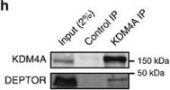
Western Blot: Lysine (K)-specific Demethylase 4A/KDM4A/JMJD2A Antibody [NB110-40585] -
Western Blot: Lysine (K)-specific Demethylase 4A/KDM4A/JMJD2A Antibody [NB110-40585] - KDM4A interacts with the mTORC1/2 complex.(a) Relative mRNA levels of negative & positive regulators of the PTEN/AKT/mTOR pathway following KDM4A depletion. Quantifications of mRNAs by RT–qPCR were normalized against beta -actin (ActB) mRNA. Asterisks denote a statistical difference between siKDM4A-treated cells & siGFP control cells, two-sided t-test P<0.05 (graph represents two independent experiences). Error bars represent standard deviation. (b) Co-immunoprecipitation of endogenous mTORC1/2 complex members with Flag-KDM4A in 293T transfected cells. (c) Comparison of mTORC1/2-associated proteins with Flag-tagged mTOR or KDM4A. The 293T cells were transfected with either Flag-eYFP, Flag-KDM4A or Flag-mTOR, & protein lysates were subjected to anti-Flag immunoprecipitation. (d) Co-immunoprecipitation of Flag-KDM4A & HA-DEPTOR in 293T cells. (e) Endogenous KDM4A co-immunoprecipitates with Flag-DEPTOR. (f) DEPTOR PDZ domain associates with endogenous KDM4A. Flag immunoprecipitation of flag-tagged full length or fragments of DEPTOR. The samples were not sonicated in this experiment to confirm that the interaction is independent of nucleus disruption. (g) Endogenous KDM4A & DEPTOR associate in 293E cells. (h) Endogenous KDM4A & DEPTOR co-immunoprecipitate in NHA-hTERT cells. Image collected & cropped by CiteAb from the following publication (https://www.nature.com/articles/ncomms12700), licensed under a CC-BY license. Not internally tested by Novus Biologicals.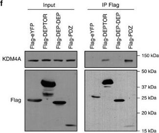
Western Blot: Lysine (K)-specific Demethylase 4A/KDM4A/JMJD2A Antibody [NB110-40585] -
Western Blot: Lysine (K)-specific Demethylase 4A/KDM4A/JMJD2A Antibody [NB110-40585] - KDM4A interacts with the mTORC1/2 complex.(a) Relative mRNA levels of negative & positive regulators of the PTEN/AKT/mTOR pathway following KDM4A depletion. Quantifications of mRNAs by RT–qPCR were normalized against beta -actin (ActB) mRNA. Asterisks denote a statistical difference between siKDM4A-treated cells & siGFP control cells, two-sided t-test P<0.05 (graph represents two independent experiences). Error bars represent standard deviation. (b) Co-immunoprecipitation of endogenous mTORC1/2 complex members with Flag-KDM4A in 293T transfected cells. (c) Comparison of mTORC1/2-associated proteins with Flag-tagged mTOR or KDM4A. The 293T cells were transfected with either Flag-eYFP, Flag-KDM4A or Flag-mTOR, & protein lysates were subjected to anti-Flag immunoprecipitation. (d) Co-immunoprecipitation of Flag-KDM4A & HA-DEPTOR in 293T cells. (e) Endogenous KDM4A co-immunoprecipitates with Flag-DEPTOR. (f) DEPTOR PDZ domain associates with endogenous KDM4A. Flag immunoprecipitation of flag-tagged full length or fragments of DEPTOR. The samples were not sonicated in this experiment to confirm that the interaction is independent of nucleus disruption. (g) Endogenous KDM4A & DEPTOR associate in 293E cells. (h) Endogenous KDM4A & DEPTOR co-immunoprecipitate in NHA-hTERT cells. Image collected & cropped by CiteAb from the following publication (https://www.nature.com/articles/ncomms12700), licensed under a CC-BY license. Not internally tested by Novus Biologicals.Applications for Lysine (K)-specific Demethylase 4A/KDM4A/JMJD2A Antibody
Application
Recommended Usage
Immunocytochemistry/ Immunofluorescence
1:10-1:500
Immunoprecipitation
2-5 ug/mg lysate
Western Blot
1:2000-1:10000
Application Notes
Use in ICC/IF reported in scientific literature (PMID 22373579), Use Use in ICC/IF reported in scientific literature ( 25645366).
Reviewed Applications
Read 2 reviews rated 5 using NB110-40585 in the following applications:
Formulation, Preparation, and Storage
Purification
Immunogen affinity purified
Formulation
TBS and 0.1% BSA
Preservative
0.09% Sodium Azide
Concentration
0.2 mg/ml
Shipping
The product is shipped with polar packs. Upon receipt, store it immediately at the temperature recommended below.
Stability & Storage
Store at 4C. Do not freeze.
Background: Lysine (K)-specific Demethylase 4A/KDM4A
Alternate Names
JHDM3A, JMJD2, JMJD2A, KDM4A, Lysine (K)specific Demethylase 4A
Gene Symbol
KDM4A
UniProt
Additional Lysine (K)-specific Demethylase 4A/KDM4A Products
Product Documents for Lysine (K)-specific Demethylase 4A/KDM4A/JMJD2A Antibody
Certificate of Analysis
To download a Certificate of Analysis, please enter a lot or batch number in the search box below.
Product Specific Notices for Lysine (K)-specific Demethylase 4A/KDM4A/JMJD2A Antibody
This product is for research use only and is not approved for use in humans or in clinical diagnosis. Primary Antibodies are guaranteed for 1 year from date of receipt.
Related Research Areas
Citations for Lysine (K)-specific Demethylase 4A/KDM4A/JMJD2A Antibody
 Powered by Bioz
Customer Reviews for Lysine (K)-specific Demethylase 4A/KDM4A/JMJD2A Antibody (2)
5 out of 5
2 Customer Ratings
Have you used Lysine (K)-specific Demethylase 4A/KDM4A/JMJD2A Antibody?
Submit a review and receive an Amazon gift card!
$25/€18/£15/$25CAN/¥2500 for a review with an image
$10/€7/£6/$10CAN/¥1110 for a review without an image
Submit a review
Customer Images
Showing
1
-
2 of
2 reviews
Showing All
Filter By:
-
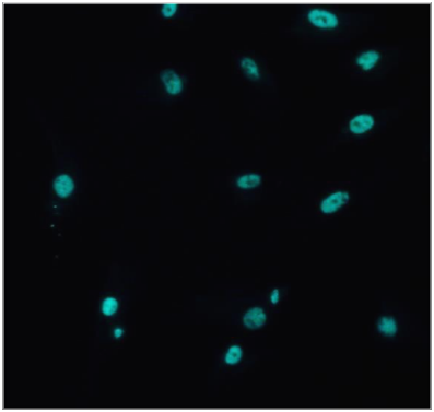
Application: Immunofluorescent stainingSample Tested: epithelial cellsSpecies: HumanVerified Customer | Posted 07/10/2018

-
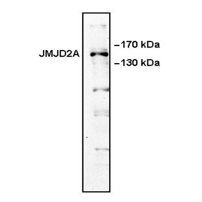
Application: Western BlotSample Tested: HeLa cell lysatesSpecies: HumanVerified Customer | Posted 01/11/2012
There are no reviews that match your criteria.
Protocols
Find general support by application which include: protocols, troubleshooting, illustrated assays, videos and webinars.
- Appropriate Fixation of IHC/ICC Samples
- Cellular Response to Hypoxia Protocols
- ClariTSA™ Fluorophore Kits
- Detection & Visualization of Antibody Binding
- ICC Cell Smear Protocol for Suspension Cells
- ICC Immunocytochemistry Protocol Videos
- ICC for Adherent Cells
- Immunocytochemistry (ICC) Protocol
- Immunocytochemistry Troubleshooting
- Immunofluorescence of Organoids Embedded in Cultrex Basement Membrane Extract
- Immunohistochemistry (IHC) and Immunocytochemistry (ICC) Protocols
- Immunoprecipitation Protocol
- Preparing Samples for IHC/ICC Experiments
- Preventing Non-Specific Staining (Non-Specific Binding)
- Primary Antibody Selection & Optimization
- Protocol for VisUCyte™ HRP Polymer Detection Reagent
- Protocol for the Fluorescent ICC Staining of Cell Smears - Graphic
- Protocol for the Fluorescent ICC Staining of Cultured Cells on Coverslips - Graphic
- Protocol for the Preparation and Fluorescent ICC Staining of Cells on Coverslips
- Protocol for the Preparation and Fluorescent ICC Staining of Non-adherent Cells
- Protocol for the Preparation and Fluorescent ICC Staining of Stem Cells on Coverslips
- Protocol for the Preparation of a Cell Smear for Non-adherent Cell ICC - Graphic
- R&D Systems Quality Control Western Blot Protocol
- TUNEL and Active Caspase-3 Detection by IHC/ICC Protocol
- The Importance of IHC/ICC Controls
- Troubleshooting Guide: Western Blot Figures
- Western Blot Conditions
- Western Blot Protocol
- Western Blot Protocol for Cell Lysates
- Western Blot Troubleshooting
- Western Blot Troubleshooting Guide
- View all Protocols, Troubleshooting, Illustrated assays and Webinars
Loading...