LYVE-1 Antibody - BSA Free
Novus Biologicals | Catalog # NB100-725
![Western Blot: LYVE-1 AntibodyBSA Free [NB100-725]](https://resources.rndsystems.com/images/products/LYVE-1-Antibody-Western-Blot-NB100-725-img0027.jpg "Western Blot: LYVE-1 AntibodyBSA Free [NB100-725]")
Key Product Details
Species Reactivity
Validated:
Human, Mouse, Rat
Cited:
Human, Mouse, Rat
Applications
Validated:
Immunohistochemistry, Immunohistochemistry-Paraffin, Immunohistochemistry-Frozen, Western Blot, Flow Cytometry, Flow (Intracellular)
Cited:
Immunohistochemistry, Immunohistochemistry-Paraffin, Immunohistochemistry-Frozen, Immunofluorescence
Label
Unconjugated
Antibody Source
Polyclonal Rabbit IgG
Format
BSA Free
Loading...
Product Specifications
Immunogen
This LYVE-1 Antibody was developed against a synthetic peptide made to a C-terminal portion of the mouse LYVE1 protein sequence (between residues 250-318). [UniProt# Q8BHC0]
Localization
Plasma membrane
Marker
Lymphatic Vessel Marker
Clonality
Polyclonal
Host
Rabbit
Isotype
IgG
Theoretical MW
35 kDa.
Disclaimer note: The observed molecular weight of the protein may vary from the listed predicted molecular weight due to post translational modifications, post translation cleavages, relative charges, and other experimental factors.
Disclaimer note: The observed molecular weight of the protein may vary from the listed predicted molecular weight due to post translational modifications, post translation cleavages, relative charges, and other experimental factors.
Scientific Data Images for LYVE-1 Antibody - BSA Free
Western Blot: LYVE-1 AntibodyBSA Free [NB100-725]
Western Blot: LYVE-1 Antibody [NB100-725] - Western Blot: [NB100-725] - Total protein from human stomach, lymph node and placenta was separated on a 7.5% gel by SDS-PAGE, transferred to PVDF membrane and blocked in 5% non-fat milk in TBST. The membrane was probed with 1.0 ug/ml anti-LYVE1 in 1% milk, and detected with an anti-rabbit HRP secondary antibody using chemiluminescence.![Immunohistochemistry-Paraffin: LYVE-1 Antibody - BSA Free [NB100-725]](https://resources.rndsystems.com/images/products/LYVE-1-Antibody-Immunohistochemistry-Paraffin-NB100-725-img0028.jpg "Immunohistochemistry-Paraffin: LYVE-1 Antibody - BSA Free [NB100-725]")
Immunohistochemistry-Paraffin: LYVE-1 Antibody - BSA Free [NB100-725]
Immunohistochemistry-Paraffin: LYVE-1 Antibody [NB100-725] - Analysis using the DyLight 488 conjugate of NB100-725. Staining of LYVE1 in paraffin embedded MDA-MB-231 breast cancer orthotopic transplantation tissue. Image courtesy of product review submitted by Luana Schito.![Immunohistochemistry: LYVE-1 Antibody - BSA Free [NB100-725]](https://resources.rndsystems.com/images/products/LYVE-1-Antibody-Immunohistochemistry-NB100-725-img0011.jpg "Immunohistochemistry: LYVE-1 Antibody - BSA Free [NB100-725]")
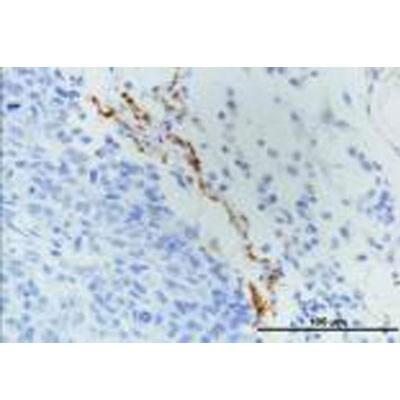
Immunohistochemistry: LYVE-1 Antibody - BSA Free [NB100-725]
Immunohistochemistry: LYVE-1 Antibody [NB100-725] - Detection of LYVE1 in endothelial cells of human lung blood vessels using NB100-725. Note presence of RBCs within vessel lumen.![Flow Cytometry: LYVE-1 Antibody - BSA Free [NB100-725]](https://resources.rndsystems.com/images/products/LYVE-1-Antibody-Flow-Cytometry-NB100-725-img0024.jpg "Flow Cytometry: LYVE-1 Antibody - BSA Free [NB100-725]")
Flow Cytometry: LYVE-1 Antibody - BSA Free [NB100-725]
Flow Cytometry: LYVE-1 Antibody [NB100-725] - Analysis using the DyLight 488 conjugate of NB100-725. Unlabeled control with labeled samples of cells dissociated mechanically and enzymatically from human skin. Image provided via product review by Patricia Redondo.![Western Blot: LYVE-1 AntibodyBSA Free [NB100-725]](https://resources.rndsystems.com/images/products/LYVE-1-Antibody-Western-Blot-NB100-725-img0018.jpg "Western Blot: LYVE-1 AntibodyBSA Free [NB100-725]")
Western Blot: LYVE-1 AntibodyBSA Free [NB100-725]
Western Blot: LYVE-1 Antibody [NB100-725] - Analysis of extracts from A549 cells using LYVE1 antibody (NB100-725, 1:100). Image from verified customer review.![Flow Cytometry: LYVE-1 Antibody - BSA Free [NB100-725]](https://resources.rndsystems.com/images/products/LYVE-1-Antibody-Flow-Cytometry-NB100-725-img0016.jpg "Flow Cytometry: LYVE-1 Antibody - BSA Free [NB100-725]")
Flow Cytometry: LYVE-1 Antibody - BSA Free [NB100-725]
Flow Cytometry: LYVE-1 Antibody [NB100-725] - LYVE1 antibody was tested at 1:400 in A549 cells using an Alexa Fluor 488 secondary (shown in green) alongside unstained cells (shown in red).![Flow Cytometry: LYVE-1 Antibody - BSA Free [NB100-725]](https://resources.rndsystems.com/images/products/LYVE-1-Antibody-Flow-Cytometry-NB100-725-img0031.jpg "Flow Cytometry: LYVE-1 Antibody - BSA Free [NB100-725]")
Flow Cytometry: LYVE-1 Antibody - BSA Free [NB100-725]
Flow Cytometry: LYVE-1 Antibody [NB100-725] - An intracellular stain was performed on A549 cells with LYVE-1 Antibody NB100-725 (blue) and a matched isotype control (orange). Cells were fixed with 4% PFA and then permeabilized with 0.1% saponin. Cells were incubated in an antibody dilution of 1.0 ug/mL for 30 minutes at room temperature, followed by Rabbit IgG (H+L) Cross-Adsorbed Secondary Antibody, Dylight 550 (SA5-10033, Thermo Fisher).![Immunohistochemistry: LYVE-1 Antibody - BSA Free [NB100-725]](https://resources.rndsystems.com/images/products/LYVE-1-Antibody-Immunohistochemistry-NB100-725-img0012.jpg "Immunohistochemistry: LYVE-1 Antibody - BSA Free [NB100-725]")
Immunohistochemistry: LYVE-1 Antibody - BSA Free [NB100-725]
Immunohistochemistry: LYVE-1 Antibody [NB100-725] - Detection of LYVE1 in endothelial cells of human bladder vasculature using NB100-725.![Immunohistochemistry: LYVE-1 Antibody - BSA Free [NB100-725]](https://resources.rndsystems.com/images/products/LYVE-1-Antibody-Immunohistochemistry-NB100-725-img0013.jpg "Immunohistochemistry: LYVE-1 Antibody - BSA Free [NB100-725]")
Immunohistochemistry: LYVE-1 Antibody - BSA Free [NB100-725]
Immunohistochemistry: LYVE-1 Antibody [NB100-725] - Detection of LYVE1 in endothelial cells of human lung blood vessels using NB100-725.![Flow Cytometry: LYVE-1 Antibody - BSA Free [NB100-725]](https://resources.rndsystems.com/images/products/LYVE-1-Antibody-Flow-Cytometry-NB100-725-img0023.jpg "Flow Cytometry: LYVE-1 Antibody - BSA Free [NB100-725]")
Flow Cytometry: LYVE-1 Antibody - BSA Free [NB100-725]
Flow Cytometry: LYVE-1 Antibody [NB100-725] - Analysis using the Alexa Fluor (R) 488 conjugate of NB100-725. Staining of LYVE1 in human dissociated skin lyve1+ using AF488 conjugated anti-LYVE1 antibody. Image from verified customer review.![Flow Cytometry: LYVE-1 Antibody - BSA Free [NB100-725]](https://resources.rndsystems.com/images/products/LYVE-1-Antibody-Flow-Cytometry-NB100-725-img0029.jpg "Flow Cytometry: LYVE-1 Antibody - BSA Free [NB100-725]")
Flow Cytometry: LYVE-1 Antibody - BSA Free [NB100-725]
Flow Cytometry: LYVE-1 Antibody [NB100-725] - An intracellular stain was performed on A549 cells with LYVE-1 Antibody NB100-725G (blue) and a matched isotype control (orange). Cells were fixed with 4% PFA and then permeabilized with 0.1% saponin. Cells were incubated in an antibody dilution of 10 ug/mL for 30 minutes at room temperature. Both antibodies were conjugated to Dylight 488.![Flow Cytometry: LYVE-1 Antibody - BSA Free [NB100-725]](https://resources.rndsystems.com/images/products/LYVE-1-Antibody-Flow-Cytometry-NB100-725-img0030.jpg "Flow Cytometry: LYVE-1 Antibody - BSA Free [NB100-725]")
Flow Cytometry: LYVE-1 Antibody - BSA Free [NB100-725]
Flow Cytometry: LYVE-1 Antibody [NB100-725] - An intracellular stain was performed on K562 cells with LYVE-1 Antibody NB100-725C (blue) and a matched isotype control (orange). Cells were fixed with 4% PFA and then permeabilized with 0.1% saponin. Cells were incubated in an antibody dilution of 2.5 ug/mL for 30 minutes at room temperature. Both antibodies were conjugated to DyLight 650.Applications for LYVE-1 Antibody - BSA Free
Application
Recommended Usage
Flow Cytometry
1:50-1:500
Immunohistochemistry
1:100-1:200
Immunohistochemistry-Frozen
1:100-1:200
Immunohistochemistry-Paraffin
1:100-1:200
Western Blot
1:500-1:2000
Application Notes
In Western Blot, a band at ~52 kDa is seen. For Immunohistochemistry citrate buffer antigen retrieval is recommended.
Reviewed Applications
Read 2 reviews rated 5 using NB100-725 in the following applications:
Flow Cytometry Panel Builder
Bio-Techne Knows Flow Cytometry
Save time and reduce costly mistakes by quickly finding compatible reagents using the Panel Builder Tool.
Advanced Features
- Spectra Viewer - Custom analysis of spectra from multiple fluorochromes
- Spillover Popups - Visualize the spectra of individual fluorochromes
- Antigen Density Selector - Match fluorochrome brightness with antigen density

Formulation, Preparation, and Storage
Purification
Immunogen affinity purified
Formulation
PBS
Format
BSA Free
Preservative
0.02% Sodium Azide
Concentration
1.0 mg/ml
Shipping
The product is shipped with polar packs. Upon receipt, store it immediately at the temperature recommended below.
Stability & Storage
Store at 4C short term. Aliquot and store at -20C long term. Avoid freeze-thaw cycles.
Background: LYVE-1
LYVE-1 has been an important marker in studies of embryonic and tumor lymphangiogenesis, as many cancers are characterized by early metastasis to the lymph nodes (1-3, 5). One study of five different vascular tumors in infants used immunohistochemical analysis and found positive LYVE-1 expression in infantile hemangioma, tufted angioma, and kaposiform hemangioendothelioma (5). LYVE-1 along with other markers such as GLUT-1, CD31, CD34, Prox-1, and WT-1 can be used to help provide immunohistologic profiles of various tumors and, when used in conjunction with clinical and histopathologic approaches, may offer better overall diagnosis and disease treatment (5).
References
1. Jackson D. G. (2019). Hyaluronan in the lymphatics: The key role of the hyaluronan receptor LYVE-1 in leucocyte trafficking. Matrix Biology : Journal of the International Society for Matrix Biology. https://doi.org/10.1016/j.matbio.2018.02.001
2. Jackson D. G. (2004). Biology of the lymphatic marker LYVE-1 and applications in research into lymphatic trafficking and lymphangiogenesis. APMIS : acta pathologica, microbiologica, et immunologica Scandinavica. https://doi.org/10.1111/j.1600-0463.2004.apm11207-0811.x
3. Jackson D. G. (2003). The lymphatics revisited: new perspectives from the hyaluronan receptor LYVE-1. Trends in Cardiovascular Medicine. https://doi.org/10.1016/s1050-1738(02)00189-5
4. Unitprot (Q9Y5Y7)
5. Johnson, E. F., Davis, D. M., Tollefson, M. M., Fritchie, K., & Gibson, L. E. (2018). Vascular Tumors in Infants: Case Report and Review of Clinical, Histopathologic, and Immunohistochemical Characteristics of Infantile Hemangioma, Pyogenic Granuloma, Noninvoluting Congenital Hemangioma, Tufted Angioma, and Kaposiform Hemangioendothelioma. The American Journal of Dermatopathology. https://doi.org/10.1097/DAD.0000000000000983
Long Name
Lymphatic Vessel Endothelial Hyaluronan Receptor 1
Alternate Names
LYVE1, XLKD1
Gene Symbol
LYVE1
Additional LYVE-1 Products
Product Documents for LYVE-1 Antibody - BSA Free
Certificate of Analysis
To download a Certificate of Analysis, please enter a lot or batch number in the search box below.
Product Specific Notices for LYVE-1 Antibody - BSA Free
This product is for research use only and is not approved for use in humans or in clinical diagnosis. Primary Antibodies are guaranteed for 1 year from date of receipt.
Related Research Areas
Citations for LYVE-1 Antibody - BSA Free
 Powered by Bioz
Customer Reviews for LYVE-1 Antibody - BSA Free (2)
5 out of 5
2 Customer Ratings
Have you used LYVE-1 Antibody - BSA Free?
Submit a review and receive an Amazon gift card!
$25/€18/£15/$25CAN/¥2500 Yen for a review with an image
$10/€7/£6/$10CAN/¥1110 Yen for a review without an image
Submit a review
Customer Images
-(01-ml)_NB100-725_8631.png)
Showing
1
-
2 of
2 reviews
Showing All
Filter By:
-
Application: Western BlotSample Tested:Species: HumanVerified Customer | Posted 07/01/2014Western blot analysis of extracts from A549 cells using LYVE1 antibody (NB100-725, 1:100).

-
Application: Immunohistochemistry-ParaffinSample Tested: MDA-MB-231 breast cancer orthotopic transplantationSpecies: HumanVerified Customer | Posted 03/19/2013
There are no reviews that match your criteria.
Protocols
View specific protocols for LYVE-1 Antibody - BSA Free (NB100-725):
Protocol for Flow Cytometry Intracellular Staining
Sample Preparation.
1. Grow cells to 60-85% confluency. Flow cytometry requires between 2 x 10⁵ and 1 x 10⁶ cells for optimal performance.
2. If cells are adherent, harvest gently by washing once with staining buffer and then scraping. Avoid using trypsin as this can disrupt certain epitopes of interest. If enzymatic harvest is required, use Accutase, Collagenase, or TrypLE Express for a less damaging option.
3. Reserve 100 uL for counting, then transfer cell volume into a 50 mL conical tube and centrifuge for 8 minutes at 400 RCF.
a. Count cells using a hemocytometer and a 1:1 trypan blue exclusion stain to determine cell viability before starting the flow protocol. If cells appear blue, do not proceed.
4. Re-suspend cells to a concentration of 1 x 10⁶ cells/mL in staining buffer (NBP2-26247).
5. Aliquot out 100 uL samples in accordance with your experimental samples.
Tip: When cell surface and intracellular staining are required in the same sample, it is advisable that the cell surface staining be performed first since the fixation and permeabilization steps might reduce the availability of surface antigens.
Intracellular Staining.
Tip: When performing intracellular staining, it is important to use appropriate fixation and permeabilization reagents based upon the target and its subcellular location. Generally, our Intracellular Flow Assay Kit (NBP2-29450) is a good place to start as it contains an optimized combination of reagents for intracellular staining as well as an inhibitor of intracellular protein transport (necessary if staining secreted proteins). Certain targets may require more gentle or transient permeabilization protocols such as the commonly employed methanol or saponin-based methods.
Protocol for Cytoplasmic Targets:
1. Fix the cells by adding 100 uL fixation solution (such as 4% PFA) to each sample for 10-15 minutes.
2. Permeabilize cells by adding 100 uL of a permeabilization buffer to every 1 x 10⁶ cells present in the sample. Mix well and incubate at room temperature for 15 minutes.
a. For cytoplasmic targets, use a gentle permeabilization solution such as 1X PBS + 0.5% Saponin or 1X PBS + 0.5% Tween-20.
b. To maintain the permeabilized state throughout your experiment, use staining buffer + 0.1% of the permeabilization reagent (i.e. 0.1% Tween-20 or 0.1% Saponin).
3. Following the 15 minute incubation, add 2 mL of the staining buffer + 0.1% permeabilizer to each sample.
4. Centrifuge for 1 minute at 400 RCF.
5. Discard supernatant and re-suspend in 100 uL of staining buffer + 0.1% permeabilizer.
6. Add appropriate amount of each antibody (eg. 1 test or 1 ug per sample, as experimentally determined).
7. Mix well and incubate at room temperature for 30 minutes- 1 hour. Gently mix samples every 10-15 minutes.
8. Following the primary/conjugate incubation, add 1-2 mL/sample of staining buffer +0.1% permeabilizer and centrifuge for 1 minute at 400 RCF.
9. Wash twice by re-suspending cells in staining buffer (2 mL for tubes or 200 uL for wells) and centrifuging at 400 RCF for 5 minutes. Discard supernatant.
10. Add appropriate amount of secondary antibody (as experimentally determined) to each sample.
11. Incubate at room temperature in dark for 20 minutes.
12. Add 1-2 mL of staining buffer and centrifuge at 400 RCF for 1 minute and discard supernatant.
13. Wash twice by re-suspending cells in staining buffer (2 mL for tubes or 200 uL for wells) and centrifuging at 400 RCF for 5 minutes. Discard supernatant.
14. Resuspend in an appropriate volume of staining buffer (usually 500 uL per sample) and proceed with analysis on your flow cytometer.
Immunohistochemistry-Paraffin Embedded Sections
Antigen Unmasking:
Bring slides to a boil in 10 mM sodium citrate buffer (pH 6.0) then maintain at a sub-boiling temperature for 10 minutes. Cool slides on bench-top for 30 minutes (keep slides in the sodium citrate buffer at all times).
Staining:
1. Wash sections in deionized water three times for 5 minutes each.
2. Wash sections in PBS for 5 minutes.
3. Block each section with 100-400 ul blocking solution (1% BSA in PBS) for 1 hour at room temperature.
4. Remove blocking solution and add 100-400 ul diluted primary antibody. Incubate overnight at 4 C.
5. Remove antibody solution and wash sections in wash buffer three times for 5 minutes each.
6. Add 100-400 ul HRP polymer conjugated secondary antibody. Incubate 30 minutes at room temperature.
7. Wash sections three times in wash buffer for 5 minutes each.
8. Add 100-400 ul DAB substrate to each section and monitor staining closely.
9. As soon as the sections develop, immerse slides in deionized water.
10. Counterstain sections in hematoxylin.
11. Wash sections in deionized water two times for 5 minutes each.
12. Dehydrate sections.
13. Mount coverslips.
Western Blot Protocol
1. Perform SDS-PAGE on samples to be analyzed, loading 10-25 ug of total protein per lane.
2. Transfer proteins to PVDF membrane according to the instructions provided by the manufacturer of the membrane and transfer apparatus.
3. Stain the membrane with Ponceau S (or similar product) to assess transfer success, and mark molecular weight standards where appropriate.
4. Rinse the blot TBS -0.05% Tween 20 (TBST).
5. Block the membrane in 5% Non-fat milk in TBST (blocking buffer) for at least 1 hour.
6. Wash the membrane in TBST three times for 10 minutes each.
7. Dilute primary antibody in blocking buffer and incubate overnight at 4C with gentle rocking.
8. Wash the membrane in TBST three times for 10 minutes each.
9. Incubate the membrane in diluted HRP conjugated secondary antibody in blocking buffer (as per manufacturer's instructions) for 1 hour at room temperature.
10. Wash the blot in TBST three times for 10 minutes each (this step can be repeated as required to reduce background).
11. Apply the detection reagent of choice in accordance with the manufacturer's instructions.
Find general support by application which include: protocols, troubleshooting, illustrated assays, videos and webinars.
- 7-Amino Actinomycin D (7-AAD) Cell Viability Flow Cytometry Protocol
- Antigen Retrieval Protocol (PIER)
- Antigen Retrieval for Frozen Sections Protocol
- Appropriate Fixation of IHC/ICC Samples
- Cellular Response to Hypoxia Protocols
- Chromogenic IHC Staining of Formalin-Fixed Paraffin-Embedded (FFPE) Tissue Protocol
- Chromogenic Immunohistochemistry Staining of Frozen Tissue
- ClariTSA™ Fluorophore Kits
- Detection & Visualization of Antibody Binding
- Extracellular Membrane Flow Cytometry Protocol
- Flow Cytometry Protocol for Cell Surface Markers
- Flow Cytometry Protocol for Staining Membrane Associated Proteins
- Flow Cytometry Staining Protocols
- Flow Cytometry Troubleshooting Guide
- Fluorescent IHC Staining of Frozen Tissue Protocol
- Graphic Protocol for Heat-induced Epitope Retrieval
- Graphic Protocol for the Preparation and Fluorescent IHC Staining of Frozen Tissue Sections
- Graphic Protocol for the Preparation and Fluorescent IHC Staining of Paraffin-embedded Tissue Sections
- Graphic Protocol for the Preparation of Gelatin-coated Slides for Histological Tissue Sections
- IHC Sample Preparation (Frozen sections vs Paraffin)
- Immunofluorescent IHC Staining of Formalin-Fixed Paraffin-Embedded (FFPE) Tissue Protocol
- Immunohistochemistry (IHC) and Immunocytochemistry (ICC) Protocols
- Immunohistochemistry Frozen Troubleshooting
- Immunohistochemistry Paraffin Troubleshooting
- Intracellular Flow Cytometry Protocol Using Alcohol (Methanol)
- Intracellular Flow Cytometry Protocol Using Detergents
- Intracellular Nuclear Staining Flow Cytometry Protocol Using Detergents
- Intracellular Staining Flow Cytometry Protocol Using Alcohol Permeabilization
- Intracellular Staining Flow Cytometry Protocol Using Detergents to Permeabilize Cells
- Preparing Samples for IHC/ICC Experiments
- Preventing Non-Specific Staining (Non-Specific Binding)
- Primary Antibody Selection & Optimization
- Propidium Iodide Cell Viability Flow Cytometry Protocol
- Protocol for Heat-Induced Epitope Retrieval (HIER)
- Protocol for Liperfluo
- Protocol for Making a 4% Formaldehyde Solution in PBS
- Protocol for VisUCyte™ HRP Polymer Detection Reagent
- Protocol for the Characterization of Human Th22 Cells
- Protocol for the Characterization of Human Th9 Cells
- Protocol for the Preparation & Fixation of Cells on Coverslips
- Protocol for the Preparation and Chromogenic IHC Staining of Frozen Tissue Sections
- Protocol for the Preparation and Chromogenic IHC Staining of Frozen Tissue Sections - Graphic
- Protocol for the Preparation and Chromogenic IHC Staining of Paraffin-embedded Tissue Sections
- Protocol for the Preparation and Chromogenic IHC Staining of Paraffin-embedded Tissue Sections - Graphic
- Protocol for the Preparation and Fluorescent IHC Staining of Frozen Tissue Sections
- Protocol for the Preparation and Fluorescent IHC Staining of Paraffin-embedded Tissue Sections
- Protocol for the Preparation of Gelatin-coated Slides for Histological Tissue Sections
- Protocol: Annexin V and PI Staining by Flow Cytometry
- Protocol: Annexin V and PI Staining for Apoptosis by Flow Cytometry
- R&D Systems Quality Control Western Blot Protocol
- TUNEL and Active Caspase-3 Detection by IHC/ICC Protocol
- The Importance of IHC/ICC Controls
- Troubleshooting Guide: Fluorokine Flow Cytometry Kits
- Troubleshooting Guide: Immunohistochemistry
- Troubleshooting Guide: Western Blot Figures
- Western Blot Conditions
- Western Blot Protocol
- Western Blot Protocol for Cell Lysates
- Western Blot Troubleshooting
- Western Blot Troubleshooting Guide
- View all Protocols, Troubleshooting, Illustrated assays and Webinars
FAQs for LYVE-1 Antibody - BSA Free
Showing
1
-
1 of
1 FAQ
Showing All
-
Q: What negative staining control should I be using for this antibody?
A:
Testing of expression of LYVE-1 in various tissues can be found at the Human Protein Atlas (https://www.proteinatlas.org/ENSG00000133800-LYVE1). This should help you find a tissue for use as a negative control.
Loading...