LYVE-1 Antibody [Alexa Fluor® 488]
Novus Biologicals | Catalog # NB100-725AF488
Key Product Details
Species Reactivity
Human, Mouse, Rat
Applications
Immunohistochemistry, Immunohistochemistry-Paraffin, Immunohistochemistry-Frozen, Western Blot, Flow Cytometry, Flow (Intracellular)
Label
Alexa Fluor 488 (Excitation = 488 nm, Emission = 515-545 nm)
Antibody Source
Polyclonal Rabbit IgG
Loading...
Product Specifications
Immunogen
This LYVE-1 Antibody was developed against a synthetic peptide made to a C-terminal portion of the mouse LYVE1 protein sequence (between residues 250-318). [UniProt# Q8BHC0]
Localization
Plasma membrane
Marker
Lymphatic Vessel Marker
Clonality
Polyclonal
Host
Rabbit
Isotype
IgG
Theoretical MW
35 kDa.
Disclaimer note: The observed molecular weight of the protein may vary from the listed predicted molecular weight due to post translational modifications, post translation cleavages, relative charges, and other experimental factors.
Disclaimer note: The observed molecular weight of the protein may vary from the listed predicted molecular weight due to post translational modifications, post translation cleavages, relative charges, and other experimental factors.
Scientific Data Images for LYVE-1 Antibody [Alexa Fluor® 488]
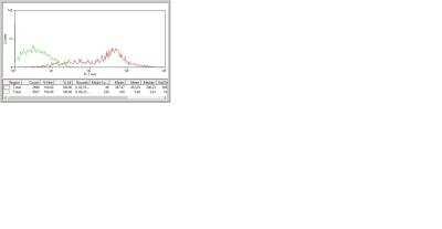
Flow Cytometry: LYVE1 Antibody [Alexa Fluor (R) 488] [NB100-725AF488] - analysis of LYVE1 in human dissociated skin lyve1+ using AF488 conjugated anti-LYVE1 antibody. Image from verified customer review.
Applications for LYVE-1 Antibody [Alexa Fluor® 488]
Application
Recommended Usage
Flow (Intracellular)
Optimal dilutions of this antibody should be experimentally determined.
Flow Cytometry
Optimal dilutions of this antibody should be experimentally determined.
Immunohistochemistry
Optimal dilutions of this antibody should be experimentally determined.
Immunohistochemistry-Frozen
Optimal dilutions of this antibody should be experimentally determined.
Immunohistochemistry-Paraffin
Optimal dilutions of this antibody should be experimentally determined.
Western Blot
Optimal dilutions of this antibody should be experimentally determined.
Application Notes
Optimal dilution of this antibody should be experimentally determined.
Reviewed Applications
Read 1 review rated 5 using NB100-725AF488 in the following applications:
Spectra Viewer
Plan Your Experiments
Use our spectra viewer to interactively plan your experiments, assessing multiplexing options. View the excitation and emission spectra for our fluorescent dye range and other commonly used dyes.
Spectra Viewer
Flow Cytometry Panel Builder
Bio-Techne Knows Flow Cytometry
Save time and reduce costly mistakes by quickly finding compatible reagents using the Panel Builder Tool.
Advanced Features
- Spectra Viewer - Custom analysis of spectra from multiple fluorochromes
- Spillover Popups - Visualize the spectra of individual fluorochromes
- Antigen Density Selector - Match fluorochrome brightness with antigen density

Formulation, Preparation, and Storage
Purification
Immunogen affinity purified
Formulation
50mM Sodium Borate
Preservative
0.05% Sodium Azide
Concentration
Please see the vial label for concentration. If unlisted please contact technical services.
Shipping
The product is shipped with polar packs. Upon receipt, store it immediately at the temperature recommended below.
Stability & Storage
Store at 4C in the dark.
Background: LYVE-1
LYVE-1 has been an important marker in studies of embryonic and tumor lymphangiogenesis, as many cancers are characterized by early metastasis to the lymph nodes (1-3, 5). One study of five different vascular tumors in infants used immunohistochemical analysis and found positive LYVE-1 expression in infantile hemangioma, tufted angioma, and kaposiform hemangioendothelioma (5). LYVE-1 along with other markers such as GLUT-1, CD31, CD34, Prox-1, and WT-1 can be used to help provide immunohistologic profiles of various tumors and, when used in conjunction with clinical and histopathologic approaches, may offer better overall diagnosis and disease treatment (5).
References
1. Jackson D. G. (2019). Hyaluronan in the lymphatics: The key role of the hyaluronan receptor LYVE-1 in leucocyte trafficking. Matrix Biology : Journal of the International Society for Matrix Biology. https://doi.org/10.1016/j.matbio.2018.02.001
2. Jackson D. G. (2004). Biology of the lymphatic marker LYVE-1 and applications in research into lymphatic trafficking and lymphangiogenesis. APMIS : acta pathologica, microbiologica, et immunologica Scandinavica. https://doi.org/10.1111/j.1600-0463.2004.apm11207-0811.x
3. Jackson D. G. (2003). The lymphatics revisited: new perspectives from the hyaluronan receptor LYVE-1. Trends in Cardiovascular Medicine. https://doi.org/10.1016/s1050-1738(02)00189-5
4. Unitprot (Q9Y5Y7)
5. Johnson, E. F., Davis, D. M., Tollefson, M. M., Fritchie, K., & Gibson, L. E. (2018). Vascular Tumors in Infants: Case Report and Review of Clinical, Histopathologic, and Immunohistochemical Characteristics of Infantile Hemangioma, Pyogenic Granuloma, Noninvoluting Congenital Hemangioma, Tufted Angioma, and Kaposiform Hemangioendothelioma. The American Journal of Dermatopathology. https://doi.org/10.1097/DAD.0000000000000983
Long Name
Lymphatic Vessel Endothelial Hyaluronan Receptor 1
Alternate Names
LYVE1, XLKD1
Gene Symbol
LYVE1
Additional LYVE-1 Products
Product Documents for LYVE-1 Antibody [Alexa Fluor® 488]
Certificate of Analysis
To download a Certificate of Analysis, please enter a lot or batch number in the search box below.
Product Specific Notices for LYVE-1 Antibody [Alexa Fluor® 488]
Alexa Fluor (R) products are provided under an intellectual property license from Life Technologies Corporation. The purchase of this product conveys to the buyer the non-transferable right to use the purchased product and components of the product only in research conducted by the buyer (whether the buyer is an academic or for-profit entity). The sale of this product is expressly conditioned on the buyer not using the product or its components, or any materials made using the product or its components, in any activity to generate revenue, which may include, but is not limited to use of the product or its components: (i) in manufacturing; (ii) to provide a service, information, or data in return for payment; (iii) for therapeutic, diagnostic or prophylactic purposes; or (iv) for resale, regardless of whether they are resold for use in research. For information on purchasing a license to this product for purposes other than as described above, contact Life Technologies Corporation, 5791 Van Allen Way, Carlsbad, CA 92008 USA or outlicensing@lifetech.com. This conjugate is made on demand. Actual recovery may vary from the stated volume of this product. The volume will be greater than or equal to the unit size stated on the datasheet.
This product is for research use only and is not approved for use in humans or in clinical diagnosis. Primary Antibodies are guaranteed for 1 year from date of receipt.
Related Research Areas
Customer Reviews for LYVE-1 Antibody [Alexa Fluor® 488] (1)
5 out of 5
1 Customer Rating
Have you used LYVE-1 Antibody [Alexa Fluor® 488]?
Submit a review and receive an Amazon gift card!
$25/€18/£15/$25CAN/¥2500 Yen for a review with an image
$10/€7/£6/$10CAN/¥1110 Yen for a review without an image
Submit a review
Customer Images
![LYVE-1 Antibody [Alexa Fluor® 488] NB100-725AF488](https://resources.rndsystems.com/images/reviews/Flow-Cytometry_LYVE1-Antibody-(NB100-725AF488)-(100-Tests)_NB100-725AF488_11741.jpg)
Showing
1
-
1 of
1 review
Showing All
Filter By:
-
Application: Flow CytometrySample Tested: human skinSpecies: HumanVerified Customer | Posted 11/05/2014Human dissociated skin lyve1+; orange line, unstained control; green line, labelled cells
![LYVE-1 Antibody [Alexa Fluor® 488] NB100-725AF488](data:image/png;base64,R0lGODlhAQABAAD/ACwAAAAAAQABAAACADs=)
There are no reviews that match your criteria.
Protocols
Find general support by application which include: protocols, troubleshooting, illustrated assays, videos and webinars.
- 7-Amino Actinomycin D (7-AAD) Cell Viability Flow Cytometry Protocol
- Antigen Retrieval Protocol (PIER)
- Antigen Retrieval for Frozen Sections Protocol
- Appropriate Fixation of IHC/ICC Samples
- Cellular Response to Hypoxia Protocols
- Chromogenic IHC Staining of Formalin-Fixed Paraffin-Embedded (FFPE) Tissue Protocol
- Chromogenic Immunohistochemistry Staining of Frozen Tissue
- ClariTSA™ Fluorophore Kits
- Detection & Visualization of Antibody Binding
- Extracellular Membrane Flow Cytometry Protocol
- Flow Cytometry Protocol for Cell Surface Markers
- Flow Cytometry Protocol for Staining Membrane Associated Proteins
- Flow Cytometry Staining Protocols
- Flow Cytometry Troubleshooting Guide
- Fluorescent IHC Staining of Frozen Tissue Protocol
- Graphic Protocol for Heat-induced Epitope Retrieval
- Graphic Protocol for the Preparation and Fluorescent IHC Staining of Frozen Tissue Sections
- Graphic Protocol for the Preparation and Fluorescent IHC Staining of Paraffin-embedded Tissue Sections
- Graphic Protocol for the Preparation of Gelatin-coated Slides for Histological Tissue Sections
- IHC Sample Preparation (Frozen sections vs Paraffin)
- Immunofluorescent IHC Staining of Formalin-Fixed Paraffin-Embedded (FFPE) Tissue Protocol
- Immunohistochemistry (IHC) and Immunocytochemistry (ICC) Protocols
- Immunohistochemistry Frozen Troubleshooting
- Immunohistochemistry Paraffin Troubleshooting
- Intracellular Flow Cytometry Protocol Using Alcohol (Methanol)
- Intracellular Flow Cytometry Protocol Using Detergents
- Intracellular Nuclear Staining Flow Cytometry Protocol Using Detergents
- Intracellular Staining Flow Cytometry Protocol Using Alcohol Permeabilization
- Intracellular Staining Flow Cytometry Protocol Using Detergents to Permeabilize Cells
- Preparing Samples for IHC/ICC Experiments
- Preventing Non-Specific Staining (Non-Specific Binding)
- Primary Antibody Selection & Optimization
- Propidium Iodide Cell Viability Flow Cytometry Protocol
- Protocol for Heat-Induced Epitope Retrieval (HIER)
- Protocol for Liperfluo
- Protocol for Making a 4% Formaldehyde Solution in PBS
- Protocol for VisUCyte™ HRP Polymer Detection Reagent
- Protocol for the Characterization of Human Th22 Cells
- Protocol for the Characterization of Human Th9 Cells
- Protocol for the Preparation & Fixation of Cells on Coverslips
- Protocol for the Preparation and Chromogenic IHC Staining of Frozen Tissue Sections
- Protocol for the Preparation and Chromogenic IHC Staining of Frozen Tissue Sections - Graphic
- Protocol for the Preparation and Chromogenic IHC Staining of Paraffin-embedded Tissue Sections
- Protocol for the Preparation and Chromogenic IHC Staining of Paraffin-embedded Tissue Sections - Graphic
- Protocol for the Preparation and Fluorescent IHC Staining of Frozen Tissue Sections
- Protocol for the Preparation and Fluorescent IHC Staining of Paraffin-embedded Tissue Sections
- Protocol for the Preparation of Gelatin-coated Slides for Histological Tissue Sections
- Protocol: Annexin V and PI Staining by Flow Cytometry
- Protocol: Annexin V and PI Staining for Apoptosis by Flow Cytometry
- R&D Systems Quality Control Western Blot Protocol
- TUNEL and Active Caspase-3 Detection by IHC/ICC Protocol
- The Importance of IHC/ICC Controls
- Troubleshooting Guide: Fluorokine Flow Cytometry Kits
- Troubleshooting Guide: Immunohistochemistry
- Troubleshooting Guide: Western Blot Figures
- Western Blot Conditions
- Western Blot Protocol
- Western Blot Protocol for Cell Lysates
- Western Blot Troubleshooting
- Western Blot Troubleshooting Guide
- View all Protocols, Troubleshooting, Illustrated assays and Webinars
Loading...