Mcl-1 Antibody - BSA Free
Novus Biologicals | Catalog # NB100-56146
![Western Blot: Mcl-1 Antibody [NB100-56146]](https://resources.rndsystems.com/images/products/MCL1-Antibody-Western-Blot-NB100-56146-img0002.jpg "Western Blot: Mcl-1 Antibody [NB100-56146]")
Loading...
Key Product Details
Species Reactivity
Validated:
Human
Cited:
Human
Applications
Validated:
Immunohistochemistry, Immunohistochemistry-Paraffin, Western Blot, Immunoprecipitation
Cited:
Western Blot, IF/IHC
Label
Unconjugated
Antibody Source
Polyclonal Rabbit IgG
Format
BSA Free
Loading...
Product Specifications
Immunogen
A synthetic peptide corresponding to amino acids 121-139 (SPEEELDGYEPEPLGKRPA) of human Mcl-1 was used as immunogen, GenBank no. NP_068779.1. The immunogen sequence is 100% conserved in Mcl-1 isoform 1 (GenBank no. NP_068779.1) and isoform 2 (GenBank no. NP_877495.1).
Clonality
Polyclonal
Host
Rabbit
Isotype
IgG
Scientific Data Images for Mcl-1 Antibody - BSA Free
Western Blot: Mcl-1 Antibody [NB100-56146]
Western Blot: MCL1 Antibody [NB100-56146] - analysis of Mcl-1 in A) Ramos and B) ThP-1 lysate using Mcl-1 antibody at 1:2000.![Immunohistochemistry-Paraffin: Mcl-1 Antibody [NB100-56146]](https://resources.rndsystems.com/images/products/MCL1-Antibody-Immunohistochemistry-Paraffin-NB100-56146-img0003.jpg "Immunohistochemistry-Paraffin: Mcl-1 Antibody [NB100-56146]")
Immunohistochemistry-Paraffin: Mcl-1 Antibody [NB100-56146]
Immunohistochemistry-Paraffin: MCL1 Antibody [NB100-56146] - Formalin-fixed paraffin-embedded tissue sections of human diffuse, aggressive Non-Hodgkin's lymphoma (NHL) of B cell lineage stained for Mcl-1 expression using Mcl-1 antibody at 1:2000. A-D, Mcl-1 staining shown as varying magnifications. Hematoxylin-eosin counterstain.
Western Blot: Mcl-1 Antibody - BSA Free [NB100-56146] -
Nuclear translocation of TCA cycle enzymes prevents doxorubicin-mediated cellular damage.a–c Enzyme activity of IDH-2 (n = 4) (a), MDH-2 (n = 3) (b), and PDHC (n = 3) (c) in nuclei isolated from cells transduced with tetracycline-inducible NLS-IDH-2 constructs. Activity is represented as the rate in nM/min or delta OD/min, p value* <0.05, p value**** = 0.0001, and lysates were probed for PCNA to detect equal nuclear protein amounts used for the assay. d Reduced apoptotic cell death as measured by Annexin:V:FITC staining in cells expressing nuclear TCA cycle dehydrogenases prior to doxorubicin DRN treatment. N = 3 p value* <0.05 (e) Quantification of troponin I released from cardiomyocytes as a marker of cardiac injury, minimum n = 4 p value** <0.007, p value* <0.05. f Cell death as measured by Cell Titer GloTM in cells expressing nuclear dehydrogenases prior to DRN treatment. minimum n = 4 p value*** = 0.0001, p value**** <0.0001. NLS-EGFP-2A-NLS-mCherry was used as a control for the 2 A linker system. g DNA damage as assessed by ɣ-H2AX foci formation. Scale bar 20 um. h, i qRT‒PCR showing the expression of anti-apoptotic genes BCL2 (h) and MCL1 (i). n = 3 P value ** <0.005, *<0.05. j–l Protein levels of anti-apoptotic proteins (j, k along with reduced caspase 9 cleavage (l) in cells expressing nuclear IDH-2 compared to cells treated with doxorubicin. Image collected and cropped by CiteAb from the following open publication (https://pubmed.ncbi.nlm.nih.gov/37468519), licensed under a CC-BY license. Not internally tested by Novus Biologicals.Applications for Mcl-1 Antibody - BSA Free
Application
Recommended Usage
Immunohistochemistry-Paraffin
1:1000-1:5000
Immunoprecipitation
1:50-1:200
Western Blot
1:1000-1:2000
Application Notes
Immunoprecipitation, Western Blot, Immunohistochemistry-Paraffin IHC (frozen): Users should optimize according to model and immunodetection system used (secondary reagents)
Reviewed Applications
Read 1 review rated 5 using NB100-56146 in the following applications:
Formulation, Preparation, and Storage
Purification
Unpurified
Formulation
Whole antisera
Format
BSA Free
Preservative
0.02% Sodium Azide
Concentration
This product is unpurified. The exact concentration of antibody is not quantifiable.
Shipping
The product is shipped with polar packs. Upon receipt, store it immediately at the temperature recommended below.
Stability & Storage
Store at 4C short term. Aliquot and store at -20C long term. Avoid freeze-thaw cycles.
Background: Mcl-1
Long Name
Myeloid Cell Leukemia Sequence 1
Alternate Names
BCL2L3, Mcl1
Entrez Gene IDs
4170 (Human)
Gene Symbol
MCL1
Additional Mcl-1 Products
Product Documents for Mcl-1 Antibody - BSA Free
Certificate of Analysis
To download a Certificate of Analysis, please enter a lot or batch number in the search box below.
Product Specific Notices for Mcl-1 Antibody - BSA Free
This product is for research use only and is not approved for use in humans or in clinical diagnosis. Primary Antibodies are guaranteed for 1 year from date of receipt.
Related Research Areas
Citations for Mcl-1 Antibody - BSA Free
 Powered by Bioz
Customer Reviews for Mcl-1 Antibody - BSA Free (1)
5 out of 5
1 Customer Rating
Have you used Mcl-1 Antibody - BSA Free?
Submit a review and receive an Amazon gift card!
$25/€18/£15/$25CAN/¥2500 Yen for a review with an image
$10/€7/£6/$10CAN/¥1110 Yen for a review without an image
Submit a review
Customer Images
Showing
1
-
1 of
1 review
Showing All
Filter By:
-
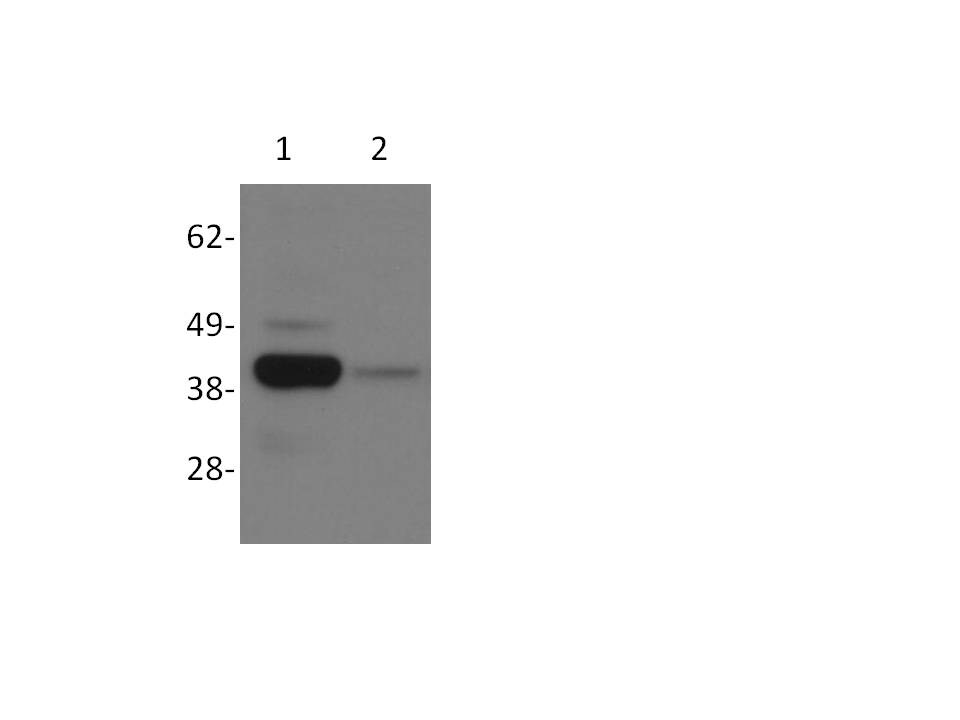
Application: Western BlotSample Tested: Whole cell lysate from UW228 cellsSpecies: HumanVerified Customer | Posted 06/15/2016UW228 cells transfected with MCL1 targeting (Lane 2) or scrambled control siRNAs (Lane 1).

There are no reviews that match your criteria.
Protocols
Find general support by application which include: protocols, troubleshooting, illustrated assays, videos and webinars.
- Antigen Retrieval Protocol (PIER)
- Antigen Retrieval for Frozen Sections Protocol
- Appropriate Fixation of IHC/ICC Samples
- Cellular Response to Hypoxia Protocols
- Chromogenic IHC Staining of Formalin-Fixed Paraffin-Embedded (FFPE) Tissue Protocol
- Chromogenic Immunohistochemistry Staining of Frozen Tissue
- ClariTSA™ Fluorophore Kits
- Detection & Visualization of Antibody Binding
- Fluorescent IHC Staining of Frozen Tissue Protocol
- Graphic Protocol for Heat-induced Epitope Retrieval
- Graphic Protocol for the Preparation and Fluorescent IHC Staining of Frozen Tissue Sections
- Graphic Protocol for the Preparation and Fluorescent IHC Staining of Paraffin-embedded Tissue Sections
- Graphic Protocol for the Preparation of Gelatin-coated Slides for Histological Tissue Sections
- IHC Sample Preparation (Frozen sections vs Paraffin)
- Immunofluorescent IHC Staining of Formalin-Fixed Paraffin-Embedded (FFPE) Tissue Protocol
- Immunohistochemistry (IHC) and Immunocytochemistry (ICC) Protocols
- Immunohistochemistry Frozen Troubleshooting
- Immunohistochemistry Paraffin Troubleshooting
- Immunoprecipitation Protocol
- Preparing Samples for IHC/ICC Experiments
- Preventing Non-Specific Staining (Non-Specific Binding)
- Primary Antibody Selection & Optimization
- Protocol for Heat-Induced Epitope Retrieval (HIER)
- Protocol for Making a 4% Formaldehyde Solution in PBS
- Protocol for VisUCyte™ HRP Polymer Detection Reagent
- Protocol for the Preparation & Fixation of Cells on Coverslips
- Protocol for the Preparation and Chromogenic IHC Staining of Frozen Tissue Sections
- Protocol for the Preparation and Chromogenic IHC Staining of Frozen Tissue Sections - Graphic
- Protocol for the Preparation and Chromogenic IHC Staining of Paraffin-embedded Tissue Sections
- Protocol for the Preparation and Chromogenic IHC Staining of Paraffin-embedded Tissue Sections - Graphic
- Protocol for the Preparation and Fluorescent IHC Staining of Frozen Tissue Sections
- Protocol for the Preparation and Fluorescent IHC Staining of Paraffin-embedded Tissue Sections
- Protocol for the Preparation of Gelatin-coated Slides for Histological Tissue Sections
- R&D Systems Quality Control Western Blot Protocol
- TUNEL and Active Caspase-3 Detection by IHC/ICC Protocol
- The Importance of IHC/ICC Controls
- Troubleshooting Guide: Immunohistochemistry
- Troubleshooting Guide: Western Blot Figures
- Western Blot Conditions
- Western Blot Protocol
- Western Blot Protocol for Cell Lysates
- Western Blot Troubleshooting
- Western Blot Troubleshooting Guide
- View all Protocols, Troubleshooting, Illustrated assays and Webinars
Loading...
Associated Pathways
