Melan-A/MART-1 Antibody (A19-P)
Novus Biologicals | Catalog # NBP1-30151
![Immunohistochemistry-Paraffin: Melan-A/MART-1 Antibody (A19-P) [NBP1-30151]](https://resources.rndsystems.com/images/products/Melan-A-MART-1-Antibody-A19-P-Immunohistochemistry-Paraffin-NBP1-30151-img0012.jpg "Immunohistochemistry-Paraffin: Melan-A/MART-1 Antibody (A19-P) [NBP1-30151]")
Loading...
Key Product Details
Species Reactivity
Validated:
Human, Mouse, Canine
Cited:
Human, Mouse, Canine, Primate - Macaca mulatta (Rhesus Macaque)
Applications
Validated:
Immunohistochemistry, Immunohistochemistry-Paraffin, Immunocytochemistry/ Immunofluorescence
Cited:
Immunohistochemistry-Paraffin, Immunohistochemistry-Frozen, Immunocytochemistry/ Immunofluorescence, IF/IHC
Label
Unconjugated
Antibody Source
Recombinant Monoclonal Rabbit IgG Clone # A19-P
Loading...
Product Specifications
Immunogen
Peptide derived from C-terminal sequence of human Melan-A/MART-1. Antibody recognizes the epitope between Glu105 - Pro115.
Epitope
Glu105 - Pro115
Reactivity Notes
Mouse reactivity reported in scientific literature (PMID: 26547111). Use in Canine reported in secitific publication PMID: 32423311
Specificity
This antibody is specific for the C terminal portions of Melan-A
Clonality
Monoclonal
Host
Rabbit
Isotype
IgG
Description
This antibody is immunoaffinity purified with immunogenic peptide as a ligand.
Scientific Data Images for Melan-A/MART-1 Antibody (A19-P)
Immunohistochemistry-Paraffin: Melan-A/MART-1 Antibody (A19-P) [NBP1-30151]
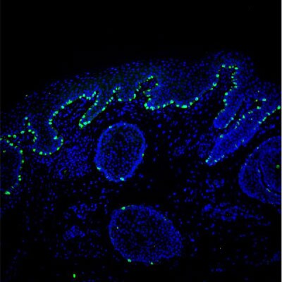
Immunohistochemistry-Paraffin: Melan-A/MART-1 Antibody (A19-P) [NBP1-30151] - Confocal microscopy image of human fetal inner ear tisue section stained with Melan-A antibody (green), 1:200 dilution, incubated over night. IHC-P image submitted by a verified customer review.![Immunohistochemistry-Paraffin: Melan-A/MART-1 Antibody (A19-P) [NBP1-30151]](https://resources.rndsystems.com/images/products/Melan-A-MART-1-Antibody-A19-P-Immunohistochemistry-Paraffin-NBP1-30151-img0011.jpg "Immunohistochemistry-Paraffin: Melan-A/MART-1 Antibody (A19-P) [NBP1-30151]")
Immunohistochemistry-Paraffin: Melan-A/MART-1 Antibody (A19-P) [NBP1-30151]
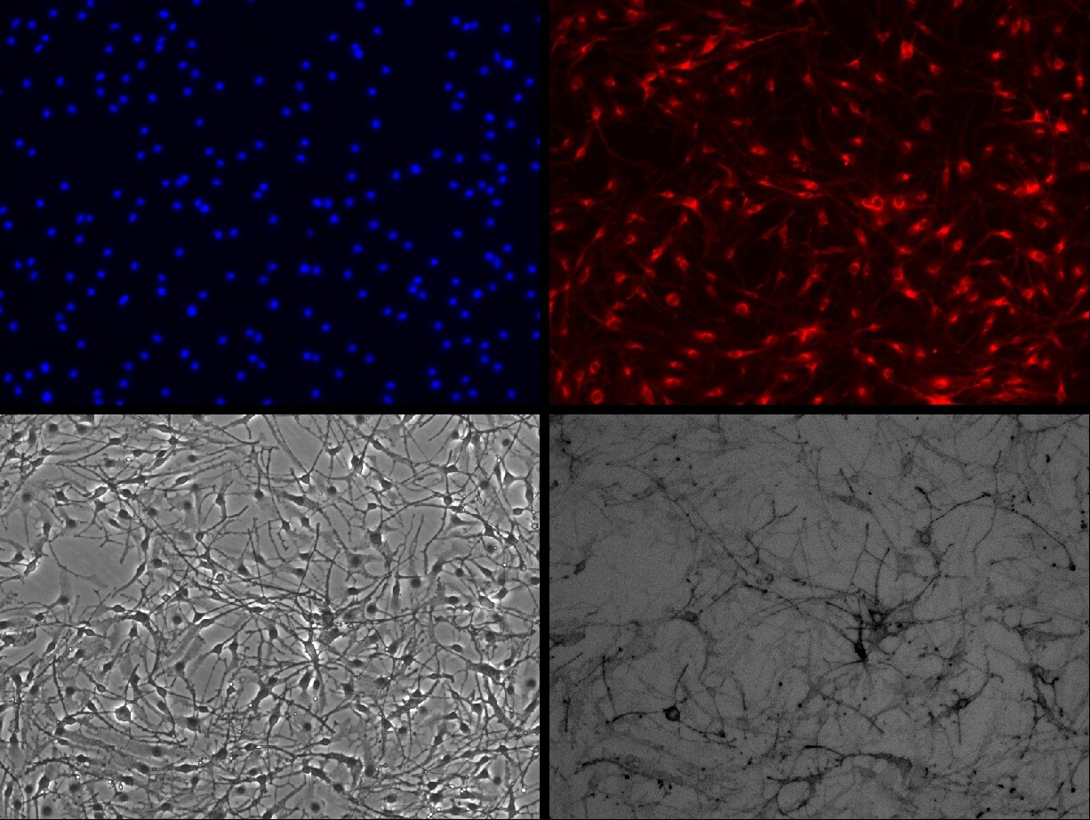
Immunohistochemistry-Paraffin: Melan-A/MART-1 Antibody (A19-P) [NBP1-30151] - Human tissue (4 um section) stained with anti - Melan A monospecific clonal antibody.Applications for Melan-A/MART-1 Antibody (A19-P)
Application
Recommended Usage
Immunohistochemistry
1:100 - 1:200
Immunohistochemistry-Paraffin
1:100 - 1:200
Reviewed Applications
Read 4 reviews rated 4.8 using NBP1-30151 in the following applications:
Formulation, Preparation, and Storage
Purification
Immunogen affinity purified
Formulation
20mM Tris-HCl (pH 8.0) and 20mg/ml BSA
Preservative
0.05% Sodium Azide
Concentration
Please see the vial label for concentration. If unlisted please contact technical services.
Shipping
The product is shipped with polar packs. Upon receipt, store it immediately at the temperature recommended below.
Stability & Storage
Store at 4C. Do not freeze.
Background: Melan-A/MART-1
Alternate Names
Antigen LB39-AA, Antigen SK29-AA, MART-1, MelanA, MLANA
Entrez Gene IDs
2315 (Human)
Gene Symbol
MLANA
UniProt
Additional Melan-A/MART-1 Products
Product Documents for Melan-A/MART-1 Antibody (A19-P)
Certificate of Analysis
To download a Certificate of Analysis, please enter a lot or batch number in the search box below.
Product Specific Notices for Melan-A/MART-1 Antibody (A19-P)
This antibody is immunoaffinity purified with immunogenic peptide as a ligand.
This product is for research use only and is not approved for use in humans or in clinical diagnosis. Primary Antibodies are guaranteed for 1 year from date of receipt.
Related Research Areas
Citations for Melan-A/MART-1 Antibody (A19-P)
 Powered by Bioz
Customer Reviews for Melan-A/MART-1 Antibody (A19-P) (4)
4.8 out of 5
4 Customer Ratings
Have you used Melan-A/MART-1 Antibody (A19-P)?
Submit a review and receive an Amazon gift card!
$25/€18/£15/$25CAN/¥2500 Yen for a review with an image
$10/€7/£6/$10CAN/¥1110 Yen for a review without an image
Submit a review
Customer Images

Showing
1
-
4 of
4 reviews
Showing All
Filter By:
-
Application: Immunohistochemistry-ParaffinSample Tested: human fetal inner earSpecies: HumanVerified Customer | Posted 10/30/2019Confocal image of melan-a rabb (NBP1-30151; in green), diluted 1:200 and incubated o/n on a section of human inner ear (5um).

-
Application: ImmunocytochemistrySample Tested: Mouse melanocyte cell lineSpecies: MouseVerified Customer | Posted 09/02/2013
-
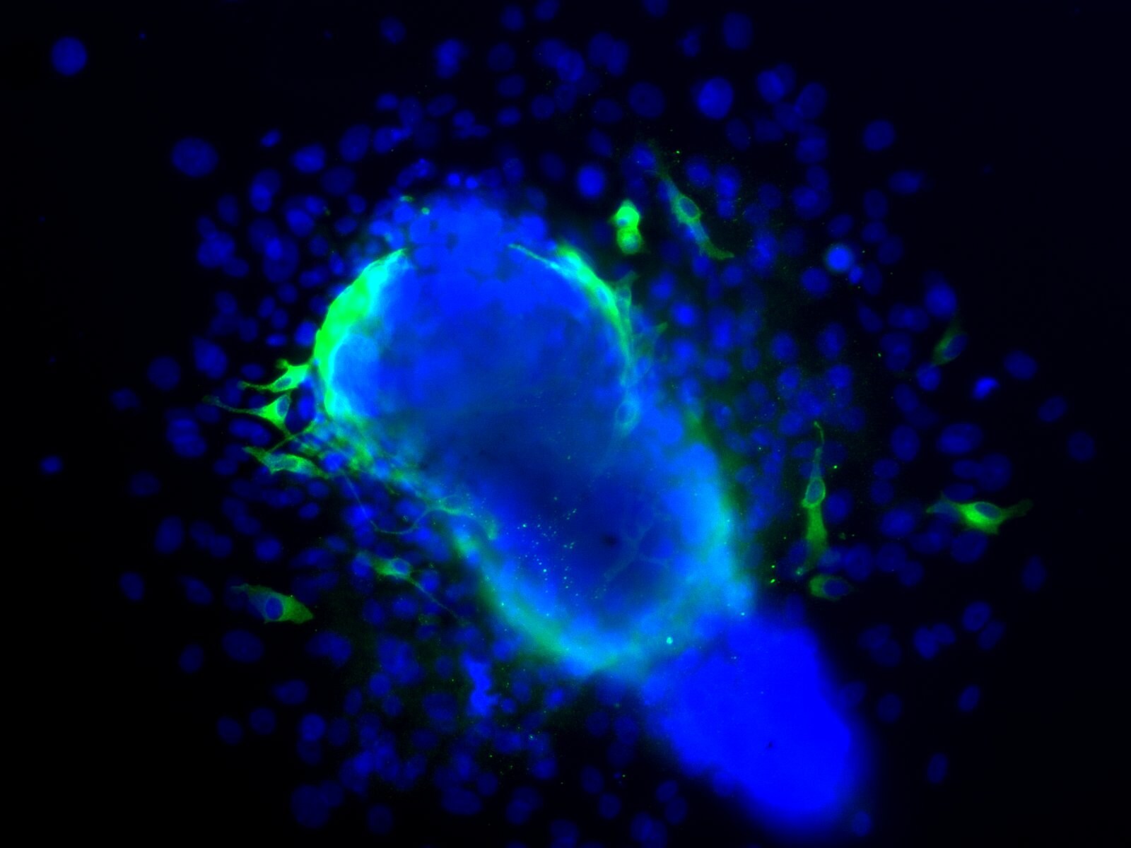
Application: ImmunocytochemistrySample Tested: Human hair follicle cultureSpecies: HumanVerified Customer | Posted 09/02/2013
-
Application: Immunohistochemistry-ParaffinSample Tested: Human adult facial skinSpecies: HumanVerified Customer | Posted 09/02/2013
There are no reviews that match your criteria.
Protocols
Find general support by application which include: protocols, troubleshooting, illustrated assays, videos and webinars.
- Antigen Retrieval Protocol (PIER)
- Antigen Retrieval for Frozen Sections Protocol
- Appropriate Fixation of IHC/ICC Samples
- Cellular Response to Hypoxia Protocols
- Chromogenic IHC Staining of Formalin-Fixed Paraffin-Embedded (FFPE) Tissue Protocol
- Chromogenic Immunohistochemistry Staining of Frozen Tissue
- ClariTSA™ Fluorophore Kits
- Detection & Visualization of Antibody Binding
- Fluorescent IHC Staining of Frozen Tissue Protocol
- Graphic Protocol for Heat-induced Epitope Retrieval
- Graphic Protocol for the Preparation and Fluorescent IHC Staining of Frozen Tissue Sections
- Graphic Protocol for the Preparation and Fluorescent IHC Staining of Paraffin-embedded Tissue Sections
- Graphic Protocol for the Preparation of Gelatin-coated Slides for Histological Tissue Sections
- ICC Cell Smear Protocol for Suspension Cells
- ICC Immunocytochemistry Protocol Videos
- ICC for Adherent Cells
- IHC Sample Preparation (Frozen sections vs Paraffin)
- Immunocytochemistry (ICC) Protocol
- Immunocytochemistry Troubleshooting
- Immunofluorescence of Organoids Embedded in Cultrex Basement Membrane Extract
- Immunofluorescent IHC Staining of Formalin-Fixed Paraffin-Embedded (FFPE) Tissue Protocol
- Immunohistochemistry (IHC) and Immunocytochemistry (ICC) Protocols
- Immunohistochemistry Frozen Troubleshooting
- Immunohistochemistry Paraffin Troubleshooting
- Preparing Samples for IHC/ICC Experiments
- Preventing Non-Specific Staining (Non-Specific Binding)
- Primary Antibody Selection & Optimization
- Protocol for Heat-Induced Epitope Retrieval (HIER)
- Protocol for Making a 4% Formaldehyde Solution in PBS
- Protocol for VisUCyte™ HRP Polymer Detection Reagent
- Protocol for the Fluorescent ICC Staining of Cell Smears - Graphic
- Protocol for the Fluorescent ICC Staining of Cultured Cells on Coverslips - Graphic
- Protocol for the Preparation & Fixation of Cells on Coverslips
- Protocol for the Preparation and Chromogenic IHC Staining of Frozen Tissue Sections
- Protocol for the Preparation and Chromogenic IHC Staining of Frozen Tissue Sections - Graphic
- Protocol for the Preparation and Chromogenic IHC Staining of Paraffin-embedded Tissue Sections
- Protocol for the Preparation and Chromogenic IHC Staining of Paraffin-embedded Tissue Sections - Graphic
- Protocol for the Preparation and Fluorescent ICC Staining of Cells on Coverslips
- Protocol for the Preparation and Fluorescent ICC Staining of Non-adherent Cells
- Protocol for the Preparation and Fluorescent ICC Staining of Stem Cells on Coverslips
- Protocol for the Preparation and Fluorescent IHC Staining of Frozen Tissue Sections
- Protocol for the Preparation and Fluorescent IHC Staining of Paraffin-embedded Tissue Sections
- Protocol for the Preparation of Gelatin-coated Slides for Histological Tissue Sections
- Protocol for the Preparation of a Cell Smear for Non-adherent Cell ICC - Graphic
- TUNEL and Active Caspase-3 Detection by IHC/ICC Protocol
- The Importance of IHC/ICC Controls
- Troubleshooting Guide: Immunohistochemistry
- View all Protocols, Troubleshooting, Illustrated assays and Webinars
Loading...