
Key Product Details
Species Reactivity
Validated:
Mouse
Cited:
Mouse, Transgenic Mouse, Xenograft
Applications
Validated:
Immunohistochemistry, Flow Cytometry, Immunoprecipitation, Cell depletion, Costimulation of T Cells, Inhibition of T Cell Function, CyTOF-ready
Cited:
Immunohistochemistry, Immunohistochemistry-Frozen, Neutralization, Flow Cytometry, Immunocytochemistry/ Immunofluorescence, Cell Selection, In vivo assay
Label
Unconjugated
Antibody Source
Monoclonal Rat IgG2B Clone # GK1.5
Loading...
Product Specifications
Immunogen
Mouse CTL clone V4
Specificity
Reacts with mouse CD4, an antigen co-receptor on the T cell surface which interacts with MHC II molecules on antigen presenting cells.
Clonality
Monoclonal
Host
Rat
Isotype
IgG2B
Endotoxin Level
<0.10 EU per 1 μg of the antibody by the LAL method.
Scientific Data Images for Mouse CD4 Antibody (GK1.5)
CD4 in Mouse Intestine.
CD4 was detected in immersion fixed frozen sections of mouse intestine using Rat Anti-Mouse CD4 Monoclonal Antibody (Catalog # MAB554) at 1 µg/mL overnight at 4 °C. Tissue was stained using the Anti-Rat HRP-DAB Cell & Tissue Staining Kit (brown; Catalog # CTS017) and counterstained with hematoxylin (blue). Specific staining was localized to plasma membranes of lymphocytes. View our protocol for Chromogenic IHC Staining of Frozen Tissue Sections.
Detection of CD4 by Flow Cytometry
T cells and Con A-induced hepatitis. Isotype, CD4 and/or CD8 mAbs (100 µg per mouse) was administrated i.p. to Balb/c mice (n = 8 per group) 24h before Con A injection (20 mg kg–1). The effect of anti-CD4 orCD-8 antibodies administration on depleting these cells in the liver of mice prior injecting Con A was determined by FACS. Representative results were shown in (A). Sera were collected 24 h after Con A injection. Serum ALT (B) and AST (C) levels were measured. The results were presented as the mean ± SD of three separate experiments. ***, p<0.001 vs control. (D) The livers were removed 24 h later. Paraffin sections were stained with hematoxylin and eosin (H&E) staining. Representative liver sections were shown for each group, original magnification: ×200. Image collected and cropped by CiteAb from the following open publication (https://pubmed.ncbi.nlm.nih.gov/23118999), licensed under a CC-BY license. Not internally tested by R&D Systems.
Detection of CD4 by Immunohistochemistry
T cells and Con A-induced hepatitis. Isotype, CD4 and/or CD8 mAbs (100 µg per mouse) was administrated i.p. to Balb/c mice (n = 8 per group) 24h before Con A injection (20 mg kg–1). The effect of anti-CD4 orCD-8 antibodies administration on depleting these cells in the liver of mice prior injecting Con A was determined by FACS. Representative results were shown in (A). Sera were collected 24 h after Con A injection. Serum ALT (B) and AST (C) levels were measured. The results were presented as the mean ± SD of three separate experiments. ***, p<0.001 vs control. (D) The livers were removed 24 h later. Paraffin sections were stained with hematoxylin and eosin (H&E) staining. Representative liver sections were shown for each group, original magnification: ×200. Image collected and cropped by CiteAb from the following open publication (https://pubmed.ncbi.nlm.nih.gov/23118999), licensed under a CC-BY license. Not internally tested by R&D Systems.Applications for Mouse CD4 Antibody (GK1.5)
Application
Recommended Usage
Cell depletion
Dialynas, D.P. et al. (1983) Immunol. Rev. 74:29; Martin, P. et al. (2000) Blood 96:2511.
Costimulation of T Cells
Bosselut, R. et al. (1999) J. Exp. Med. 190:1517.
CyTOF-ready
Ready to be labeled using established conjugation methods. No BSA or other carrier proteins that could interfere with conjugation.
Flow Cytometry
2.5 µg/106 cells
Sample: Mouse splenocytes
Sample: Mouse splenocytes
Immunohistochemistry
1-25 µg/mL
Sample: Immersion fixed frozen sections of mouse intestine
Sample: Immersion fixed frozen sections of mouse intestine
Immunoprecipitation
Dialynas, D.P. et al. (1983) J. Immunol. 131:2445.
Inhibition of T Cell Function
Dialynas, D.P. et al. (1983) J. Immunol. 131:2445; Dialynas, D.P. et al. (1983) Immunol. Rev. 74:29; Kruman, I.I. et al. (1996) Cell. Immunol. 173:236; Grakoui, A. et al. (1999) Science 285:221.
Reviewed Applications
Read 2 reviews rated 4.5 using MAB554 in the following applications:
Flow Cytometry Panel Builder
Bio-Techne Knows Flow Cytometry
Save time and reduce costly mistakes by quickly finding compatible reagents using the Panel Builder Tool.
Advanced Features
- Spectra Viewer - Custom analysis of spectra from multiple fluorochromes
- Spillover Popups - Visualize the spectra of individual fluorochromes
- Antigen Density Selector - Match fluorochrome brightness with antigen density

Formulation, Preparation, and Storage
Purification
Protein A or G purified from hybridoma culture supernatant
Reconstitution
Reconstitute at 0.5 mg/mL in sterile PBS. For liquid material, refer to CoA for concentration.
Loading...
Formulation
Lyophilized from a 0.2 μm filtered solution in PBS with Trehalose. *Small pack size (SP) is supplied either lyophilized or as a 0.2 µm filtered solution in PBS.
Shipping
Lyophilized product is shipped at ambient temperature. Liquid small pack size (-SP) is shipped with polar packs. Upon receipt, store immediately at the temperature recommended below.
Stability & Storage
Use a manual defrost freezer and avoid repeated freeze-thaw cycles.
- 12 months from date of receipt, -20 to -70 °C as supplied.
- 1 month, 2 to 8 °C under sterile conditions after reconstitution.
- 6 months, -20 to -70 °C under sterile conditions after reconstitution.
Calculators
Background: CD4
Alternate Names
CD4
Entrez Gene IDs
Gene Symbol
CD4
Additional CD4 Products
Product Documents for Mouse CD4 Antibody (GK1.5)
Certificate of Analysis
To download a Certificate of Analysis, please enter a lot or batch number in the search box below.
Note: Certificate of Analysis not available for kit components.
Product Specific Notices for Mouse CD4 Antibody (GK1.5)
For research use only
Citations for Mouse CD4 Antibody (GK1.5)
 Powered by Bioz
Customer Reviews for Mouse CD4 Antibody (GK1.5) (2)
4.5 out of 5
2 Customer Ratings
Have you used Mouse CD4 Antibody (GK1.5)?
Submit a review and receive an Amazon gift card!
$25/€18/£15/$25CAN/¥2500 Yen for a review with an image
$10/€7/£6/$10CAN/¥1110 Yen for a review without an image
Submit a review
Customer Images
Showing
1
-
2 of
2 reviews
Showing All
Filter By:
-
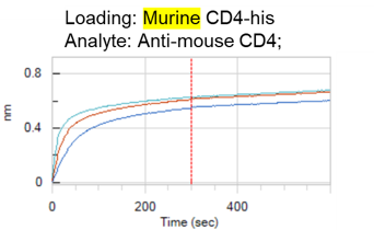
Application: Protein:Protein interactionSample Tested: Recombinant proteinSpecies: MouseVerified Customer | Posted 08/24/2021Binds to murine CD4-his. Immobilize CD4-his on Penta-his sensors, and flow 3 conc of Anti-murine CD4 (100, 50, 25nM). Specific binding of Anti-murine CD4 to murine CD4, no binding to human CD4.

-
Application: Flow CytometrySample Tested: peritoneal granulocytesSpecies: MouseVerified Customer | Posted 05/30/2017
There are no reviews that match your criteria.
Protocols
Find general support by application which include: protocols, troubleshooting, illustrated assays, videos and webinars.
- 7-Amino Actinomycin D (7-AAD) Cell Viability Flow Cytometry Protocol
- Antigen Retrieval Protocol (PIER)
- Antigen Retrieval for Frozen Sections Protocol
- Appropriate Fixation of IHC/ICC Samples
- Cellular Response to Hypoxia Protocols
- Chromogenic IHC Staining of Formalin-Fixed Paraffin-Embedded (FFPE) Tissue Protocol
- Chromogenic Immunohistochemistry Staining of Frozen Tissue
- ClariTSA™ Fluorophore Kits
- Detection & Visualization of Antibody Binding
- Extracellular Membrane Flow Cytometry Protocol
- Flow Cytometry Protocol for Cell Surface Markers
- Flow Cytometry Protocol for Staining Membrane Associated Proteins
- Flow Cytometry Staining Protocols
- Flow Cytometry Troubleshooting Guide
- Fluorescent IHC Staining of Frozen Tissue Protocol
- Graphic Protocol for Heat-induced Epitope Retrieval
- Graphic Protocol for the Preparation and Fluorescent IHC Staining of Frozen Tissue Sections
- Graphic Protocol for the Preparation and Fluorescent IHC Staining of Paraffin-embedded Tissue Sections
- Graphic Protocol for the Preparation of Gelatin-coated Slides for Histological Tissue Sections
- IHC Sample Preparation (Frozen sections vs Paraffin)
- Immunofluorescent IHC Staining of Formalin-Fixed Paraffin-Embedded (FFPE) Tissue Protocol
- Immunohistochemistry (IHC) and Immunocytochemistry (ICC) Protocols
- Immunohistochemistry Frozen Troubleshooting
- Immunohistochemistry Paraffin Troubleshooting
- Immunoprecipitation Protocol
- Intracellular Flow Cytometry Protocol Using Alcohol (Methanol)
- Intracellular Flow Cytometry Protocol Using Detergents
- Intracellular Nuclear Staining Flow Cytometry Protocol Using Detergents
- Intracellular Staining Flow Cytometry Protocol Using Alcohol Permeabilization
- Intracellular Staining Flow Cytometry Protocol Using Detergents to Permeabilize Cells
- Preparing Samples for IHC/ICC Experiments
- Preventing Non-Specific Staining (Non-Specific Binding)
- Primary Antibody Selection & Optimization
- Propidium Iodide Cell Viability Flow Cytometry Protocol
- Protocol for Heat-Induced Epitope Retrieval (HIER)
- Protocol for Liperfluo
- Protocol for Making a 4% Formaldehyde Solution in PBS
- Protocol for VisUCyte™ HRP Polymer Detection Reagent
- Protocol for the Characterization of Human Th22 Cells
- Protocol for the Characterization of Human Th9 Cells
- Protocol for the Preparation & Fixation of Cells on Coverslips
- Protocol for the Preparation and Chromogenic IHC Staining of Frozen Tissue Sections
- Protocol for the Preparation and Chromogenic IHC Staining of Frozen Tissue Sections - Graphic
- Protocol for the Preparation and Chromogenic IHC Staining of Paraffin-embedded Tissue Sections
- Protocol for the Preparation and Chromogenic IHC Staining of Paraffin-embedded Tissue Sections - Graphic
- Protocol for the Preparation and Fluorescent IHC Staining of Frozen Tissue Sections
- Protocol for the Preparation and Fluorescent IHC Staining of Paraffin-embedded Tissue Sections
- Protocol for the Preparation of Gelatin-coated Slides for Histological Tissue Sections
- Protocol: Annexin V and PI Staining by Flow Cytometry
- Protocol: Annexin V and PI Staining for Apoptosis by Flow Cytometry
- TUNEL and Active Caspase-3 Detection by IHC/ICC Protocol
- The Importance of IHC/ICC Controls
- Troubleshooting Guide: Fluorokine Flow Cytometry Kits
- Troubleshooting Guide: Immunohistochemistry
- View all Protocols, Troubleshooting, Illustrated assays and Webinars


