Mouse glial cell line-derived neurotrophic factor (GDNF) family receptor alpha 3 (GFR alpha -3) is a member of the GDNF family of receptors. It is one of four known members, all of which contain three conserved cysteine repeats (1, 3, 4). It is synthesized as a 397 aa precursor, with a hydrophobic signal sequence of 28 aa, a 343 aa mature segment, and a propeptide segment of 26 aa that is removed in the mature protein. The mature globular membrane glycoprotein has an approximate molecular weight of 50 kDa (5), contains three potential N-linked glycosylation sites, and a hydrophobic stretch of residues at its COOH terminus that comprises a GPI linkage sequence (2, 3, 5). Mouse GFR alpha -3 shares 81% aa sequence identity with human GFR alpha -3. High-level expression of GFR alpha -3 is observed only during early stages of neurogenesis in the central nervous system and in developing and adult peripheral nerves, organs, and ganglia (2, 3, 5, 7). The expression and proportions of GFR alpha -3 are closely linked to those of the Ret receptor tyrosine kinase, particularly in the trigeminal ganglion, pituitary gland, thymus, lung, and duodenum (3). GFR alpha -3 associates with Ret to form the receptor complex for the GDNF family ligand artemin, which, in turn, activates the complex (1, 6, 7). The activated complex’s signal is required for the development and maintenance of superior cervical ganglion (SCG), specifically the rostral migration of SCG precursors between embryonic days 11.5 and 14.5, and the survival of SCG neurons after birth (8).
Mouse GFR alpha-3/GDNF R alpha-3 Antibody
R&D Systems | Catalog # AF2645

Key Product Details
Species Reactivity
Validated:
Mouse
Cited:
Mouse, Rat
Applications
Validated:
Immunohistochemistry, Western Blot
Cited:
Immunohistochemistry, Immunohistochemistry-Frozen, Western Blot, Neutralization, Immunocytochemistry
Label
Unconjugated
Antibody Source
Polyclonal Goat IgG
Loading...
Product Specifications
Immunogen
S. frugiperda insect ovarian cell line Sf 21-derived recombinant mouse GFR alpha -3/GDNF R alpha -3
Glu34-Arg379
Accession # AAB70931
Glu34-Arg379
Accession # AAB70931
Specificity
Detects mouse GFR alpha -3/GDNF R alpha -3 in direct ELISAs and Western blots. In direct ELISAs, approximately 10% cross-reactivity with recombinant human GFR alpha -3 is observed.
Clonality
Polyclonal
Host
Goat
Isotype
IgG
Scientific Data Images for Mouse GFR alpha-3/GDNF R alpha-3 Antibody
GFR alpha ‑3/GDNF R alpha ‑3 in Mouse Embryo.
GFRa‑3/GDNF Ra‑3 was detected in immersion fixed frozen sections of mouse embryo (15 d.p.c.) using 15 µg/mL Goat Anti-Mouse GFRa‑3/GDNF Ra‑3 Antigen Affinity-purified Polyclonal Antibody (Catalog # AF2645) overnight at 4 °C. Tissue was stained with the Anti-Goat HRP-DAB Cell & Tissue Staining Kit (brown; Catalog # CTS008) and counterstained with hematoxylin (blue). Specific labeling was localized to developing dorsal root ganglion and spinal cord. View our protocol for Chromogenic IHC Staining of Frozen Tissue Sections.Applications for Mouse GFR alpha-3/GDNF R alpha-3 Antibody
Application
Recommended Usage
Immunohistochemistry
5-15 µg/mL
Sample: Immersion fixed frozen sections of mouse embryo (15 d.p.c.)
Sample: Immersion fixed frozen sections of mouse embryo (15 d.p.c.)
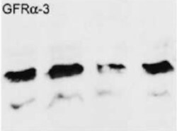
Western Blot
0.1 µg/mL
Sample: Recombinant Mouse GFR alpha ‑3/GDNF R alpha ‑3 Fc Chimera (Catalog # 2645-FR)
Sample: Recombinant Mouse GFR alpha ‑3/GDNF R alpha ‑3 Fc Chimera (Catalog # 2645-FR)
Reviewed Applications
Read 2 reviews rated 5 using AF2645 in the following applications:
Formulation, Preparation, and Storage
Purification
Antigen Affinity-purified
Reconstitution
Reconstitute at 0.2 mg/mL in sterile PBS. For liquid material, refer to CoA for concentration.
Loading...
Formulation
Lyophilized from a 0.2 μm filtered solution in PBS with Trehalose. *Small pack size (SP) is supplied either lyophilized or as a 0.2 µm filtered solution in PBS.
Shipping
Lyophilized product is shipped at ambient temperature. Liquid small pack size (-SP) is shipped with polar packs. Upon receipt, store immediately at the temperature recommended below.
Stability & Storage
Use a manual defrost freezer and avoid repeated freeze-thaw cycles.
- 12 months from date of receipt, -20 to -70 °C as supplied.
- 1 month, 2 to 8 °C under sterile conditions after reconstitution.
- 6 months, -20 to -70 °C under sterile conditions after reconstitution.
Calculators
Background: GFR alpha-3/GDNF R alpha-3
References
- Xinquan, W. et al. (2006) Structure 14:1083.
- Worby, C. et al. (1998) J. Biol. Chem. 273:3502.
- Naveilhan, P. et al. (1998) Proc. Natl. Acad. Sci. USA 95:1295.
- Nomoto, S. et al. (1998) Biochem. Biophys. Res. Commun. 244:849.
- Baloh, R. et al. (1998) Proc. Natl. Acad. Sci. USA 95:5801.
- Saarma, M. (2000) Eur. J. Biochem. 267:6968.
- Baloh, R. et al. (1998) Neuron 21:1291.
- Nishino, J et al. (1999) Neuron 23:725.
Long Name
Glial Cell line-derived Neurotrophic Factor Receptor alpha 3
Alternate Names
GDNF R alpha-3, GFR alpha3, GFRa-3, GFRA3
Gene Symbol
GFRA3
UniProt
Additional GFR alpha-3/GDNF R alpha-3 Products
Product Documents for Mouse GFR alpha-3/GDNF R alpha-3 Antibody
Certificate of Analysis
To download a Certificate of Analysis, please enter a lot or batch number in the search box below.
Note: Certificate of Analysis not available for kit components.
Product Specific Notices for Mouse GFR alpha-3/GDNF R alpha-3 Antibody
For research use only
Related Research Areas
Citations for Mouse GFR alpha-3/GDNF R alpha-3 Antibody
 Powered by Bioz
Customer Reviews for Mouse GFR alpha-3/GDNF R alpha-3 Antibody (2)
5 out of 5
2 Customer Ratings
Have you used Mouse GFR alpha-3/GDNF R alpha-3 Antibody?
Submit a review and receive an Amazon gift card!
$25/€18/£15/$25CAN/¥2500 Yen for a review with an image
$10/€7/£6/$10CAN/¥1110 Yen for a review without an image
Submit a review
Customer Images
Showing
1
-
2 of
2 reviews
Showing All
Filter By:
-
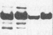
Application: Western BlotSample Tested: Dorsal root ganglionSpecies: MouseVerified Customer | Posted 07/21/2021

-
Application: Western BlotSample Tested: Brain (hippocampus)Species: MouseVerified Customer | Posted 04/28/2021
There are no reviews that match your criteria.
Protocols
Find general support by application which include: protocols, troubleshooting, illustrated assays, videos and webinars.
- Antigen Retrieval Protocol (PIER)
- Antigen Retrieval for Frozen Sections Protocol
- Appropriate Fixation of IHC/ICC Samples
- Cellular Response to Hypoxia Protocols
- Chromogenic IHC Staining of Formalin-Fixed Paraffin-Embedded (FFPE) Tissue Protocol
- Chromogenic Immunohistochemistry Staining of Frozen Tissue
- ClariTSA™ Fluorophore Kits
- Detection & Visualization of Antibody Binding
- Fluorescent IHC Staining of Frozen Tissue Protocol
- Graphic Protocol for Heat-induced Epitope Retrieval
- Graphic Protocol for the Preparation and Fluorescent IHC Staining of Frozen Tissue Sections
- Graphic Protocol for the Preparation and Fluorescent IHC Staining of Paraffin-embedded Tissue Sections
- Graphic Protocol for the Preparation of Gelatin-coated Slides for Histological Tissue Sections
- IHC Sample Preparation (Frozen sections vs Paraffin)
- Immunofluorescent IHC Staining of Formalin-Fixed Paraffin-Embedded (FFPE) Tissue Protocol
- Immunohistochemistry (IHC) and Immunocytochemistry (ICC) Protocols
- Immunohistochemistry Frozen Troubleshooting
- Immunohistochemistry Paraffin Troubleshooting
- Preparing Samples for IHC/ICC Experiments
- Preventing Non-Specific Staining (Non-Specific Binding)
- Primary Antibody Selection & Optimization
- Protocol for Heat-Induced Epitope Retrieval (HIER)
- Protocol for Making a 4% Formaldehyde Solution in PBS
- Protocol for VisUCyte™ HRP Polymer Detection Reagent
- Protocol for the Preparation & Fixation of Cells on Coverslips
- Protocol for the Preparation and Chromogenic IHC Staining of Frozen Tissue Sections
- Protocol for the Preparation and Chromogenic IHC Staining of Frozen Tissue Sections - Graphic
- Protocol for the Preparation and Chromogenic IHC Staining of Paraffin-embedded Tissue Sections
- Protocol for the Preparation and Chromogenic IHC Staining of Paraffin-embedded Tissue Sections - Graphic
- Protocol for the Preparation and Fluorescent IHC Staining of Frozen Tissue Sections
- Protocol for the Preparation and Fluorescent IHC Staining of Paraffin-embedded Tissue Sections
- Protocol for the Preparation of Gelatin-coated Slides for Histological Tissue Sections
- R&D Systems Quality Control Western Blot Protocol
- TUNEL and Active Caspase-3 Detection by IHC/ICC Protocol
- The Importance of IHC/ICC Controls
- Troubleshooting Guide: Immunohistochemistry
- Troubleshooting Guide: Western Blot Figures
- Western Blot Conditions
- Western Blot Protocol
- Western Blot Protocol for Cell Lysates
- Western Blot Troubleshooting
- Western Blot Troubleshooting Guide
- View all Protocols, Troubleshooting, Illustrated assays and Webinars
Loading...