
Key Product Details
Species Reactivity
Validated:
Mouse
Cited:
Human, Mouse
Applications
Validated:
Immunohistochemistry, Western Blot
Cited:
Immunohistochemistry, Immunohistochemistry-Paraffin, Western Blot, Immunocytochemistry, Immunoprecipitation
Label
Unconjugated
Antibody Source
Polyclonal Goat IgG
Loading...
Product Specifications
Immunogen
Mouse myeloma cell line NS0-derived recombinant mouse Lumican
Gln19-Asn338, predicted
Accession # P51885
Gln19-Asn338, predicted
Accession # P51885
Specificity
Detects mouse Lumican in direct ELISAs and Western blots.
Clonality
Polyclonal
Host
Goat
Isotype
IgG
Scientific Data Images for Mouse Lumican Antibody
Lumican in Mouse Intestine.
Lumican was detected in perfusion fixed frozen sections of mouse intestine using Goat Anti-Mouse Lumican Antigen Affinity-purified Polyclonal Antibody (Catalog # AF2745) at 15 µg/mL overnight at 4 °C. Tissue was stained using the Anti-Goat HRP-DAB Cell & Tissue Staining Kit (brown; CTS008) and counterstained with hematoxylin (blue). Lower panel shows a lack of labeling if primary antibodies are omitted and tissue is stained only with secondary antibody followed by incubation with detection reagents. View our protocol for Chromogenic IHC Staining of Frozen Tissue Sections.
Detection of Mouse Lumican by Immunohistochemistry
Immunoperoxidase for lumican. (A) Ovx group: A weak immunoreaction is observed in the endometrial stroma, especially under the luminal epithelium (arrow) and in the deep stroma, as well as in the EML and connective tissue between muscle layers of the myometrium; (B) E2-group: the reaction is present in the whole endometrial stroma and in both layers of the myometrium; (C) MPA-group: the reaction is present in the whole stroma, however it is weak in the subepithelial stroma (asterisk). In the myometrium, deposition of lumican is observed in the EML and connective tissue between layers; (D) E2+MPA-group: the immunoreaction is strong in the whole endometrial stroma. In the myometrium, a weak staining is observed in both IML and EML, being strong in the connective tissue between them. L = uterine lumen; G = endometrial gland; SS = superficial stroma; DS = deep stroma; V = blood vessel; IML = internal muscle layer; EML = external muscle layer. Scale bar: 50 μm. Image collected and cropped by CiteAb from the following publication (https://rbej.biomedcentral.com/articles/10.1186/1477-7827-9-22), licensed under a CC-BY license. Not internally tested by R&D Systems.
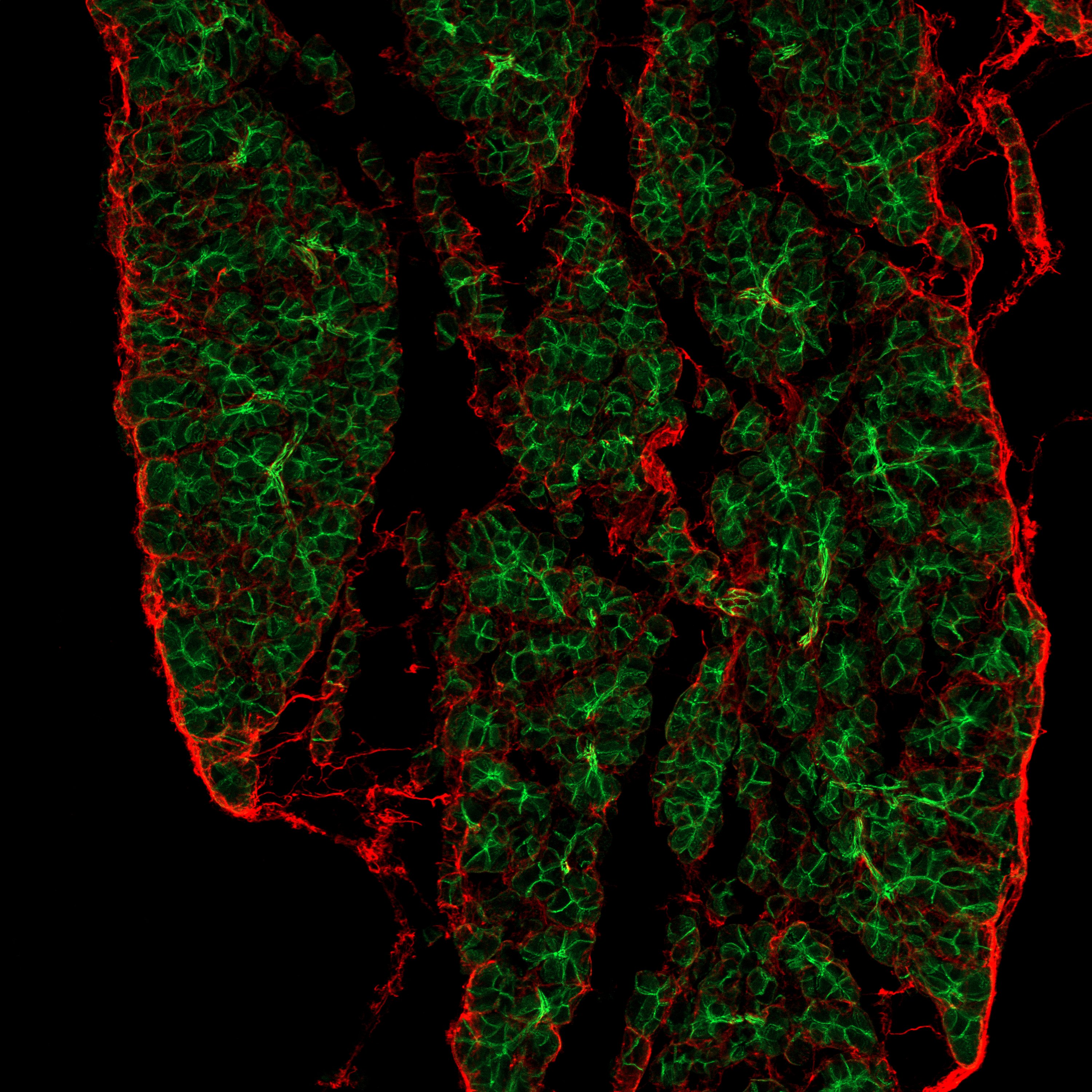
Detection of Mouse Lumican by Immunohistochemistry.
Adult mouse pancreas stained with CD324 (green) and Lumican (red). Image from a verified customer reviewApplications for Mouse Lumican Antibody
Application
Recommended Usage
Immunohistochemistry
5-15 µg/mL
Sample: Perfusion fixed frozen sections of mouse intestine, liver, lung, thymus, and spleen
Sample: Perfusion fixed frozen sections of mouse intestine, liver, lung, thymus, and spleen
Western Blot
0.1 µg/mL
Sample: Recombinant Mouse Lumican (Catalog # 2745-LU)
Sample: Recombinant Mouse Lumican (Catalog # 2745-LU)
Reviewed Applications
Read 1 review rated 5 using AF2745 in the following applications:
Formulation, Preparation, and Storage
Purification
Antigen Affinity-purified
Reconstitution
Reconstitute at 0.2 mg/mL in sterile PBS. For liquid material, refer to CoA for concentration.
Loading...
Formulation
Lyophilized from a 0.2 μm filtered solution in PBS with Trehalose. *Small pack size (SP) is supplied either lyophilized or as a 0.2 µm filtered solution in PBS.
Shipping
Lyophilized product is shipped at ambient temperature. Liquid small pack size (-SP) is shipped with polar packs. Upon receipt, store immediately at the temperature recommended below.
Stability & Storage
Use a manual defrost freezer and avoid repeated freeze-thaw cycles.
- 12 months from date of receipt, -20 to -70 °C as supplied.
- 1 month, 2 to 8 °C under sterile conditions after reconstitution.
- 6 months, -20 to -70 °C under sterile conditions after reconstitution.
Calculators
Background: Lumican
References
- Nikitovic, D. et al. (2008) IUBMB Life 60:818.
- Blochberger, T.C. et al. (1992) J. Biol. Chem. 267:347.
- Chakravarti, S. et al. (1998) J. Cell Biol. 141:1277.
- Chakravarti, S. et al. (2000) Invest. Ophthalmol. Vis. Sci. 41:3365.
- Jepsen, K.J. et al. (2002) J. Biol. Chem. 277:35532.
- Chakravarti, S. et al. (2003) Invest. Ophthalmol. Vis. Sci. 44:2422.
- Vuillermoz, B. et al. (2004) Exp. Cell Res. 296:294
- Nikitovic, D. et al. (2008) FEBS J. 275:350.
- D’Onofrio, M.F. et al. (2008) Biochem. Biophys. Res. Commun. 365:266.
- Ishiwata, T. et al. (2007) Oncol. Rep. 18:537.
Alternate Names
LDC, LUM, SLRR2D
Gene Symbol
LUM
UniProt
Additional Lumican Products
Product Documents for Mouse Lumican Antibody
Certificate of Analysis
To download a Certificate of Analysis, please enter a lot or batch number in the search box below.
Note: Certificate of Analysis not available for kit components.
Product Specific Notices for Mouse Lumican Antibody
For research use only
Related Research Areas
Citations for Mouse Lumican Antibody
 Powered by Bioz
Customer Reviews for Mouse Lumican Antibody (1)
5 out of 5
1 Customer Rating
Have you used Mouse Lumican Antibody?
Submit a review and receive an Amazon gift card!
$25/€18/£15/$25CAN/¥2500 Yen for a review with an image
$10/€7/£6/$10CAN/¥1110 Yen for a review without an image
Submit a review
Customer Images
Showing
1
-
1 of
1 review
Showing All
Filter By:
-
Application: ImmunohistochemistrySample Tested: Adult pancreasSpecies: MouseVerified Customer | Posted 10/14/2025Mouse pancreas stained with CD324 (green) and Lumican (red)

There are no reviews that match your criteria.
Protocols
Find general support by application which include: protocols, troubleshooting, illustrated assays, videos and webinars.
- Antigen Retrieval Protocol (PIER)
- Antigen Retrieval for Frozen Sections Protocol
- Appropriate Fixation of IHC/ICC Samples
- Cellular Response to Hypoxia Protocols
- Chromogenic IHC Staining of Formalin-Fixed Paraffin-Embedded (FFPE) Tissue Protocol
- Chromogenic Immunohistochemistry Staining of Frozen Tissue
- ClariTSA™ Fluorophore Kits
- Detection & Visualization of Antibody Binding
- Fluorescent IHC Staining of Frozen Tissue Protocol
- Graphic Protocol for Heat-induced Epitope Retrieval
- Graphic Protocol for the Preparation and Fluorescent IHC Staining of Frozen Tissue Sections
- Graphic Protocol for the Preparation and Fluorescent IHC Staining of Paraffin-embedded Tissue Sections
- Graphic Protocol for the Preparation of Gelatin-coated Slides for Histological Tissue Sections
- IHC Sample Preparation (Frozen sections vs Paraffin)
- Immunofluorescent IHC Staining of Formalin-Fixed Paraffin-Embedded (FFPE) Tissue Protocol
- Immunohistochemistry (IHC) and Immunocytochemistry (ICC) Protocols
- Immunohistochemistry Frozen Troubleshooting
- Immunohistochemistry Paraffin Troubleshooting
- Preparing Samples for IHC/ICC Experiments
- Preventing Non-Specific Staining (Non-Specific Binding)
- Primary Antibody Selection & Optimization
- Protocol for Heat-Induced Epitope Retrieval (HIER)
- Protocol for Making a 4% Formaldehyde Solution in PBS
- Protocol for VisUCyte™ HRP Polymer Detection Reagent
- Protocol for the Preparation & Fixation of Cells on Coverslips
- Protocol for the Preparation and Chromogenic IHC Staining of Frozen Tissue Sections
- Protocol for the Preparation and Chromogenic IHC Staining of Frozen Tissue Sections - Graphic
- Protocol for the Preparation and Chromogenic IHC Staining of Paraffin-embedded Tissue Sections
- Protocol for the Preparation and Chromogenic IHC Staining of Paraffin-embedded Tissue Sections - Graphic
- Protocol for the Preparation and Fluorescent IHC Staining of Frozen Tissue Sections
- Protocol for the Preparation and Fluorescent IHC Staining of Paraffin-embedded Tissue Sections
- Protocol for the Preparation of Gelatin-coated Slides for Histological Tissue Sections
- R&D Systems Quality Control Western Blot Protocol
- TUNEL and Active Caspase-3 Detection by IHC/ICC Protocol
- The Importance of IHC/ICC Controls
- Troubleshooting Guide: Immunohistochemistry
- Troubleshooting Guide: Western Blot Figures
- Western Blot Conditions
- Western Blot Protocol
- Western Blot Protocol for Cell Lysates
- Western Blot Troubleshooting
- Western Blot Troubleshooting Guide
- View all Protocols, Troubleshooting, Illustrated assays and Webinars
Loading...